|
|
| (8 intermediate revisions by the same user not shown) |
| Line 9: |
Line 9: |
|
| |
|
|
| |
|
| [[Media:BGDA Lecture 20189 - Development of the Nervous System.pdf|'''2019 Lecture PDF''']] ) ''Link to be updated - (Notice removed when completed)''
| | |
|
| |
|
| |} | | |} |
| Line 155: |
Line 155: |
|
| |
|
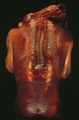
| * Common sites of neural tube defects. | | * Common sites of neural tube defects. |
| | |
| | ====Folate and Neural Development==== |
| | |
| | <br> |
| | {| class="wikitable mw-collapsible mw-collapsed" |
| | ! Folate Requirement |
| | |- |
| | | [[Abnormal Development - Folic Acid and Neural Tube Defects]] |
| | |
| | {| |
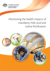
| | | [[File:Monitoring the health impacts of mandatory folic acid and iodine fortification 2016.jpg|200px]] |
| | | Monitoring the health impacts of mandatory folic acid and iodine fortification 2016<ref name=“PHE208”>AIHW 2016. '''Monitoring the health impacts of mandatory folic acid and iodine fortification 2016'''. [http://www.aihw.gov.au/publication-detail/?id=60129555435 Cat. no. '''PHE 208''']. Canberra: AIHW. [http://www.aihw.gov.au/WorkArea/DownloadAsset.aspx?id=60129555568 PDF]</ref> |
| | |
| | Mandatory fortification of bread with folic acid (in Australia) and iodine (in Australia and New Zealand) was introduced in 2009 |
| | * Overall decrease in the rate of neural tube defects (NTDs) by 14.4% |
| | * Teenagers the rate of NTDs decreased by almost 55% |
| | * Aboriginal and Torres Strait Islander women the rate of NTDs decreased by 74% |
| | |} |
| | |
| | {| |
| | |-bgcolor="FFCC00" |
| | ! {{ICD-11}} |
| | |-bgcolor="FEF9E7" |
| | | |
| | {{ICD11weblink}}439233336 5B5E Folate deficiency] - ''Between days 21 and 27 post-conception, the neural plate closes to form what will eventually be the spinal cord and cranium. Spina bifida, anencephaly, and other similar conditions are collectively called NTDs. They result from improper closure of the spinal cord and cranium, respectively, and are the most common congenital abnormalities associated with folate deficiency.'' |
| | |
| | {{ICD11weblink}}215057274 '''LA00-LA0Z''' Structural developmental anomalies of the nervous system] - {{ICD11weblink}}1292761836 LA00.0 Anencephaly] {{ICD11weblink}}1558931335 LA00.1 Iniencephaly] {{ICD11weblink}}546224466 LA00.2 Acephaly] {{ICD11weblink}}154698183 LA00.3 Amyelencephaly] {{ICD11weblink}}2036217905 LA02 Spina bifida] - {{ICD11weblink}}979482551 LA02.0 Spina bifida cystica] {{ICD11weblink}}182894151 LA02.00 Myelomeningocele with hydrocephalus] {{ICD11weblink}}1008004337 LA02.01 Myelomeningocele without hydrocephalus] {{ICD11weblink}}863949070 LA02.02 Myelocystocele] {{ICD11weblink}}187581000 LA02.1 Spina bifida aperta] |
| | |} |
| | |} |
| | |
| ===Neural Crest=== | | ===Neural Crest=== |
| {| | | {| |
| Line 370: |
Line 400: |
| |} | | |} |
|
| |
|
| | ====Thyroid and Neural Development==== |
| | [[File:Human thyroid system and neural development.jpg|600px]] |
| | <br> |
| {| class="wikitable mw-collapsible mw-collapsed" | | {| class="wikitable mw-collapsible mw-collapsed" |
| ! Iodine deficiency | | ! Iodine deficiency |
| Line 415: |
Line 448: |
| Myelination process occurs both in the CNS (from neural tube glia) and also in peripheral nerves (from neural crest Schwann cells). | | Myelination process occurs both in the CNS (from neural tube glia) and also in peripheral nerves (from neural crest Schwann cells). |
| |} | | |} |
| | |
| ==Postnatal== | | ==Postnatal== |
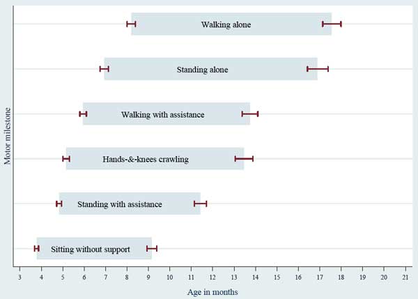
| [[File:WHO motor development milestones.jpg|alt=WHO motor development milestones|link=Neural Exam Movies|600px]] | | [[File:WHO motor development milestones.jpg|alt=WHO motor development milestones|link=Neural Exam Movies|600px]] |
|
| |
|
| [[Neural Exam Movies]] | | [[Neural Exam Movies]] |
| | |
| | {| class="wikitable mw-collapsible mw-collapsed" |
| | ! Additional Information - Multiple Sclerosis |
| | |- |
| | | Only humans spontaneously develop {{multiple sclerosis}} (MS), a chronic demyelinating immune-mediated disease. This disease has an onset generally coinciding with the end of the long-term myelination process and incidence has been recently increasing in female/male (F/M) ratio and occurring in women of childbearing age. |
| | |} |
| | |
| ==Movies== | | ==Movies== |
| {| class="wikitable mw-collapsible mw-collapsed" | | {| class="wikitable mw-collapsible mw-collapsed" |
| Line 477: |
Line 518: |
| | {{Prenatal diagnosis}} | | | {{Prenatal diagnosis}} |
| |} | | |} |
| | <br> |
| | {{BGDA - Neural Development Interactive}} |
| | <br> |
| ==Terms== | | ==Terms== |
| {{Neural terms}} | | {{Neural terms}} |
Introduction
|
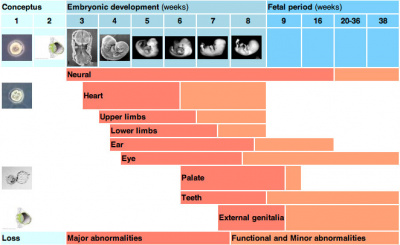
Neural development is a complex and ongoing process that commences in week 3 and continues through into the postnatal period. This lecture will introduce concepts about the timing, origin and abnormalities of the nervous system.
Final lecture content will be added to this current page, the linked online textbook chapters are available as pre-reading for this lecture.
|
Aim
To develop an understanding of the development of the nervous system and the consequences of abnormal development.
Textbooks
| Textbooks
|
2018 PDF
UNSW Embryology
The Developing Human: Clinically Oriented Embryology
Moore, K.L., Persaud, T.V.N. & Torchia, M.G. (2015). The developing human: clinically oriented embryology (10th ed.). Philadelphia: Saunders. (links only function with UNSW connection)
Larsen's Human Embryology
Schoenwolf, G.C., Bleyl, S.B., Brauer, P.R., Francis-West, P.H. & Philippa H. (2015). Larsen's human embryology (5th ed.). New York; Edinburgh: Churchill Livingstone.(links only function with UNSW connection)
Neuroscience
More Textbooks?
|
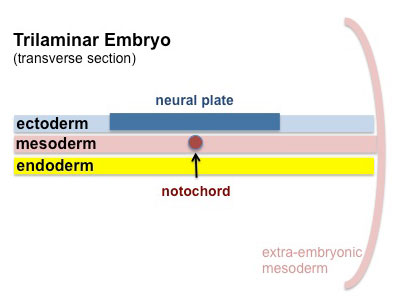
Week 3
|
<html5media height="520" width="320">File:Neuralplate_001.mp4</html5media>
|
Ectoderm
- neural plate - midline (columnar cells)
- neural crest - outside lateral edges of neural plate
- surface ectoderm - lateral (cuboidal cells)
- head - sensory and anterior pituitary (placodes)
- integument - epidermis of skin, hair, glands, teeth enamel
Neural Plate
- extends from buccopharyngeal membrane (oral membrane) to primitive node (Hensen's node)
- forms above notochord and paraxial mesoderm
- neuroectodermal cells - neural plate, neural crest
- rostrocaudal width
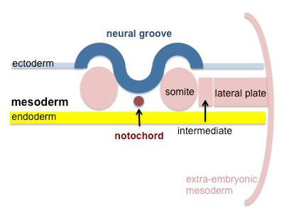
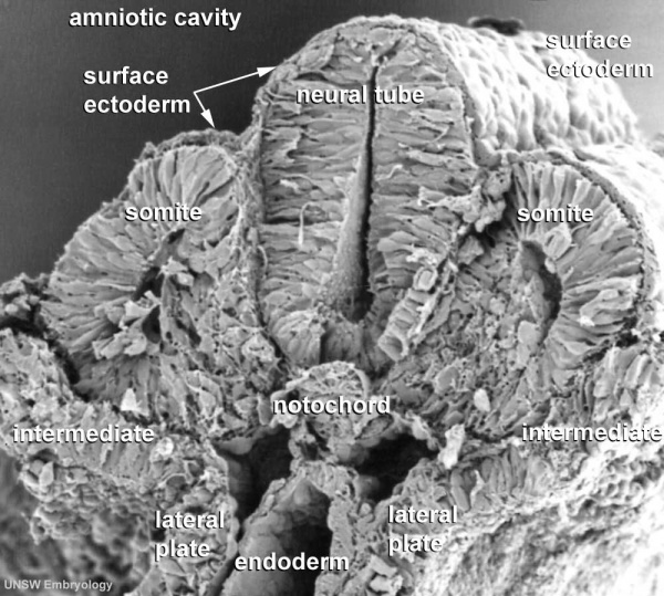
Week 4
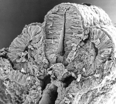
Neural Tube
| neural groove
|
neural tube and neural crest
|
|

|
| <html5media height="480" width="480">File:Neuraltube_001.mp4</html5media>
|
<html5media height="440" width="380">File:Mouse neural tube 01.mp4</html5media>
|
Neuropores
Cranial neuropore (cephalic, rostral or anterior) closes about 24 days post-fertilization.
Caudal neuropore (posterior) closes about 28 days post-fertilization.
- Common sites of neural tube defects.
Folate and Neural Development
| Folate Requirement
|
Abnormal Development - Folic Acid and Neural Tube Defects
|
Monitoring the health impacts of mandatory folic acid and iodine fortification 2016[1]
Mandatory fortification of bread with folic acid (in Australia) and iodine (in Australia and New Zealand) was introduced in 2009
- Overall decrease in the rate of neural tube defects (NTDs) by 14.4%
- Teenagers the rate of NTDs decreased by almost 55%
- Aboriginal and Torres Strait Islander women the rate of NTDs decreased by 74%
|
|
Neural Crest
|
|
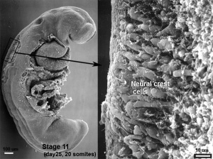
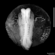
Human embryo neural crest cells (Week 4, Stage 11)
Neural crest (acoustico-facial primordium)
|
| <html5media height="380" width="410">File:Chicken-neural crest migration 01.mp4</html5media>
Chicken neural crest cell migration into pharyngeal arches.
|
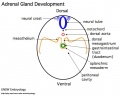
| <html5media height="400" width="400">File:Adrenal medulla.mp4</html5media>
|
Cartoon shows example of some neural crest medial migration and structures formed at the level of the body.
- Cells staying dorsal to neural tube - dorsal root ganglia (DRG)
- Cells migrating ventral to neural tube - sympathetic ganglia
- Cells migrating peritoneal cavity wall - adrenal medulla
- Cells migrate into GIT wall - enteric nervous system
|
|
Neural Crest Origin
| System
|
Cell Type
|
| Peripheral Nervous System (PNS)
|
Neurons - sensory ganglia, sympathetic and parasympathetic ganglia, enteric nervous system, and plexuses
Glia (neuroglial cells) - Schwann cells[2], satellite cells, olfactory ensheathing cells[3]
|
| endocrine
|
Adrenal medulla
Calcitonin-secreting cells
Carotid body type I cells
|
| integumentary
|
Epidermal pigment cells melanocyte
|
| Facial cartilage and bone
|
Facial and anterior ventral skull cartilage and bones
|
| Sensory
|
inner ear, cornea endothelium and stroma
|
| Connective tissue
|
tooth odontoblast
smooth muscle, and adipose tissue of skin in head and neck
Connective tissue of meninges, salivary, lachrymal, thymus, thyroid, and pituitary glands
Connective tissue and smooth muscle in arteries of aortic arch origin
|
| Links: neural crest | Category:Neural Crest | Neural Crest collapsible table
|
neural crest
Primary Brain Vesicles
Traditional vesicle description (simplified name and alternate neuromere description in brackets)
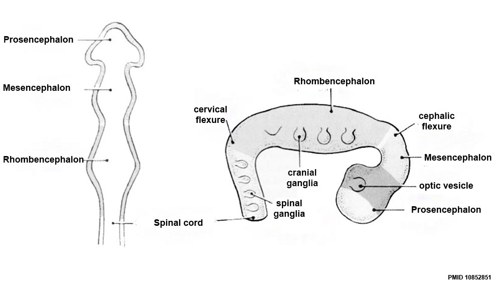
Brain
- Prosencephalon (forebrain, prosomeres)
- Mesencephalon (midbrain, mesomeres)
- Rhombencephalon (hindbrain, rhombomeres)
Spinal Cord

| Neural Tube Regions
|
| Neural Tube Early Structures
|
| Neural Tube (stage 11)
|
Regions
|
Anatomical location
|
Patterning region
|
|
roof plate
|
dorsal
|
surface ectoderm
|
| alar plate
|
dorsal lateral
|
surface ectoderm
|
| basal plate
|
ventral lateral
|
notochord and floor plate
|
| floor plate
|
ventral
|
notochord
|
Table above shows the future transient regions that develop from the early neural tube.
|
Links: Spinal Cord
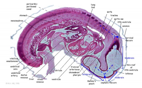
Week 5
Secondary Brain Vesicles
Brain Flexures
Rapid growth folds the neural tube forming 3 brain flexures (cranial to caudal)
- cephalic flexure - (mesencephalic) pushes mesencephalon upwards
- pontine flexure - generates 4th ventricle (cerebellum will grow into this space)
- cervical flexure - between brain stem and spinal cord
|
|
Ventricles
- cavity within neural tube will form the contiguious space of the ventricules of the brain and central canal of spinal cord
- space is filled initially with amniotic fluid, later with CerebroSpinal Fluid (CSF)
- CSF is secreted by
- chorioid plexus modified vascular structures lying within the ventricles
- floor of lateral ventricle and roof of the third and fourth ventricles
- ventricular ependymal cells and cells lining the subarachnoid space
- CSF also fills the subarachnoid space (between arachnoid mater and pia mater).
| Adult Ventricular Structures
|
Brain four ventricles and several foramina (openings that connect ventricular spaces)
- 2 lateral ventricles (right and left)
- interventricular foramina (foramina of Monro)
- third ventricle
- cerebral aqueduct (Sylvius)
- fourth ventricle
- median aperture (Magendie) subarachnoid space via the cisterna magna
- right and left lateral aperture (Luschka) subarachnoid space via the cistern of great cerebral vein
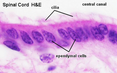
Spinal cord
- central canal lined with ependymal cells
|
|
| Adult Meninges Layers
|
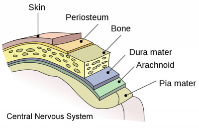
|
|
- Links: Neural - Ventricular System Development
|
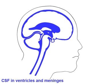
CSF-filled spaces in adult brain.
|
Week 6
| <html5media height="600" width="520">File:Human embryo tomography Carnegie stage 17.mp4</html5media>
Movie
|
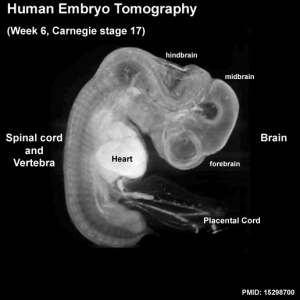
Note the shape and size of the different regions of the brain and spinal cord.
- Telencephalon (cerebrum) has begun to expand and will eventually cover the midbrain region.
- Dorsal root ganglia are visible outside the spinal cord.
|
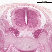
Week 8
The human MRI movie below (head, sagittal plane, left to right) shows the central nervous system (CNS) development at the end of the embryonic period (week 8; GA week 10).
<html5media height="500" width="550">File:Stage23 MRI S01.mp4</html5media>
Cortex
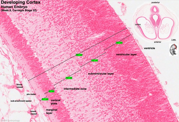
| Week 8 Developing Cortex
|
| Human embryo, Week 8, (GA week 10) Carnegie stage 22 section from the neural tube at the level of the developing cortex. Inset (upper right) shows whole section overview and approximate level of section (red line). Grey box shows detailed image region of developing cerebrum layer thicknesses are shown in microns.
Developing Cortex will form from the thin outer layer called the cortical plate. The underlying layers transient structures that continue to supply cells to the cortex through fetal period, most of these layers will eventually be lost, except for a thin ventricular layer. Cells migrate out along radial glia that establish the initial columnar and layered structure of the cortex. Layers are named according to the nervous system revised terminology (1970)[4]
Developing Vascular blood vessels can also be seen spanning the developing layers. In the adult, these vessels will be lined with non-fenestrated endothelial cells that together with other vascular cells (pericytes and vascular smooth muscle cells), glial cells (astrocytes and microglia) and neurons will form the "blood-brain barrier".
Developing Ventricular Space is cerebrospinal fluid (CSF) filled and the lateral ventricles form within the cortical region. The inset image shows lying within the lateral ventricles, the choroid plexus the modified vascular structure that forms and secretes the CSF.
Developing Meninges layers lie outside the neural tube. The thin pia mater that closely covers the entire brain. The mesh-like arachnoid mater and the sub-arachnoid space that will also be CSF filled. The dense dura mater lies outside these 2 layers and under the skull, it cannot be seen in the enlarged image.
|
Spinal Cord

| Week 8 Developing Spinal Cord (virtual slide)
|
|
|
These listed features link to zoomed views of the virtual slide with the named feature generally in the centre of the view.
Use the (-) at the top left of the screen to see where this feature is located.
|
| Spinal Cord Features
|
Other Features
|
Fetal
Second Trimester
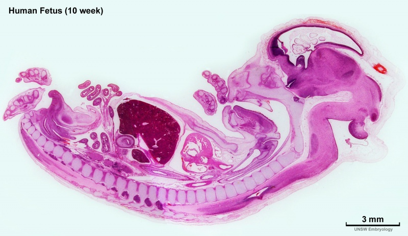
Human week 10 fetus
|

|
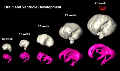
| Brain and Ventricular Development[5]
|
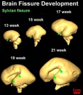
Brain Fissure Development[5]
|
| Sylvian Fissure Development
|
| <html5media height="600" width="600">File:Neural_-_Sylvian_fissure.mp4</html5media>
|
Thyroid and Neural Development
| Iodine deficiency
|
Abnormal Development - Iodine Deficiency
| ICD-11
|
|
5B5K.3 Iodine deficiency - Iodine deficiency disorders (IDD), caused mainly by a low dietary supply of iodine, refer to all of the consequences of iodine deficiency in a population that can be prevented by ensuring that the population has an adequate intake of iodine. Iodine deficiency is the most frequent cause of preventable brain damage in childhood.
5A00.04 Congenital hypothyroidism due to iodine deficiency - Hypothyroidism is a condition which arises at birth where the thyroid gland produces too little or no thyroid hormone and it can be induced by iodine-deficiency.
|
|
Third Trimester
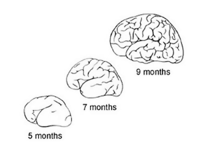
The brain goes from a smooth surface to begin to fold.
- Folds occur as millions of cells push into the cortex, increasing the surface area.
- groove - fissure (plural, fissures).
- fold - gyrus (plural, gyri).
|
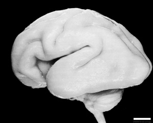
Human Fetus (CRL 240mm) Brain
|
| Human Brain Growth
|
| Embryonic
|
| Table below shows a direct comparison of brain growth in size between week 4 to 8 (GA 6-10)
|
| Fetal
|
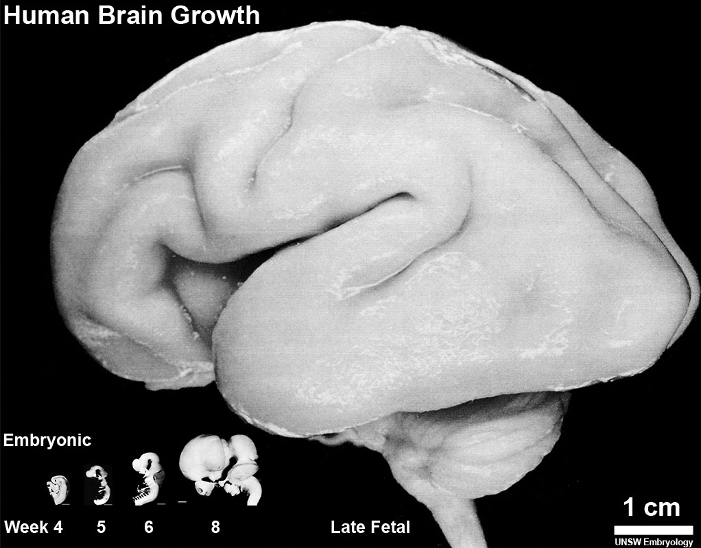
|
| Adult
|
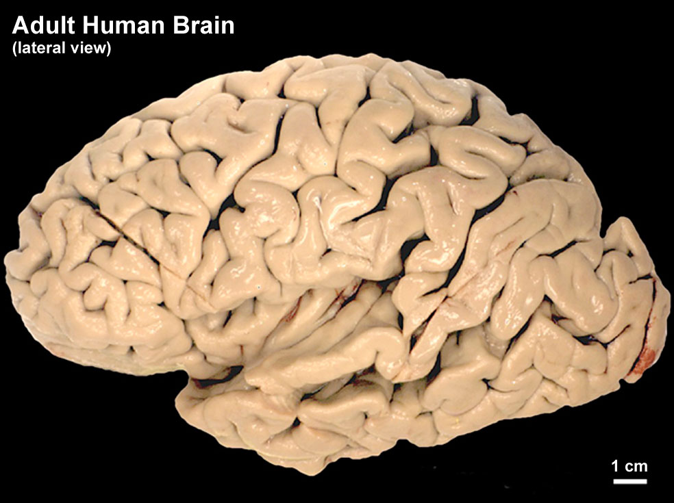
|
| Adult CNS Structures
|
Neural Tube Development
| Neural Tube
|
Primary Vesicles
|
Secondary Vesicles
|
Adult Structures
|
| week 3
|
week 4
|
week 5
|
adult
|
neural plate
neural groove
neural tube
Brain
|
prosencephalon (forebrain)
|
telencephalon
|
Rhinencephalon, Amygdala, hippocampus, cerebrum (cortex), hypothalamus, pituitary | Basal Ganglia, lateral ventricles
|
| diencephalon
|
epithalamus, thalamus, Subthalamus, pineal, posterior commissure, pretectum, third ventricle
|
| mesencephalon (midbrain)
|
mesencephalon
|
tectum, Cerebral peduncle, cerebral aqueduct, pons
|
| rhombencephalon (hindbrain)
|
metencephalon
|
cerebellum
|
| myelencephalon
|
medulla oblongata, isthmus
|
| spinal cord, pyramidal decussation, central canal
|
|
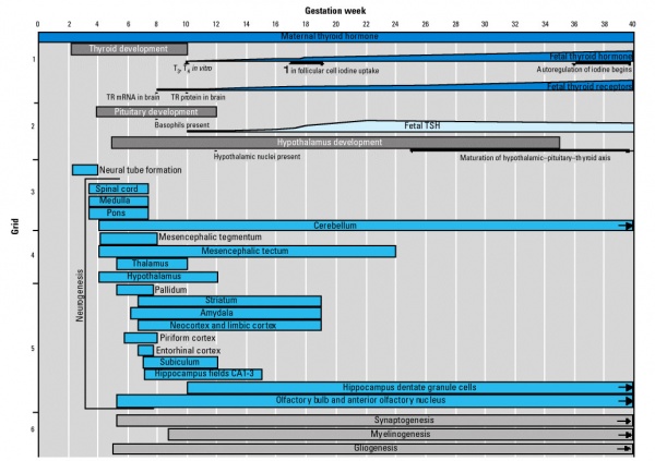
Fetal Timeline
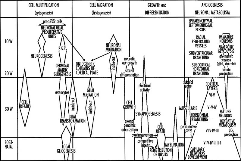
| Electrical Activity
|
Myelination
|
- Cerebral cortex has no neuronal connections at end of first trimester GA 12 weeks.
- Electroencephalogram (EEG) activity first seen in third trimester GA 7 months.
|
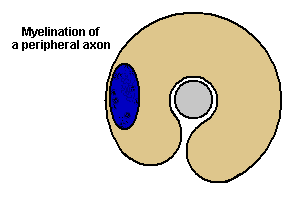
Myelination process occurs both in the CNS (from neural tube glia) and also in peripheral nerves (from neural crest Schwann cells).
|
Postnatal
Neural Exam Movies
| Additional Information - Multiple Sclerosis
|
| Only humans spontaneously develop multiple sclerosis (MS), a chronic demyelinating immune-mediated disease. This disease has an onset generally coinciding with the end of the long-term myelination process and incidence has been recently increasing in female/male (F/M) ratio and occurring in women of childbearing age.
|
Movies
Abnormalities
| There are a large number of different neural abnormalities associated with genetic, environmental and unknown causes. These can also involve several different systems including: neural tube, neural crest, sensory development, ventricular and vascular system development.
It would be difficult to cover all in this current lecture so a few examples are given and students should explore the topic more widely themselves.
- Links: neural abnormalities | Neural Crest Abnormalities | Ventricular Abnormalities
|
|
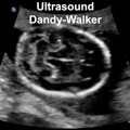
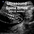
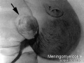
spina bifida and anencephaly
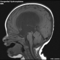
Congenital hydrocephalus (MRI)
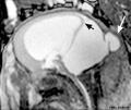
Dandy Walker malformation (MRI)
Environmental
|
The long time course of neural development (week 3 through to postnatal) also means that a large number of different environmental factors, including dietary deficiency, can impact upon its development and also have a range of different effects.
|
Postnatal Neural Assessment - there are several basic clinical motor assessments that can identify normal and abnormal development.
Neural Development Interactive Component
| Attempt the Quiz - Neural Development
|

Here are a few simple Quiz questions that relate to Neural development and abnormalities from the lecture. Some questions may require some additional research.
|
Terms
| Neural Terms
|
Neural Development
- 3DMRI - Three-dimensional magnetic resonance imaging. A new technique that allows 3D analysis of embryonic structures. (More? Magnetic Resonance Imaging)
- 3rd ventricle - a fluid-filled space formed from neural tube lumen, located within the diencephalon (from the primary vesicle prosencephalon, forebrain).
- 4th ventricle - a fluid-filled space formed from neural tube lumen, located within the rhombencephalon (from the primary vesicle, hindbrain).
- adenohypophysis - (anterior pituitary) = 3 parts pars distalis, pars intermedia, pars tuberalis.
- afferent - refers to the direction of conduction from the periphery toward the central nervous system. Efferent is in the opposite direction.
- alar plate - embryonic dorsolateral region of the neural tube forming at spinal cord level dorsal horns (afferent) and brain level different structures.
- anlage - (German = primordium) structure or cells that will form a future adult structure.
- arachnoid mater - (G.) spider web-like used in reference to the middle layer of the brain meninges.
- astrocytes - cells named by their "star-like" branching appearance, are the most abundant glial cells in the brain, important for the blood-brain barrier.
- basal ganglia - (basal nuclei) neural structure derived from the secondary vesicle telencephalon (endbrain) structure from the earlier primary vesicle prosencephalon (forebrain).
- basal plate - embryonic ventrolateral region of the neural tube forming at spinal cord level ventral horns (efferent) and brain level different structures.
- brachial plexus - mixed spinal nerves innervating the upper limb form a complex meshwork (crossing).
- brain - general term for the central nervous system formed from 3 primary vesicles.
- buccopharyngeal membrane - (oral membrane) at cranial (mouth) end of gastrointestinal tract (GIT) where surface ectoderm and GIT endoderm meet. (see also cloacal membrane).
- cauda equina - (horse's tail) caudal extension of the mature spinal cord.
- central canal - lumen, cavity of neural tube within the spinal cord. Space is continuous with ventricular system of the brain.
- central cerebral sulcus - (central fissure, fissure of Rolando, Rolandic fissure) fold in the cerebral cortex associated with the sensorimotor cortex.
- cerebral aqueduct - ventricular cavity within the mesencephalon.
- cervical flexure - most caudal brain flexure (of 3) between spinal cord and rhompencephalon.
- choroid plexus - specialized vascular plexus responsible for secreting ventricular fluid that with further additions becomes cerebrospinal fluid (CSF).
- cloacal membrane - at caudal (anal) end of gastrointestinal tract (GIT) where surface ectoderm and GIT endoderm meet forms the openings for GIT, urinary, reproductive tracts. (see also buccopharyngeal membrane).
- connectome - term describing the detailed map of neural connections in the central nervous system.
- cortex - - CNS structure derived from the secondary vesicle telencephalon (endbrain) from the earlier primary vesicle prosencephalon (forebrain).
- cortical plate - outer neural tube region which post-mitotic neuroblasts migrate too along radial glia to form adult cortical layers.
- cranial flexure - (=midbrain flexure) most cranial brain flexure (of 3) between mesencephalon and prosencephalon.
- diencephalon - the caudal portion of forebrain after it divides into 2 parts in the 5 secondary vesicle brain (week 5). (cavity- 3rd ventricle) Forms the thalmus and other nuclei in the adult brain. (sc-My-Met-Mes-Di-Tel)
- dorsal root ganglia - (spinal ganglia) sensory ganglia derived from the neural crest lying laterally paired and dorsally to the spinal cord (in the embryo found ventral to the spinal cord). Connects centrally with the dorsal horn of the spinal cord.
- dura mater- "tough" (Latin, mater = mother) used in reference to the tough outer layer of the brain meninges.
- efferent - refers to the direction of conduction from the central nervous system toward the periphery. Afferent is in the opposite direction.
- ependyma - epithelia of remnant cells after neurons and glia have been generated and left the ventricular zone.
- floorplate - early forming thin region of neural tube closest to the notochord.
- ganglia - (pl. of ganglion) specialized neural cluster within either the CNS or PNS.
- glia - supporting, non-neuronal cells of the nervous system. Generated from the same neuroepithelial stem cells that form neurons in ventricular zone of neural tube. Form astrocytes, oligodendrocytes.
- grey matter - neural regions containing cell bodies (somas) of neurons. In the brain it is the outer layer, in the spinal cord it is inner layer. (see white matter white matter).
- growth factor - usually a protein or peptide that will bind a cell membrane receptor and then activates an intracellular signaling pathway. The function of the pathway will be to alter the cell directly or indirectly by changing gene expression. (eg SHH).
- HOX - (homeobox) family of transcription factors that bind DNA and activate gene expression. Expression of different Hox genes along neural tube defines rostral-caudal axis and segmental levels.
- hydrocephalus - abnormality as the result of an imbalance between the rate at which the CSF is being formed and the rate at which the CSF is passing through the arachnoidal villi back into the blood (hydrocephalus rate is a function of the degree of imbalance in these two). Very small imbalance exhibit subtle, if any, symptoms. Large imbalances will have rapidly evolving symptoms of unmistakable import.
- isthmus- (G. narrow passage).
- lamina terminalis - anterior region of brain where cranial neuropore closes.
- lumbar plexus - mixed spinal nerves innervating the lower limb form a complex meshwork (crossing).
- mantle layer - layer of cells generated by first neuroblasts migrating from the ventricular zone of the neural tube. Layers are rearranged during development of the brain and spinal cord. (Ven-Man-Mar-CP)
- marginal zone - layer of processes from neuroblasts in mantle layer. (Ven-Man-Mar-CP)
- mater - (Latin, mater = mother) used in relation to the 3 layers of the meninges.
- meninges - mesenchyme surrounding neural tube forms 3 layer (Dura-, pia-, arachnoid- mater) connective tissue sheath of nervous system. (D-P-A-cns)
- mesencephalon - (midbrain), the middle portion of the 3 primary vesicle brain (week 4). (sc-R-M-P)
- metencephalon - the cranial portion of hindbrain after it divides into 2 parts in the 5 secondary vesicle brain (week 5). Forms the pons and cerebellum in the adult brain. (sc-My-Met-Mes-Di-Tel)
- microglia - CNS innate immune cells that have a macrophage function, derive from yolk sac progenitor cells migrating into the CNS. microglia
- myelencephalon - the caudal portion of hindbrain after it divides into 2 parts in the 5 secondary vesicle brain (week 5). Forms the medulla in the adult brain. (sc-My-Met-Mes-Di-Tel)
- neural tube - neural plate region of ectoderm pinched off to form hollow ectodermal tube above notochord in mesoderm.
- neural tube defect - (NTD) any developmental abnormality that affects neural tube development. Commonly failure of neural tube closure.
- neuroblast - undifferentiated neuron found in ventricular layer of neural tube.
- neurohypophysis - (posterior pituitary; pas nervosa)
- neuromere - (prosomere) the model units for segmental brain development regions based upon a series of neural tube transverse subunits.
- neuron - The cellur "unit" of the nervous system, transmitting signals between neurons and other cells. The post-mitotic cells generated from neuroepithelial stem cells (neuroblasts) in ventricular zone of neural tube.
- neuropore - opening at either end of neural tube cranial (rostral, anterior) neuropore closes (day 25) about 2 days before caudal (posterior) that closes at somite level 32 to 34. Neural Tube Defects (NTDs) can be due to failure of these two neuropores to close.
- notochord - rod of cells lying in mesoderm layer ventral to the neural tube, induces neural tube and secretes sonic hedgehog which "ventralizes" the neural tube.
- olfactory bulb - (cranial nerve I, CN I) bipolar neurons from nasal epithelium project axons through cribiform palate into olfactory bulb of the brain associated with smell.
- optic nerve - (cranial nerve II, CN II) retinal ganglion neurons project from the retina as a tract into the brain (at the level of the diencephalon) associated with vision.
- optic vesicle - diencephalon region of neural tube outgrowth that forms the primordia of the retina associated with vision.
- opercularization - during fetal development of the sensorimotor cortex, the insula (located deep within the lateral sulcus) begins to invaginate from the surface of the immature cerebrum, until at term, the opercula completely cover the insula.
- otocyst - (otic vesicle) sensory placode that sinks into mesoderm to form spherical vesicle (stage 13/14 embryo) that will form components of the inner ear associated with hearing.
- pharyngeal arch - (branchial arch, Gk. gill) form the main structures of the head and neck. Humans have 5 arches appearing in week 4 that form 4 external swellings, each arch has a pouch, membrane and cleft.
- pharynx - uppermost end of GIT, beginning at the buccopharyngeal membrane and at the level of the pharyngeal arches.
- pia mater - (G.) (L. pius = soft, faithful + mater = mother) delicate vascular membrane which adheres to surface of brain and spinal cord, faithfully following their contours, the inner layer of the brain meninges.
- placode - specialized regions of ectoderm which form components of the sensory apparatus.
- pontine flexure - middle brain flexure (of 3) between cervical and cranial flexure in opposite direction, also generates thin roof of rhombencephalon and divides it into myelencephalon and metencephalon. ( sc-^V^ )
- posterior insula - during sensorimotor cortex development this region is composed of the anterior and posterior long insular gyri and the postcentral insular sulcus, which separates them.
- prosencephalon - (forebrain), the most cranial portion of the 3 primary vesicle brain (week 4). (sc-R-M-P)
- prosomere - (neuromere) a model for segmental brain development based upon a series of neural tube transverse subunits. PMID 12948657
- Rathke's pouch - a portion of the roof of the pharynx pushes upward towards the floor of the brain forming the anterior pituitary (adenohypophysis, pars distalis, pars tuberalis pars intermedia). Where it meets a portion of the brain pushing downward forming the posterior pituitary (neurohypophysis, pars nervosa). Rathke's pouch eventually looses its connection with the pharynx.
- rhombencephalon - (hindbrain), the most caudal portion of the 3 primary vesicle brain (week 4). (sc-R-M-P)
- rhombic lip - metencephalon posterior part extending from the roof of the fourth ventricle to dorsal neuroepithelial cells that contributes to the cerebellum.
- roofplate - early forming thin region of neural tube closest to the overlying ectoderm.
- spinal cord - caudal end of neural tube that does not contribute to brain. Note: the process of secondary neuralation contributes the caudal end of the spinal cord.
- spinal ganglia - (dorsal root ganglia, drg) sensory ganglia derived from the neural crest lying laterally paired and dorsally to the spinal cord (in the embryo found ventral to the spinal cord). Connects centrally with the dorsal horn of the spinal cord.
- spinal nerve - mixed nerve (motor and sensory) arising as latera pairs at each vertebral segmental level.
- sonic hedgehog - (shh) secreted growth factor that binds patched (ptc) receptor on cell membrane. SHH function is different for different tissues in the embryo. In the nervous system, it is secreted by the notochord, ventralizes the neural tube, inducing the floor plate and motor neurons.
- sulcus - (L. furrow) groove.
- sulcus limitans - longitudinal lateral groove in neural tube approx. midway between roofplate and floorplate. Groove divides alar (dorsal) and basal (ventral) plate regions.
- telencephalon - the cranial portion of forebrain after it divides into 2 parts in the 5 secondary vesicle brain (week 5). (cavity- lateral ventricles and some of 3rd ventricle) Forms the cerebral hemispheres in the adult brain. (sc-My-Met-Mes-Di-Tel)
- thalamus - (G. thalamos= bedchamber) cns nucleus, lateral to 3rd ventricle, paired (pl thalami).
- thyroid hormone - hormone required for brain development. T3 (3,5,3′-triiodothyronine) binding to nuclear receptors then act as a transcription factor in both neurons and glial cells. iodine deficiency
- transcription factor - a factor (protein or protein with steroid) that binds to DNA to alter gene expression, usually to activate. (eg steroid hormone+receptor, Retinoic acid+Receptor, Hox, Pax, Lim, Nkx-2.2)
- trigeminal ganglion - (cranial nerve V, CN V) first arch ganglion, very large and has 3 portions.
- vagal ganglion - (cranial nerve X, CN X) fourth and sixth arch ganglion, innervates the viscera and heart.
- ventricles - the fluid-filled interconnected cavity system with the brain. Fluid (cerebrospinal fluid, CSF) is generated by the specialized vascular network, the choroid plexus. The ventricles are directly connected to the spinal canal (within the spinal cord).
- ventricular zone - Neuroepithelial cell layer of neural tube closest to lumen. Neuroepithelial cells generate neurons, glia and ependymal cells. (Ven-Man-Mar-CP)
- vestibulocochlear nerve - (cranial nerve VIII, CN VIII, also called statoacoustic)
- white matter - - neural regions containing processes (axons) of neurons. In the brain it is the inner layer, in the spinal cord it is outer layer. (see grey matter).
|
- ↑ AIHW 2016. Monitoring the health impacts of mandatory folic acid and iodine fortification 2016. Cat. no. PHE 208. Canberra: AIHW. PDF
- ↑ Woodhoo A & Sommer L. (2008). Development of the Schwann cell lineage: from the neural crest to the myelinated nerve. Glia , 56, 1481-90. PMID: 18803317 DOI.
- ↑ Barraud P, Seferiadis AA, Tyson LD, Zwart MF, Szabo-Rogers HL, Ruhrberg C, Liu KJ & Baker CV. (2010). Neural crest origin of olfactory ensheathing glia. Proc. Natl. Acad. Sci. U.S.A. , 107, 21040-5. PMID: 21078992 DOI.
- ↑ . (1970). Embryonic vertebrate central nervous system: revised terminology. The Boulder Committee. Anat. Rec. , 166, 257-61. PMID: 5414696 DOI.
- ↑ 5.0 5.1 Huang H, Xue R, Zhang J, Ren T, Richards LJ, Yarowsky P, Miller MI & Mori S. (2009). Anatomical characterization of human fetal brain development with diffusion tensor magnetic resonance imaging. J. Neurosci. , 29, 4263-73. PMID: 19339620 DOI.

BGDA: Lecture 1 | Lecture 2 | Practical 3 | Practical 6 | Practical 12 | Lecture Neural | Practical 14 | Histology Support - Female | Male | Tutorial
Glossary Links
- Glossary: A | B | C | D | E | F | G | H | I | J | K | L | M | N | O | P | Q | R | S | T | U | V | W | X | Y | Z | Numbers | Symbols | Term Link
Cite this page: Hill, M.A. (2024, June 5) Embryology BGDA Lecture - Development of the Nervous System. Retrieved from https://embryology.med.unsw.edu.au/embryology/index.php/BGDA_Lecture_-_Development_of_the_Nervous_System
- What Links Here?
- © Dr Mark Hill 2024, UNSW Embryology ISBN: 978 0 7334 2609 4 - UNSW CRICOS Provider Code No. 00098G


.jpg)


