Pharyngeal arches
| Embryology - 18 Apr 2026 |
|---|
| Google Translate - select your language from the list shown below (this will open a new external page) |
|
العربية | català | 中文 | 中國傳統的 | français | Deutsche | עִברִית | हिंदी | bahasa Indonesia | italiano | 日本語 | 한국어 | မြန်မာ | Pilipino | Polskie | português | ਪੰਜਾਬੀ ਦੇ | Română | русский | Español | Swahili | Svensk | ไทย | Türkçe | اردو | ייִדיש | Tiếng Việt These external translations are automated and may not be accurate. (More? About Translations) |
Introduction

The pharyngeal arches (branchial arch, Greek, branchial = gill) are a series of externally visible anterior tissue bands lying under the early brain that give rise to the structures of the head and neck. Each arch though initially formed from similar components will differentiate to form different head and neck structures. In humans, five arches form (1, 2, 3, 4 and 6) but only four are externally visible on the embryo.
Each arch has initially identical structures: an internal endodermal pouch, a mesenchymal (mesoderm and neural crest) core, a membrane (endoderm and ectoderm) and external cleft (ectoderm). Each arch mesenchymal core also contains similar components: blood vessel, nerve, muscular, cartilage.
Both the endocrine organs thymus and parathyroid have developmental origins from the pharyngeal pouches. Cranial nerves are also associated with specific cranial arches.
The arch arteries undergo extensive remodelling during development of the vascular system, in general the inferior arteries have major contributions and superior arteries have minor contributions. The endothelium of arch arteries 1 and 2 has been shown to have different embryonic origin from 3-6 (second heart field).[1]
Note is a draft page and this topic is currently covered in more detail on the head Development page.
- Links: pharyngeal arch | head | neural crest | endocrine
| Historic Head Embryology |
|---|
| 1909 Head Malformations | 1922 Aortic-Arch System | 1925 Pharyngeal Diverticula | 1926 Precervical Sinus |
Some Recent Findings

|
| More recent papers |
|---|
 This table allows an automated computer search of the external PubMed database using the listed "Search term" text link.
More? References | Discussion Page | Journal Searches | 2019 References | 2020 References Search term: Pharyngeal Arch | Pharyngeal Pouch | Pharyngeal Cleft | Pharyngeal Membrane | Pharyngeal Arch Artery | Pharyngeal Arch Cartilage |
| Older papers |
|---|
| These papers originally appeared in the Some Recent Findings table, but as that list grew in length have now been shuffled down to this collapsible table.
See also the Discussion Page for other references listed by year and References on this current page.
|
Pharyngeal Arch Development


- branchial arch (Gk. branchia= gill)
- arch consists of all 3 trilaminar embryo layers
- ectoderm - outside
- mesoderm - core of mesenchyme
- endoderm - inside
Pharyngeal Arch Components
This table gives an overview of what each arch will contribute to the embryo.
| Pharyngeal Arch | Nerve | Artery | Neural Crest (Skeletal Structures) |
Muscles | Ligaments |
|---|---|---|---|---|---|
| 1 (maxillary/mandibular) |
trigeminal (CN V) | maxillary artery (terminal branches) | mandible, maxilla, malleus, incus | muscles of mastication, mylohyoid, tensor tympanic, ant. belly digastric | ant lig of malleus, sphenomandibular ligament |
| 2 (hyoid) |
facial (CN VII) | stapedial (embryonic) corticotympanic (adult) |
stapes, styloid process, lesser cornu of hyoid, upper part of body of hyoid bone | muscles of facial expression, stapedius, stylohyoid, post. belly digastric | stylohyoid ligament |
| 3 | glossopharyngeal (CN IX) | common carotid, internal carotid arteries | greater cornu of hyoid, lower part of body of hyoid bone | stylopharyngeus | |
| 4 | vagus (CN X) superior laryngeal branch | part of aortic arch (left), part right subclavian artery (right) | thyroid, cricoid, arytenoid, corniculate and cuneform cartilages | crycothyroid, soft palate levator veli palatini (not tensor veli palatini) | |
| 6 | vagus (CN X) recurrent laryngeal branch | part of left pulmonary artery (left), part of right pulmonary artery (right) | thyroid, cricoid, arytenoid, corniculate and cuneform cartilages | larynx intrinsic muscles (not cricothyroid muscle) |
| Pharyngeal Arch Derivatives | |||||
|---|---|---|---|---|---|
| Pharyngeal Arch | Nerve | Artery | Neural Crest (Skeletal Structures) |
Muscles | Ligaments |
| 1 (maxillary/mandibular) |
trigeminal (V) | maxillary artery (terminal branches) | mandible, maxilla, malleus, incus | muscles of mastication, mylohyoid, tensor tympanic, ant. belly digastric | ant lig of malleus, sphenomandibular ligament |
| 2 (hyoid) |
facial (VII) | stapedial (embryonic) corticotympanic (adult) |
stapes, styloid process, lesser cornu of hyoid, upper part of body of hyoid bone | muscles of facial expression, stapedius, stylohyoid, post. belly digastric | stylohyoid ligament |
| 3 | glossopharyngeal (IX) | common carotid, internal carotid arteries | greater cornu of hyoid, lower part of body of hyoid bone | stylopharyngeus | |
| 4 | vagus (X) superior laryngeal branch | part of aortic arch (left), part right subclavian artery (right) | thyroid, cricoid, arytenoid, corniculate and cuneform cartilages | crycothyroid, soft palate levator veli palatini (not tensor veli palatini) | |
| 6 | vagus (X) recurrent laryngeal branch | part of left pulmonary artery (left), part of right pulmonary artery (right) | thyroid, cricoid, arytenoid, corniculate and cuneform cartilages | larynx intrinsic muscles (not cricothyroid muscle) | |
Neural Crest
Cranial neural crest-derived mesenchymal cells (ectomesenchyme) migrate into pharyngeal arches from midbrain and hindbrain region forming:
- neurons
- Schwann cells
- smooth muscle cells
- osteoblasts
- chondrocytes
- odontoblasts
SHH secreted from pharyngeal arch 1 epithelium is necessary for early mandibular arch cell survival and later cartilage (Meckel's cartilage) condensation differentiation.[7]
- Links: cranial neural crest | neural crest
Arch Features
Each arch contains: artery, cartilage, nerve, muscular component
Arches and Phanynx Form the face, tongue, lips, jaws, palate, pharynx and neck cranial nerves, sense organ components, glands
- Humans have 5 arches - 1, 2, 3, 4, 6 (Arch 5 does not form or regresses rapidly)
- from in rostro-caudal sequence, Arch 1 to 6 from week 4 onwards
- arch 1 and 2 appear at time of closure of cranial neuropore
- Face - mainly arch 1 and 2
- Neck components - arch 3 and 4 (arch 4 and 6 fuse)
Arch Features
- arch
- groove
- externally separates each arch
- also called a cleft
- only first pair persist as external auditory meatus
- externally separates each arch
- pouch
- internally separates each arch
- pockets from the pharynx
- membrane
- ectoderm and endoderm contact regions
- only first pair persist as tympanic membrane
- Pharyngeal Arch 1 (Mandibular Arch) has 2 prominances
- smaller upper- maxillary forms maxilla, zygomatic bone and squamous part of temporal
- larger lower- mandibular, forms mandible
- Pharyngeal Arch 2 (Hyoid Arch)
- forms most of hyoid bone
- Arch 3 and 4
- neck structures
Arch Arteries
Endothelium in the pharyngeal arches 3, 4 and 6 is derived from the second heart field[1]
- "Oxygenated blood from the heart is directed into the systemic circulation through the aortic arch arteries (AAAs). The AAAs arise by remodeling of three symmetrical pairs of pharyngeal arch arteries (PAAs), which connect the heart with the paired dorsal aortae at mid-gestation. Aberrant PAA formation results in defects frequently observed in patients with lethal congenital heart disease. How the PAAs form in mammals is not understood. The work presented in this manuscript shows that the second heart field (SHF) is the major source of progenitors giving rise to the endothelium of the pharyngeal arches 3 - 6, while the endothelium in the pharyngeal arches 1 and 2 is derived from a different source. During the formation of the PAAs 3 - 6, endothelial progenitors in the SHF extend cellular processes toward the pharyngeal endoderm, migrate from the SHF and assemble into a uniform vascular plexus. This plexus then undergoes remodeling, whereby plexus endothelial cells coalesce into a large PAA in each pharyngeal arch."
Embryo Week: Week 1 | Week 2 | Week 3 | Week 4 | Week 5 | Week 6 | Week 7 | Week 8 | Week 9
- Carnegie Stages: 1 | 2 | 3 | 4 | 5 | 6 | 7 | 8 | 9 | 10 | 11 | 12 | 13 | 14 | 15 | 16 | 17 | 18 | 19 | 20 | 21 | 22 | 23 | About Stages | Timeline
Pharyngeal Arch 1
cranial nerve CN V trigeminal
See Meckel's cartilage, facial canal and associated structures by Richany (1956)[8]
- Historic: 1956 first branchial arch
Pharyngeal Arch 2
cranial nerve CN VII facial
See Reichert's cartilage, facial canal and associated structures by Anson (1956).[9]
- Historic: 1956 second branchial arch
Pharyngeal Arch 3
cranial nerve CN IX glossopharyngeal
Pouch 3
Pharyngeal pouch III is the embryonic origin of endodermal component of the thymus (that also has a neural crest contribution).
Pharyngeal Arch 4
cranial nerve CN X vagus - superior laryngeal branch
Pharyngeal Arch 6
cranial nerve CN X vagus - recurrent laryngeal branch
part of right pulmonary artery (right)
References
- ↑ 1.0 1.1 1.2 Wang X, Chen D, Chen K, Jubran A, Ramirez A & Astrof S. (2017). Endothelium in the pharyngeal arches 3, 4 and 6 is derived from the second heart field. Dev. Biol. , 421, 108-117. PMID: 27955943 DOI.
- ↑ 2.0 2.1 Hasten E & Morrow BE. (2019). Tbx1 and Foxi3 genetically interact in the pharyngeal pouch endoderm in a mouse model for 22q11.2 deletion syndrome. PLoS Genet. , 15, e1008301. PMID: 31412026 DOI.
- ↑ Abe M, Cox TC, Firulli AB, Kanai SM, Dahlka J, Lim KC, Engel JD & Clouthier DE. (2021). GATA3 is essential for separating patterning domains during facial morphogenesis. Development , 148, . PMID: 34383890 DOI.
- ↑ Mao A, Zhang M, Li L, Liu J, Ning G, Cao Y & Wang Q. (2021). Pharyngeal pouches provide a niche microenvironment for arch artery progenitor specification. Development , 148, . PMID: 33334861 DOI.
- ↑ Gordon J. (2018). Hox genes in the pharyngeal region: how Hoxa3 controls early embryonic development of the pharyngeal organs. Int. J. Dev. Biol. , 62, 775-783. PMID: 30604847 DOI.
- ↑ Jin S, O J, Stellabotte F & Choe CP. (2018). Foxi1 promotes late-stage pharyngeal pouch morphogenesis through ectodermal Wnt4a activation. Dev. Biol. , 441, 12-18. PMID: 29932895 DOI.
- ↑ Billmyre KK & Klingensmith J. (2015). Sonic hedgehog from pharyngeal arch 1 epithelium is necessary for early mandibular arch cell survival and later cartilage condensation differentiation. Dev. Dyn. , 244, 564-76. PMID: 25626636 DOI.
- ↑ Richany SF. Bast TH. and Anson BJ. The development of the first branchial arch in man and the fate of Meckel's cartilage. (1956) Q Bull Northwest Univ Med Sch. 30(4):331-55. PMID: 13408429.
- ↑ {{Ref-AnsonBastRichany1956}
Additional Images
Historic Images
| Historic Disclaimer - information about historic embryology pages |
|---|
|
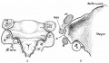
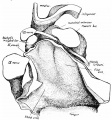
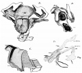
Frazer JE. The second visceral arch and groove in the tubo-tympanic region. (1914) J Anat Physiol. 48(4): 391-408. PMID 17233005
- Second Pharyngeal Arch
12 mm embryo
16 mm embryo
27mm embryo
35mm embryo


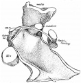
Frazer JE. Development of the larynx. (1910) J Anat. 44: 156-191. PMID 17232839
- Embryonic Larynx Development
embryo 5 mm
embryo 6 mm
embryo 7.5 mm
embryo 8.5 mm
embryo 12 mm
embryo 6.6 mm
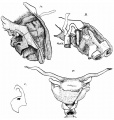
embryo 22 mm
embryo 35 mm
embryo 9.5 mm


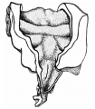
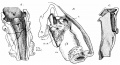
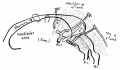
Keibel F. and Mall FP. Manual of Human Embryology II. (1912) J. B. Lippincott Company, Philadelphia.
- Pharynx
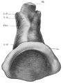
Fig 314 Pharynx embryo Klb (Kromer-Pfannenstiel
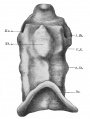
Fig 315 Pharynx embryo Rob. Meyer No. 335
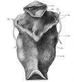
Fig 316 Pharynx embryo Hah
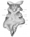
Fig 317 Pharynx embryo Rob. Meyer No. 300
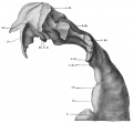
Fig 318 model shown in Fig. 317
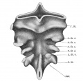
Fig 319 Pharyngeal pouches embryo Hah
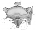
Fig 320 Pharyngeal region embryo BR
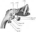
Fig 321 Branchial derivatives embryo 11.7 mm
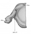
Fig 322 Pharynx embryo 21 mm
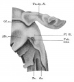
Fig 323 Pharynx embryo 24.4 mm
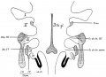
Fig 324 Branchiogenic derivatives in man
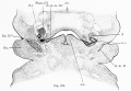
Fig 325 Section through embryo BR
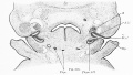
Fig 326 Section through embryo BR
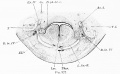
Fig 327 Section through embryo BR
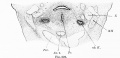
Fig 328 Section through embryo BR
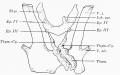
Fig 329 branchiogenic organs embryo 26 mm
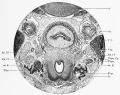
Fig 330 Laryngeal region embryo Nat2


Terms
| Head Terms (expand to view) |
|---|
|
| Other Terms Lists |
|---|
| Terms Lists: ART | Birth | Bone | Cardiovascular | Cell Division | Endocrine | Gastrointestinal | Genital | Genetic | Head | Hearing | Heart | Immune | Integumentary | Neonatal | Neural | Oocyte | Palate | Placenta | Radiation | Renal | Respiratory | Spermatozoa | Statistics | Tooth | Ultrasound | Vision | Historic | Drugs | Glossary |
Glossary Links
- Glossary: A | B | C | D | E | F | G | H | I | J | K | L | M | N | O | P | Q | R | S | T | U | V | W | X | Y | Z | Numbers | Symbols | Term Link
Cite this page: Hill, M.A. (2026, April 18) Embryology Pharyngeal arches. Retrieved from https://embryology.med.unsw.edu.au/embryology/index.php/Pharyngeal_arches
- © Dr Mark Hill 2026, UNSW Embryology ISBN: 978 0 7334 2609 4 - UNSW CRICOS Provider Code No. 00098G