Musculoskeletal System Development
| Embryology - 21 Jun 2026 |
|---|
| Google Translate - select your language from the list shown below (this will open a new external page) |
|
العربية | català | 中文 | 中國傳統的 | français | Deutsche | עִברִית | हिंदी | bahasa Indonesia | italiano | 日本語 | 한국어 | မြန်မာ | Pilipino | Polskie | português | ਪੰਜਾਬੀ ਦੇ | Română | русский | Español | Swahili | Svensk | ไทย | Türkçe | اردو | ייִדיש | Tiếng Việt These external translations are automated and may not be accurate. (More? About Translations) |
Introduction


The mesoderm forms nearly all the connective tissues of the musculoskeletal system. Each tissue (cartilage, bone, and muscle) goes through many different mechanisms of differentiation.
The musculoskeletal system consists of skeletal muscle, bone, and cartilage and is mainly mesoderm in origin with some neural crest contribution.
The intraembryonic mesoderm can be broken into paraxial, intermediate and lateral mesoderm relative to its midline position. During the 3rd week the paraxial mesoderm undergoes somitogenesis and forms into paired "balls" of somites either side of the neural groove.
Somites appear bilaterally as pairs at the same time and form earliest at the cranial (rostral,brain) end of the neural groove and add sequentially at the caudal end. This addition occurs so regularly that embryos are staged according to the number of somites that are present. Different regions of the somite differentiate into dermomyotome (dermal and muscle component) and sclerotome (forms vertebral column). An example of a specialized musculoskeletal structure can be seen in the development of the limbs.
Skeletal muscle forms by fusion of mononucleated myoblasts to form mutinucleated myotubes. Bone is formed through a lengthy process involving ossification of a cartilage formed from mesenchyme. Two main forms of ossification occur in different bones, intramembranous (eg skull) and endochondrial (eg limb long bones) ossification. Ossification continues postnatally, through puberty until mid 20s. Early ossification occurs at the ends of long bones.
Musculoskeletal and limb abnormalities are one of the largest groups of congenital abnormalities.
Some Recent Findings
|
| More recent papers |
|---|
 This table allows an automated computer search of the external PubMed database using the listed "Search term" text link.
More? References | Discussion Page | Journal Searches | 2019 References | 2020 References Search term: Musculoskeletal Development | Musculoskeletal Embryology |


| Older papers |
|---|
| These papers originally appeared in the Some Recent Findings table, but as that list grew in length have now been shuffled down to this collapsible table.
See also the Discussion Page for other references listed by year and References on this current page.
|
Textbooks

|
Hill, M.A. (2020). UNSW Embryology (20th ed.) Retrieved Haziran 21, 2026, from https://embryology.med.unsw.edu.au
| ||||

|
Moore, K.L. & Persuad, T.V.N. (2008). The Developing Human: clinically oriented embryology (8th ed.). Philadelphia: Saunders.
| ||||

|
Schoenwolf, G.C., Bleyl, S.B., Brauer, P.R. and Francis-West, P.H. (2009). Larsen’s Human Embryology (4th ed.). New York; Edinburgh: Churchill Livingstone.
| ||||
| Earlier Textbooks |
|
Objectives
- Identify the components of a somite and the adult derivatives of each component.
- Give examples of sites of endochondral and intramembranous ossification and to compare these two processes.
- Identify the general times of formation of primary and of formation of secondary ossification centres, and of fusion of such centres with each other.
- Briefly summarise the development of the limbs.
- Describe the developmental abnormalities responsible for the following malformations: selected growth plate disorders; congenital dislocation of the hip; scoliosis; arthrogryposis; and limb reduction deformities.
Development Overview
Bone is a connective tissue and develops from mesoderm except in the head where neural crest also contributes. Below is a very brief cartoon overview using simple figures of 3 aspects of early musculoskeletal development. More detailed overviews are shown on other notes pages Mesoderm and Somite, Vertebral Column, Limb in combination with serial sections and Carnegie images.
Mesoderm Development

|
Cells migrate through the primitive streak to form mesodermal layer. Extraembryonic mesoderm lies adjacent to the trilaminar embryo totally enclosing the amnion, yolk sac and forming the connecting stalk. |

|
Paraxial mesoderm accumulates under the neural plate with thinner mesoderm laterally. This forms 2 thickened streaks running the length of the embryonic disc along the rostrocaudal axis. In humans, during the 3rd week, this mesoderm begins to segment. The neural plate folds to form a neural groove and folds. |

|
Segmentation of the paraxial mesoderm into somites continues caudally at 1 somite/90minutes and a cavity (intraembryonic coelom) forms in the lateral plate mesoderm separating somatic and splanchnic mesoderm.
Note intraembryonic coelomic cavity communicates with extraembryonic coelom through portals (holes) initially on lateral margin of embryonic disc. |

|
Somites continue to form. The neural groove fuses dorsally to form a tube at the level of the 4th somite and "zips up cranially and caudally and the neural crest migrates into the mesoderm. |
Somite Development

|
Mesoderm beside the notochord (axial mesoderm, blue) thickens, forming the paraxial mesoderm as a pair of strips along the rostro-caudal axis. |

|
Paraxial mesoderm towards the rostral end, begins to segment forming the first somite. Somites are then sequentially added caudally. The somitocoel, is a cavity forming in early somites, which is lost as the somite matures. |

|
Cells in the somite differentiate medially to form the sclerotome (forms vertebral column) and dorsolaterally to form the dermomyotome. |

|
The dermomyotome then forms the dermotome (forms dermis) and myotome (forms muscle).
Neural crest cells migrate beside and through somite. |

|
The myotome differentiates to form 2 components dorsally the epimere and ventrally the hypomere, which in turn form epaxial and hypaxial muscles respectively. The bulk of the trunk and limb muscle coming from the Hypaxial mesoderm. Different structures will be contributed depending upon the somite level. |
Somite Links: 1 paraxial | 2 early somite | 3 sclerotome and dermomyotome | 4 dermatome and myotome | 5 somite spreading | SEM image - Human Embryo (week 4) showing somites | Movie - somitogenesis Hes expression
Limb Development

|
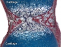
Mesoderm within the developing limb bud differentiates to initially form cartilage which later ossifies by endochondral ossification.
Hypaxial somitic mesoderm from somites at the levels of limb bud formation, migrates into the bud. These cells within the bud proliferate in regions of muscle formation, fuse to form myotubes and then differentiate to form skeletal muscle cells. |
Shoulder and Pelvis

The skeletal shoulder consists of: the clavicle (collarbone), the scapula (shoulder blade), and the humerus. Development of his region occurs through both forms of ossification processes.
The skeletal pelvis consists of: the sacrum and coccyx (axial skeleton), and pelvic girdle formed by a pair of hip bones (appendicular skeleton). Before puberty, he pelvic girdle also consists of three unfused bones: the ilium, ischium, and pubis. In chicken, the entire pelvic girdle originates from the somatopleure mesoderm (somite levels 26 to 35) and the ilium, but not of the pubis and ischium, depends on somitic and ectodermal signals.[3]
- Links: Shoulder Development | Pelvis Development
Sternum
For details on sternum development see axial skeleton notes. The sternum and sternal ribs derive from the somatic layer of the lateral plate mesoderm.[4][5]
- Links: sternum
References
- ↑ Langen UH, Pitulescu ME, Kim JM, Enriquez-Gasca R, Sivaraj KK, Kusumbe AP, Singh A, Di Russo J, Bixel MG, Zhou B, Sorokin L, Vaquerizas JM & Adams RH. (2017). Cell-matrix signals specify bone endothelial cells during developmental osteogenesis. Nat. Cell Biol. , 19, 189-201. PMID: 28218908 DOI.
- ↑ Pourquié O. (2011). Vertebrate segmentation: from cyclic gene networks to scoliosis. Cell , 145, 650-63. PMID: 21620133 DOI.
- ↑ Malashichev Y, Christ B & Pröls F. (2008). Avian pelvis originates from lateral plate mesoderm and its development requires signals from both ectoderm and paraxial mesoderm. Cell Tissue Res. , 331, 595-604. PMID: 18087724 DOI.
- ↑ Sadler TW. (2000). Embryology of the sternum. Chest Surg. Clin. N. Am. , 10, 237-44, v. PMID: 10803330
- ↑ Mekonen HK, Hikspoors JP, Mommen G, Köhler SE & Lamers WH. (2015). Development of the ventral body wall in the human embryo. J. Anat. , 227, 673-85. PMID: 26467243 DOI.
Online Textbooks
- Developmental Biology by Gilbert, Scott F. Sunderland (MA): Sinauer Associates, Inc.; c2000 Paraxial and intermediate mesoderm | Myogenesis: The Development of Muscle | Osteogenesis: The Development of Bones | Figure 14.10. Conversion of myoblasts into muscles in culture
- Molecular Biology of the Cell Alberts, Bruce; Johnson, Alexander; Lewis, Julian; Raff, Martin; Roberts, Keith; Walter, Peter New York and London: Garland Science; c2002 Search Molecular Biology of the CellBone Is Continually Remodeled by the Cells Within ItImage: Figure 22-52. Deposition of bone matrix by osteoblasts.Image: Figure 22-56. The development of a long bone.
Reviews
Pourquié O. (2011). Vertebrate segmentation: from cyclic gene networks to scoliosis. Cell , 145, 650-63. PMID: 21620133 DOI.
Piróg KA & Briggs MD. (2010). Skeletal dysplasias associated with mild myopathy-a clinical and molecular review. J. Biomed. Biotechnol. , 2010, 686457. PMID: 20508815 DOI.
Sayer AA & Cooper C. (2005). Fetal programming of body composition and musculoskeletal development. Early Hum. Dev. , 81, 735-44. PMID: 16081228 DOI.
Walker JM. (1991). Musculoskeletal development: a review. Phys Ther , 71, 878-89. PMID: 1946622
Articles
Applegate KE. (2004). Can MR imaging be used to characterize fetal musculoskeletal development?. Radiology , 233, 305-6. PMID: 15516609 DOI.
Ryu JK, Cho JY & Choi JS. (2003). Prenatal sonographic diagnosis of focal musculoskeletal anomalies. Korean J Radiol , 4, 243-51. PMID: 14726642 DOI.
Search PubMed
Search Pubmed: Musculoskeletal System Development | Musculoskeletal Development
NCBI - Policies and Guidelines | PubMed | Help:Reference Tutorial
Additional Images

Adult axial skeleton

Adult appendicular skeleton

Bone structure
Developing vertebra

Endochondral bone

Fetal head lateral (12 weeks)

Fetal head medial (12 weeks)

Fetal head section (12 weeks)


| System Links: Introduction | Cardiovascular | Coelomic Cavity | Endocrine | Gastrointestinal Tract | Genital | Head | Immune | Integumentary | Musculoskeletal | Neural | Neural Crest | Placenta | Renal | Respiratory | Sensory | Birth |
External Links
External Links Notice - The dynamic nature of the internet may mean that some of these listed links may no longer function. If the link no longer works search the web with the link text or name. Links to any external commercial sites are provided for information purposes only and should never be considered an endorsement. UNSW Embryology is provided as an educational resource with no clinical information or commercial affiliation.
- UNSW Virtual Slides Musculoskeletal Development Histology (requires login)
- Embryo Images - Musculoskeletal Development | Early Week 4 | Late Week 4
Glossary Links
- Glossary: A | B | C | D | E | F | G | H | I | J | K | L | M | N | O | P | Q | R | S | T | U | V | W | X | Y | Z | Numbers | Symbols | Term Link
Cite this page: Hill, M.A. (2026, Haziran 21) Embryology Musculoskeletal System Development. Retrieved from https://embryology.med.unsw.edu.au/embryology/index.php/Musculoskeletal_System_Development
- © Dr Mark Hill 2026, UNSW Embryology ISBN: 978 0 7334 2609 4 - UNSW CRICOS Provider Code No. 00098G