Category:Neural
From Embryology
This Embryology category shows pages and media related to Neural System Development. This includes related topics and undergraduate classes as well as pages and sub-categories describing specific components formed from the original ectoderm neural tube.
Subcategories
This category has the following 12 subcategories, out of 12 total.
Pages in category 'Neural'
The following 200 pages are in this category, out of 934 total.
(previous page) (next page)2
A
- Abnormal Development - Anencephaly
- Abnormal Development - Congenital Hydrocephalus
- Abnormal Development - Fetal Alcohol Syndrome
- Abnormal Development - Folic Acid and Neural Tube Defects
- Abnormal Development - Iodine Deficiency
- Template:Abnormal Newborn Neural Exam Table
- Template:Adult brain movie
- Template:Adult Hypothalamus Hormones table
- AE Practical - Neural Histology
- Template:AEB Neural Histology2012
- Alagille Syndrome
- Template:Alcohol
- Template:Amin1914 figures
- Template:Amygdala
- ANAT2241 Nervous Tissue
- ANAT2511 Nervous Tissue
- ANAT3411 Neuroanatomy
- Template:Anencephaly
- Template:Arachnoid mater
- Template:Arachnoid villi
- Template:Astroglia
- Atlas of the Development of Man 2 - Neural
B
- Template:Bardeen1906 figures
- Template:Bartelmez1922 figures
- Template:Bartelmez1923 figures
- Template:Basal ganglia
- BGDA - Neural Development Interactive
- Template:BGDA - Neural Development Interactive
- BGDA Lecture - Development of the Nervous System
- Talk:BGDA Lecture - Development of the Nervous System
- BGDA Practical 7 - Week 4
- BGDA Practical 7 - Week 5
- Book - A History of Science 19
- Book - A History of Science 20
- Book - A Laboratory Manual and Text-book of Embryology 13
- Book - An Atlas of the Medulla and Midbrain
- Talk:Book - An Atlas of the Medulla and Midbrain
- Book - An Atlas of the Medulla and Midbrain - Figures
- Book - An Atlas of the Medulla and Midbrain - Reconstruction General Summary
- Book - An Atlas of the Medulla and Midbrain - References
- Book - An Atlas of the Medulla and Midbrain 1
- Book - An Atlas of the Medulla and Midbrain 2
- Book - An Atlas of the Medulla and Midbrain 3
- Book - An Atlas of the Medulla and Midbrain 4
- Book - An Atlas of the Medulla and Midbrain 5
- Book - An Atlas of the Medulla and Midbrain 6
- Book - An Atlas of the Medulla and Midbrain 7
- Book - An Atlas of the Medulla and Midbrain 8
- Book - An Atlas of the Medulla and Midbrain 9
- Book - Comparative Study of the Sensory Areas of the Human Cortex
- Book - Comparative Study of the Sensory Areas of the Human Cortex 1
- Book - Comparative Study of the Sensory Areas of the Human Cortex 2
- Book - Comparative Study of the Sensory Areas of the Human Cortex 3
- Book - Comparative Study of the Sensory Areas of the Human Cortex Figures
- Book - Contributions to Embryology Carnegie Institution No.11
- Book - Contributions to Embryology Carnegie Institution No.14
- Book - Contributions to Embryology Carnegie Institution No.22
- Book - Contributions to Embryology Carnegie Institution No.30
- Book - Contributions to Embryology Carnegie Institution No.33
- Book - Contributions to Embryology Carnegie Institution No.47
- Book - Contributions to Embryology Carnegie Institution No.59
- Book - Contributions to the Development of the Human Brain (1919)
- History:Book - Contributions to the Development of the Human Brain (1919)
- Book - Developmental Anatomy 1924-13
- Book - Human Embryology and Morphology 15
- Book - Manual of Human Embryology 14
- Book - Manual of Human Embryology 14-1
- Book - Manual of Human Embryology 14-2
- Book - Manual of Human Embryology 14-3
- Book - Manual of Human Embryology 14-4
- Book - Text-Book of Embryology 17
- Book - Text-Book of Embryology 18
- Book - Text-Book of the Embryology of Man and Mammals 16-1
- Book - The brain of the tiger salamander
- Book - The brain of the tiger salamander 1
- Book - The brain of the tiger salamander 10
- Book - The brain of the tiger salamander 11
- Book - The brain of the tiger salamander 12
- Book - The brain of the tiger salamander 13
- Book - The brain of the tiger salamander 14
- Book - The brain of the tiger salamander 15
- Book - The brain of the tiger salamander 2
- Book - The brain of the tiger salamander 23
- Book - The brain of the tiger salamander 4
- Book - The brain of the tiger salamander 5
- Book - The brain of the tiger salamander 9
- Book - The comparative anatomy of the nervous system of vertebrates including man - 1
- Book - The comparative anatomy of the nervous system of vertebrates including man 1
- Book - The comparative anatomy of the nervous system of vertebrates including man 2
- Book - The Elements of Embryology - Mammalian 3
- Book - The Nervous System of Vertebrates (1907)
- Template:Bradley OC.
- Template:Brain
- Brain Awareness Week 2012
- Talk:Brain Awareness Week 2012
- Template:Brain Growth table
- Template:Brain Histology
- Template:Brain Vascular System gallery
- Template:Brain Vascular System table1
C
- Template:Cajal lectures 1899
- Carnegie Stage 17 Neural Movie
- Template:Carnegie stages CNS images table
- Template:Central canal
- Template:Central nervous system
- Central Nervous System 3D stage 22 Movie
- Template:Cerebellar Nuclei table
- Template:Cerebellum
- Template:Cerebral aqueduct
- Template:Cerebral Arterial Timeline table
- Template:Cerebral cortex
- Template:Cerebrospinal fluid
- Template:Cerebrum
- Template:Choroid plexus
- Template:CN I
- Template:CN II
- Template:CN III
- Template:CN IV
- Template:CN IX
- Template:CN V
- Template:CN VI
- Template:CN VII
- Template:CN VIII
- Template:CN X
- Template:CN XI
- Template:CN XII
- Template:CNS
- Computed Tomography
- Template:Cortex
- Template:Cranial nerve
- Template:Cranial nerve neural crest
- Template:CS10
- Template:CS11
- Template:CS12
- Template:CS13
- Template:CS9
- Template:CSF
D
- Template:Dandy-Walker
- Developmental Signals - basic Helix-Loop-Helix
- Developmental Signals - Fox
- Developmental Signals - Homeobox
- Developmental Signals - LIM-homeodomain
- Developmental Signals - Nerve Growth Factor
- Developmental Signals - Nodal
- Developmental Signals - Notch
- Developmental Signals - Pax
- Developmental Signals - Retinoic acid
- Developmental Signals - Six
- Developmental Signals - Sonic hedgehog
- Developmental Signals - Tbx
- Template:Diencephalon
- Template:Dorsal root ganglia
- Template:Dura mater
- Template:Dural venous sinuses
E
- Template:Early Neural Timeline table
- Ectoderm
- Talk:Embryo Serial Sections
- Embryology History - Albert Kuntz
- Embryology History - G. Carl Huber
- Embryology History - Orlando Charnock Bradley
- Embryology History - Robert Remak
- Embryology History - Santiago Ramón y Cajal
- Embryology History - Viktor Hamburger
- Template:Embryonic Ventricular Timeline collapsetable
- Template:Embryonic Ventricular Timeline table
- Template:Encephalocele
- Endocrine - Hypothalamus Development
- Template:Epithalamus
- Template:Eye
F
H
- Template:Hamburger V.
- Hearing - Neural Pathway
- Template:Hearing neural
- Template:Herrick CL.
- Template:Herrick1948 footer
- Template:Heuser1913 table1
- Template:Heuser1913 table2
- Template:Heuser1913 table3
- Template:Hewer1935 table 2
- Template:Hind-brain
- Template:Hindbrain
- Template:Hippocampus
- Template:Historic Cortex
Media in category 'Neural'
The following 200 files are in this category, out of 1,070 total.
(previous page) (next page) 03mo 01.jpg 320 × 240; 9 KB
 03mo 02.jpg 320 × 240; 8 KB
 03mo 03.jpg 320 × 240; 10 KB
 03mo 04.jpg 320 × 240; 11 KB
 03mo 05.jpg 320 × 240; 11 KB
 10wkcerebellumB.jpg 347 × 284; 21 KB
 1899 Cajal 01.jpg 307 × 1,200; 121 KB
 1899 Cajal 02.jpg 1,200 × 646; 177 KB
 1899 Cajal 03.jpg 952 × 1,000; 204 KB
 1899 Cajal 04.jpg 909 × 1,000; 253 KB
 1899 Cajal 05.jpg 683 × 1,000; 164 KB
 1899 Cajal 06.jpg 812 × 1,000; 163 KB
 1899 Cajal 07.jpg 837 × 1,000; 175 KB
 1899 Cajal 08.jpg 329 × 1,000; 101 KB
 2017BGDALecture-Neural.mp4 ; 52.32 MB
 Abnormal81-92-neuron.png 481 × 344; 9 KB
 Adult brain 01.mov ; 1.59 MB
- Adult brain 02.mov ; 434 KB
 Adult brain animation 01.gif 280 × 224; 396 KB
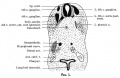
 Adult cochlea cartoon 01.jpg 986 × 800; 123 KB
 Adult cochlea nerve glia cartoon.jpg 1,000 × 725; 85 KB
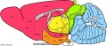
 Adult diencephalon.jpg 470 × 376; 20 KB
 Adult human brain movie icon.jpg 717 × 575; 28 KB
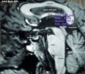 Adult human brain MRI01.jpg 700 × 607; 81 KB
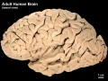 Adult human brain.jpg 984 × 735; 104 KB
 Adult mouse brain - prosomeric model.jpg 964 × 414; 91 KB
- AEB Histology Prac 171012-3 Ganglion.mp3 ; 1.05 MB
- AEB Histology Prac 171012-5 Nerve.mp3 ; 1.07 MB
 Amin1914 fig01.jpg 1,000 × 652; 120 KB
 Amin1914 fig02.jpg 1,000 × 681; 152 KB
 Amin1914 fig03.jpg 1,000 × 689; 134 KB
 Amin1914 fig04.jpg 1,000 × 727; 139 KB
 Amin1914 fig05.jpg 1,000 × 646; 91 KB
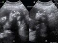 Anencephaly ultrasound.jpg 900 × 658; 108 KB
 Astrocytes and neonatal hypoxia ischemia.jpg 484 × 705; 309 KB
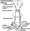 Auditory neural pathway.jpg 450 × 457; 46 KB
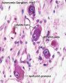 Autonomic ganglion histology 01.jpg 641 × 800; 56 KB
 Baboon- fetal brain.jpg 1,000 × 733; 127 KB
 Bailey358.jpg 854 × 560; 66 KB
 Bailey359.jpg 708 × 572; 46 KB
 Bailey360.jpg 543 × 404; 23 KB
 Bailey361.jpg 815 × 662; 86 KB
 Bailey362.jpg 801 × 354; 49 KB
 Bailey363.jpg 876 × 373; 46 KB
 Bailey364.jpg 809 × 465; 53 KB
 Bailey365.jpg 847 × 595; 94 KB
 Bailey366.jpg 558 × 633; 97 KB
 Bailey367.jpg 1,034 × 440; 101 KB
 Bailey368.jpg 1,074 × 523; 134 KB
 Bailey369.jpg 529 × 446; 33 KB
 Bailey370.jpg 975 × 1,084; 242 KB
 Bailey371.jpg 687 × 997; 97 KB
 Bailey372.jpg 803 × 690; 79 KB
 Bailey373.jpg 894 × 426; 100 KB
 Bailey374.jpg 880 × 490; 75 KB
 Bailey375.jpg 929 × 785; 130 KB
 Bailey376.jpg 922 × 862; 70 KB
 Bailey377.jpg 1,032 × 904; 90 KB
 Bailey378.jpg 1,199 × 700; 69 KB
 Bailey379-382.jpg 671 × 988; 199 KB
 Bailey383.jpg 859 × 455; 118 KB
 Bailey384.jpg 1,532 × 770; 245 KB
 Bailey385.jpg 787 × 683; 165 KB
 Bailey386.jpg 491 × 410; 53 KB
 Bailey387.jpg 507 × 442; 50 KB
 Bailey388.jpg 514 × 438; 68 KB
 Bailey389.jpg 829 × 561; 65 KB
 Bailey390.jpg 583 × 667; 50 KB
 Bailey391.jpg 633 × 485; 56 KB
 Bailey392.jpg 949 × 632; 113 KB
 Bailey393.jpg 680 × 527; 89 KB
 Bailey394.jpg 614 × 563; 70 KB
 Bailey395.jpg 787 × 741; 94 KB
 Bailey396.jpg 829 × 845; 123 KB
 Bailey397.jpg 779 × 995; 162 KB
 Bailey398.jpg 872 × 581; 78 KB
 Bailey399.jpg 832 × 704; 65 KB
 Bailey400.jpg 724 × 749; 66 KB
 Bailey401.jpg 788 × 718; 61 KB
 Bailey402.jpg 640 × 483; 116 KB
 Bailey403.jpg 495 × 617; 118 KB
 Bailey404.jpg 627 × 695; 84 KB
 Bailey405.jpg 745 × 820; 141 KB
 Bailey406.jpg 868 × 763; 116 KB
 Bailey407.jpg 793 × 695; 117 KB
 Bailey408.jpg 862 × 732; 124 KB
 Bailey409.jpg 448 × 408; 32 KB
 Bailey410.jpg 1,412 × 802; 169 KB
 Bailey411.jpg 790 × 809; 104 KB
 Bailey412.jpg 723 × 251; 21 KB
 Bailey413.jpg 674 × 412; 47 KB
 Bailey414.jpg 900 × 717; 123 KB
 Bailey415.jpg 935 × 723; 147 KB
 Bailey416.jpg 815 × 606; 142 KB
 Bailey417.jpg 940 × 494; 116 KB
 Bailey418.jpg 232 × 583; 30 KB
 Bailey419.jpg 805 × 388; 43 KB
 Bailey420.jpg 504 × 348; 34 KB
 Bailey421.jpg 625 × 538; 81 KB
 Bailey422.jpg 495 × 568; 95 KB
 Bailey423.jpg 670 × 581; 56 KB
 Bailey424.jpg 804 × 534; 61 KB
 Bailey425.jpg 957 × 490; 84 KB
 Bailey426.jpg 948 × 758; 80 KB
 Bailey427.jpg 907 × 527; 72 KB
 Bailey428.jpg 756 × 557; 67 KB
 Bailey429.jpg 844 × 484; 81 KB
 Bailey430.jpg 1,072 × 791; 121 KB
 Bailey431.jpg 832 × 535; 75 KB
 Bailey432.jpg 794 × 635; 92 KB
 Bailey433.jpg 723 × 536; 77 KB
 Bailey434.jpg 426 × 436; 44 KB
 Bailey435.jpg 788 × 456; 48 KB
 Bailey436.jpg 552 × 443; 40 KB
 Bailey437.jpg 526 × 397; 50 KB
 Bailey438.jpg 778 × 530; 70 KB
 Bailey439.jpg 932 × 1,016; 125 KB
 Bailey440.jpg 820 × 434; 54 KB
 Bailey441.jpg 786 × 520; 70 KB
 Bailey442.jpg 525 × 504; 38 KB
 Bailey443.jpg 666 × 539; 49 KB
 Bailey444.jpg 777 × 590; 127 KB
 Bailey445.jpg 900 × 655; 89 KB
 Bailey446.jpg 709 × 437; 40 KB
 Bailey447.jpg 795 × 423; 45 KB
 Bailey448.jpg 722 × 416; 55 KB
 Bailey449.jpg 777 × 374; 45 KB
 Bailey450.jpg 680 × 419; 45 KB
 Bailey451-452.jpg 753 × 862; 151 KB
 Bailey453.jpg 461 × 740; 69 KB
 Bailey454.jpg 732 × 527; 119 KB
 Bailey455.jpg 744 × 547; 124 KB
 Bailey456.jpg 591 × 168; 20 KB
 Bailey457.jpg 659 × 335; 40 KB
 Bailey458-459.jpg 741 × 386; 42 KB
 Bailey460.jpg 718 × 423; 55 KB
 Bailey461.jpg 751 × 394; 54 KB
 Bailey462.jpg 463 × 411; 45 KB
 Bailey463.jpg 679 × 345; 52 KB
 Bailey464.jpg 869 × 592; 67 KB
 Bailey465.jpg 806 × 931; 142 KB
 Bailey466.jpg 859 × 683; 139 KB
 Bailey467.jpg 946 × 515; 159 KB
 Bailey468.jpg 774 × 519; 60 KB
 Bailey469.jpg 484 × 401; 50 KB
 Bailey470.jpg 537 × 644; 88 KB
 Bailey475.jpg 1,302 × 852; 213 KB
 Bailey476.jpg 688 × 648; 110 KB
 Bailey477.jpg 593 × 509; 57 KB
 Bailey478.jpg 809 × 620; 81 KB
 Bailey479.jpg 742 × 363; 56 KB
 Bailey480.jpg 799 × 553; 61 KB
 Bailey481.jpg 842 × 752; 109 KB
 Bailey482.jpg 709 × 419; 57 KB
 Baileytable08.jpg 968 × 570; 76 KB
 Baileytable09.jpg 606 × 150; 18 KB
 Bardeen1906-fig02.jpg 1,598 × 1,183; 228 KB
 Bardeen1906-fig03.jpg 1,598 × 1,166; 231 KB
 Bardeen1906-plate01.jpg 1,565 × 2,322; 238 KB
 Bardeen1906-plate02.jpg 1,719 × 2,302; 512 KB
 Bardeen1906-plate06.jpg 1,568 × 2,299; 379 KB
 Bardeen1906-plate31.jpg 1,571 × 2,330; 257 KB
 Bardeen1906-plate32.jpg 1,588 × 2,341; 292 KB
 Bardeen1906-plate41.jpg 1,555 × 2,323; 261 KB
 Bardeen1906-plate42.jpg 1,570 × 2,331; 240 KB
 Bardeen1906-plate51.jpg 1,570 × 2,330; 392 KB
 Bardeen1906-plate52.jpg 1,596 × 2,350; 404 KB
 Bartelmez1922-fig01.jpg 900 × 770; 131 KB
 Bartelmez1922-fig02.jpg 1,203 × 1,700; 317 KB
 Bartelmez1922-fig03.jpg 885 × 1,000; 169 KB
 Bartelmez1922-fig04.jpg 1,300 × 750; 108 KB
 Bartelmez1922-fig05.jpg 800 × 750; 164 KB
 Bartelmez1922-fig06.jpg 749 × 1,000; 179 KB
 Bartelmez1922-fig07.jpg 1,200 × 620; 116 KB
 Bartelmez1922-fig08.jpg 1,000 × 1,255; 251 KB
 Bartelmez1922-fig09.jpg 1,200 × 1,558; 287 KB
 Bartelmez1922-fig10.jpg 1,000 × 1,136; 209 KB
 Bartelmez1923 fig01.jpg 1,312 × 1,919; 245 KB
 Bartelmez1923 fig02.jpg 1,295 × 2,189; 218 KB
 Bartelmez1923 fig03.jpg 1,105 × 1,350; 202 KB
 Bartelmez1923 fig04.jpg 1,416 × 1,155; 170 KB
 Bartelmez1923 fig05.jpg 1,102 × 815; 195 KB
 Bartelmez1923 fig06.jpg 1,280 × 809; 136 KB
 Bat - neural development 01.jpg 733 × 498; 33 KB
 BAW icon 2012.jpg 200 × 280; 17 KB
 Blood-brain barrier cartoon.jpg 765 × 1,000; 75 KB
 Blood-brain barrier EM01.jpg 1,656 × 810; 250 KB
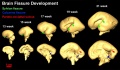
 Brain fissure development 01.jpg 1,157 × 502; 47 KB
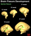
 Brain fissure development 02.jpg 1,000 × 583; 52 KB
 Brain fissure development 03.jpg 600 × 691; 33 KB
 Brain growth and birth size.jpg 800 × 492; 70 KB
 Brain histology 01.jpg 480 × 600; 125 KB
 Brain histology 02.jpg 480 × 600; 51 KB
 Brain stem subdivisions 01.jpg 1,530 × 520; 168 KB
 Brain tract development 01.jpg 720 × 1,023; 76 KB
 Brain tract development 02.jpg 1,000 × 424; 29 KB
 Brain tract development 06.jpg 1,000 × 605; 35 KB


