Week 1
| Embryology - 4 Jul 2026 |
|---|
| Google Translate - select your language from the list shown below (this will open a new external page) |
|
العربية | català | 中文 | 中國傳統的 | français | Deutsche | עִברִית | हिंदी | bahasa Indonesia | italiano | 日本語 | 한국어 | မြန်မာ | Pilipino | Polskie | português | ਪੰਜਾਬੀ ਦੇ | Română | русский | Español | Swahili | Svensk | ไทย | Türkçe | اردو | ייִדיש | Tiếng Việt These external translations are automated and may not be accurate. (More? About Translations) |
Introduction

Key Events of Human Development during the first week (week 1) following fertilization or clinical gestational age GA week 3, based upon the last menstrual period.
The first week of human development begins with fertilization of the egg by sperm forming the first cell, the zygote. Cell division leads to a ball of cells, the morula. Further cell division and the formation of a cavity in the ball of cells forms the blastocyst. These notes also cover events before fertilization formation of both the egg and sperm, gametogenesis.
Initially, there is a halving of chromosomal content in the gametes (spermatozoa and oocyte) by the process called gametogenesis. Chromosomal content is then restored by fertilization, allowing genetic recombination to occur. This is then followed by a series of cell divisions without cytoplasmic growth. During this first week the egg, then zygote, morula then the blastula is moving along the uterine horn into the uterus for implantation in the uterine wall.
Implantation also begins in this first week, but will be covered in Week 2 notes, as the implantation process is completed by the end of the second week.
| Week 1 Links: stage 1 | stage 2 | stage 3 | menstrual cycle | fertilization | zygote | morula | blastocyst | Lecture - Fertilization | meiosis | mitosis | Lecture - Week 1 and 2 | menstrual cycle | oocyte | spermatozoa | twinning | Genetic risk maternal age | Trisomy 21 | Trisomy 18 | Trisomy 13 | hydatidiform mole | GA week 3 |
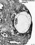
Carnegie Stages
| Week: | 1 | 2 | 3 | 4 | 5 | 6 | 7 | 8 |
| Carnegie stage: | 1 2 3 4 | 5 6 | 7 8 9 | 10 11 12 13 | 14 15 | 16 17 | 18 19 | 20 21 22 23 |
Historic Carnegie Stages 1 to 4.
| Stage 2 Links: Week 1 | Morula | Blastocyst | Mitosis | Zona pellucida | Lecture | Medicine Practical | Science Practical | Next Stage 3 |
| Historic Papers: 1956 |
| Stage 3 Links: Week 1 | zona pellucida | blastocyst | trophoblast | mitosis | Lecture | Medicine Practical | Science Practical | Blastocyst Day 3-6 Movie | Next Stage 4 |
| Historic Papers: 1954 | 1956 |
| Stage 4 Links: Week 2 | Implantation | | Trophoblast | Human Chorionic Gonadotropin | Lecture | Practical | Category:Carnegie Stage 4 | Next Stage 5 |
|
|
|
| |
| Stage 1 | Stage 2 | Stage 3 | Stage 4 | Stage 5 |
Some Recent Findings
|
Reading

- Human Embryology (2nd ed.) Larson Chapter 1 pp1-32
- The Developing Human: Clinically Oriented Embryology (6th ed.) Moore and Persaud
- Before we Are Born (5th ed.) Moore and Persaud Chapter 2 pp14-33
- Essentials of Human Embryology Larson Chapter 1 pp1-16
- Human Embryology Fitzgerald and Fitzgerald Chapter 2 pp8-14
Week 1 Movies
|
|
|
|
|
|
|
Human Blastocyst
|
|
|
|
|
|
Human blastocyst week 1 movies, 3 above movies together in single table.
Mouse Zygote
|
|
|
|
|
Models
|
|
- Links: Movies
Zygote
- male and female pronuclei, 2 nuclei approach each other and nuclear membranes break down
- DNA replicates, first mitotic division
- sperm contributes centriole which organizes mitotic spindle

|

|
| Human zygote pronuclei | Mouse zygote pronuclei[2] |
Movie - Pronuclear Fusion | Movie - Parental Genomes
Conceptus - term refers to all material derived from this fertilized zygote and includes both the embryo and the non-embryonic tissues (placenta, fetal membranes).
Epigenetics

Within the early zygote, at the 2 pronuclei stage, the male pronucleus is "reprogrammed" by the demethylation of the paternal genome. Image sequence shows the mouse zygote at pronuclear stages[2], where the male pronucleus initially contains methylcytosine (5mC, red) oxidises to form hydroxymethylcytosine (5hmC, green).
- PN1/PN2 (Early)
- PN3 (Mid)
- PN4/PN5 (Late)
5mC - 5-methylcytosine (red). 5hmC - 5-hydroxymethylcytosine (green) formed by enzymatic oxidation of 5mC.
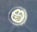
Cleavage of Zygote
Mouse zygote mitosis[2]

|

|
| First metaphase | First anaphase |
Cleavage of the zygote forms 2 blastomeres and is cleavage with no cytoplasm synthesis.
- special "embryonic" cell cycle S phases and M phases alternate without any intervening G1 or G2 phases (MSMSMSMS, adult MG1SG2) therefore individual cell volume decreases
Cell division within these cells is initially synchronous (at the same time), then becomes asynchronously (at different times).
- slow- centre cells, larger fast- peripheral cells
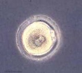
Morula

- about day 4 is a solid ball of 16-20 cells with peripheral cells flattened against zona pellucida
- compaction occurs forming a cavity and leading to the next blastocyst stage
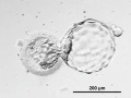
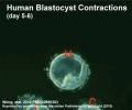
Blastocyst
- about day 5 have 2 identifiable cell types and a fluid-filled cavity (blastoceol)
- trophoblast layer - peripheral flattened cells, forms the placenta and placental membranes
- inner cell mass - embryoblast, mass of rounder cells located on one wall of the blastocoel, forms entire embryo
Blastula Cell Communication
Two forms of cellular junctions Figure 21-69. The blastula
- gap junctions, allow electrically couple cells of epithelium surrounding a fluid-filled cavity
- tight junctions, close to outer surface create a seal, isolates interior of embryo from external medium
Blastocyst Hatching - zona pellucida lost, ZP has sperm entry site, and entire ZP broken down by uterine secretions and possibly blastula secretions. Uterine Glands - secretions required for blastocyst motility and nutrition
- Links: Blastocyst Development | Carnegie stage 3
Summary of the first week following fertilization
<html5media height="260" width="660">File:Week1_001.mp4</html5media> Click Here to play on mobile device
| |||
|
Molecular Changes
There are several important changes that occur in this new diploid cell beginning the very first mitotic cell divisions and expressing a new genome.
The oocyte arrested in meiosis is initially quiescent in terms of gene expression, and many other animal models of development have shown maternal RNAs and proteins to be important for early functions.
The new zygote gene expression is about cycles of mitosis and maintaining the toptipotency of the stem cell offspring cells.
The morula gene expression supports the formation of two populations of cells the trophoblast (trophectoderm) and embryoblast (inner cell mass), each having different roles in development, while maintaining the toptipotency of these populations.
Current research is now also pointing to non-genetic mechanisms or epigenetics as an additional mechanism in play in these processes.
Genome Expression
The following figure is from a recent study[4] using video and genetic analysis of in vitro human development during week 1 following fertilization.

- EGA - embryonic genome activation
- ESSP - embryonic stage–specific pattern, four unique embryonic stage–specific patterns (1-4)
Telomere Length
A recent paper has measured telomere length in human oocyte (GV, germinal vesicle), morula and blastocyst and found changes in this length in preimplantation embryos.[5] Telomeres are the regions found at the ends of each chromosome and involved in cellular ageing and the capacity for division. The regions consist of repeated sequences protecting the ends of chromosomes and harbour DNA repair proteins. In the absence of the enzyme telomerase, these regions shorten during each cell division and becoming critically short, cell senescence occurs.

|

|
| Early human telomeres[5] | Early human telomere length[5] |
Timeline
| Event | ||
| Secretory Phase Stage 1 |  Fertilization, Zygote, Secretory Phase Fertilization, Zygote, Secretory Phase
| |
| Stage 2 |  | |

| ||
| Stage 3 | 
Blastocyst Hatching (zona pellucida lost) Blastocyst (free floating) | |
| Stage 4 | Adplantation | |
| Stage 5 |  |
Week 1 Movies
| Ovulation | Fertilization | Pronuclear Fusion | Week 1 | Ovulation in the rabbit | Fertilization in the mouse |
Week 1 Abnormalities
Dizygotic Twinning
Dizygotic twins (fraternal, non-identical) arise from separate fertilization events involving two separate oocyte (egg, ova) and spermatozoa (sperm).
Monoygotic Twinning
Monozygotic twins (identical) produced from a single fertilization event (one fertilised egg and a single spermatazoa, form a single zygote), these twins therefore share the same genetic makeup. Occurs in approximately 3-5 per 1000 pregnancies, more commonly with aged mothers. The later the twinning event, the less common are initially separate placental membranes and finally resulting in conjoined twins.
| Week | Week 1 | Week 2 | |||||||||||||
| Day | 0 | 1 | 2 | 3 | 4 | 5 | 6 | 7 | 8 | 9 | 10 | 11 | 12 | 13 | 14 |
| Cell Number | 1 | 1 | 2 | 16 | 32 | 128 | bilaminar | ||||||||
| Event | Ovulation | fertilization | First cell division | Morula | Early blastocyst | Late blastocyst
Hatching |
Implantation starts | X inactivation | |||||||
|
|

|
|
|||||||||||||
| Monoygotic
Twin Type |
Diamniotic
Dichorionic |
Diamniotic
Monochorionic |
Monoamniotic
Monochorionic |
Conjoined | |||||||||||
Table based upon: Twinning. Hall JG. [6]
References
- ↑ He K, Zhao H, Wang Q & Pan Y. (2010). A comparative genome analysis of gene expression reveals different regulatory mechanisms between mouse and human embryo pre-implantation development. Reprod. Biol. Endocrinol. , 8, 41. PMID: 20459759 DOI.
- ↑ 2.0 2.1 2.2 Iqbal K, Jin SG, Pfeifer GP & Szabó PE. (2011). Reprogramming of the paternal genome upon fertilization involves genome-wide oxidation of 5-methylcytosine. Proc. Natl. Acad. Sci. U.S.A. , 108, 3642-7. PMID: 21321204 DOI.
- ↑ Zhang P, Zucchelli M, Bruce S, Hambiliki F, Stavreus-Evers A, Levkov L, Skottman H, Kerkelä E, Kere J & Hovatta O. (2009). Transcriptome profiling of human pre-implantation development. PLoS ONE , 4, e7844. PMID: 19924284 DOI.
- ↑ Wong CC, Loewke KE, Bossert NL, Behr B, De Jonge CJ, Baer TM & Reijo Pera RA. (2010). Non-invasive imaging of human embryos before embryonic genome activation predicts development to the blastocyst stage. Nat. Biotechnol. , 28, 1115-21. PMID: 20890283 DOI.
- ↑ 5.0 5.1 5.2 Turner S, Wong HP, Rai J & Hartshorne GM. (2010). Telomere lengths in human oocytes, cleavage stage embryos and blastocysts. Mol. Hum. Reprod. , 16, 685-94. PMID: 20573647 DOI.
- ↑ Hall JG. (2003). Twinning. Lancet , 362, 735-43. PMID: 12957099 DOI.
Niakan KK & Eggan K. (2013). Analysis of human embryos from zygote to blastocyst reveals distinct gene expression patterns relative to the mouse. Dev. Biol. , 375, 54-64. PMID: 23261930 DOI.
Embryo Week: Week 1 | Week 2 | Week 3 | Week 4 | Week 5 | Week 6 | Week 7 | Week 8 | Week 9
- Carnegie Stages: 1 | 2 | 3 | 4 | 5 | 6 | 7 | 8 | 9 | 10 | 11 | 12 | 13 | 14 | 15 | 16 | 17 | 18 | 19 | 20 | 21 | 22 | 23 | About Stages | Timeline
Glossary Links
- Glossary: A | B | C | D | E | F | G | H | I | J | K | L | M | N | O | P | Q | R | S | T | U | V | W | X | Y | Z | Numbers | Symbols | Term Link
Cite this page: Hill, M.A. (2026, July 4) Embryology Week 1. Retrieved from https://embryology.med.unsw.edu.au/embryology/index.php/Week_1
- © Dr Mark Hill 2026, UNSW Embryology ISBN: 978 0 7334 2609 4 - UNSW CRICOS Provider Code No. 00098G