Coelomic Cavity Development
| Embryology - 28 Jul 2026 |
|---|
| Google Translate - select your language from the list shown below (this will open a new external page) |
|
العربية | català | 中文 | 中國傳統的 | français | Deutsche | עִברִית | हिंदी | bahasa Indonesia | italiano | 日本語 | 한국어 | မြန်မာ | Pilipino | Polskie | português | ਪੰਜਾਬੀ ਦੇ | Română | русский | Español | Swahili | Svensk | ไทย | Türkçe | اردو | ייִדיש | Tiếng Việt These external translations are automated and may not be accurate. (More? About Translations) |
Introduction

- coelom - (Greek, koilma = cavity) Term used to describe a fluid-filled cavity or space. Placental vertebrate development have both extra-embryonic (outside the embryo) and intra-embryonic (inside the embryo) coeloms.
The extra-embryonic coeloms include the yolk sac, amniotic cavity and the chorionic cavity information on these spaces can also be found on placenta development pages.
The intra-embryonic coelom (coelomic cavity) forms within the lateral plate mesoderm early in embryonic development (week 3-4 (GA 5-6). This single space wall will undergo amesenchymal epithelial transition. The space then undergoes complex morphological changes of folding and partitioning during the development of the 3 major body cavities (pleural, pericardial and peritoneal). Portions of the coelomic cavity wall will also contribute to other organs (adrenal, ovary, testis).
All intra-embryonic cavities are fluid filled and developing organs push against a wall of the cavity, generating a double coat (serosal/adventital) surrounding an organ (for example the lungs). The serous membrane is the epithelium (squamous) and its associated underlying loose connective tissue.
| Coelom Links: Introduction | Lecture - Week 3 Development | Lecture - Mesoderm Development | Placenta - Membranes | Category:Coelomic Cavity |
| Historic Embryology - Coelomic Cavity |
|---|
| 1891 peritoneal | 1897 human coelom | 1910 Coelom and Diaphragm | 1924 serous |
Some Recent Findings
|
| More recent papers |
|---|
 This table allows an automated computer search of the external PubMed database using the listed "Search term" text link.
More? References | Discussion Page | Journal Searches | 2019 References | 2020 References Search term: Coelomic Cavity Development | Amniotic Cavity Development | Yolk Sac Development | Cardiac Cavity Development | Pleural Cavity Development | Peritoneal Cavity Development | mesothelium | coelomic portals |
| Older papers |
|---|
| These papers originally appeared in the Some Recent Findings table, but as that list grew in length have now been shuffled down to this collapsible table.
See also the Discussion Page for other references listed by year and References on this current page.
|
Objectives
- Describe the development of the intra- and extra-embryonic coeloms.
- Describe the processes involved in the development of the three divisions of the intra-embryonic coelom; pericardium, pleural cavities and peritoneum.
- Describe the fate of the extra embryonic coelom.
- Describe the development of the diaphragm.
Development Overview
| Coelomic Cavities | ||
| Extra-embryonic Coelom | Intra-embryonic Coelom | Other (fluid-filled spaces) |
|---|---|---|
| outside the embryo | inside the embryo | between and within embryo |
| chorionic sac | pericardial | coelomic portals - transient communication between extra and intra |
| amniotic sac | pleural | ventricular - cavities within and surrounding central nervous system |
| yolk sac | peritoneal | renal - cavities within the kidney and bladder |
Extra-embryonic Coelom


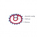
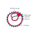
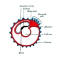
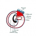
Amniotic cavity
The fluid-filled (amniotic fluid) extraembryonic coelom (cavity) formed initially by epiblast and then lined by ectoderm and surrounding extraembryonic mesoderm. In humans, it forms the innermost fetal membrane, produces amniotic fluid expanding to eventually fuse with the chorionic membrane during week 8 of development. This fluid-filled sac initially lies above the trilaminar embryo disc and with embryonic disc folding this sac is drawn ventrally to enclose (cover) the entire embryo, then fetus. The presence of this membrane led to the description of reptiles, bird, and mammals as "amniotes".
Chorionic cavity
The fluid-filled extra-embryonic coelom (cavity) formed initially from trophoblast and extra-embryonic mesoderm that forms placenta. The chorion and amnion are made by the somatopleure. The chorion becomes incorporated into placental development. The avian and reptilian chorion lies beside the egg shell and allows gas exchange. In humans, this cavity is lost during week 8 when the amniotic cavity expands and fuses with the chorion.
Yolk sac
An extra-embryonic membrane which is endoderm origin and covered with extra-embryonic mesoderm. Yolk sac lies outside the embryo connected initially by a yolk stalk to the midgut with which it is continuous with. The endodermal lining is continuous with the endoderm of the gastrointestinal tract. The extra-embryonic mesoderm differentiates to form both blood and blood vessels of the vitelline system. In reptiles and birds, the yolk sac has a function associated with nutrition. In mammals the yolk sac acts as a source of primordial germ cells and blood cells. Note that in early development (week 2) a structure called the "primitive yolk sac" forms from hypoblast, this is an entirely different structure.
Allantois

The allantois is present in reptiles, birds, acts in respiration and waste storage. In mammals, other than marsupials, associated with chorion blood vessel development and the urinary bladder.
The human allantois forms from Carnegie stage 7 forms an endoderm cloacal diverticulum adjacent to the yolk sac. It extends from the umbilicus into the connecting stalk. Vascular development occurs within the adjacent connecting stalk mesoderm associated with placental cord vessel development. Separate vascular development of the allantois occurs initially in the distal tip mesoderm.
Later septation of the cloacal has the allantois associated ventrally with the urogenital sinus. It lies at the superior end of the primitive urinary bladder and forms the urachus. Postnatally, the urachus degenerates to form a fibrous ligament (median umbilical ligament) between the urinary bladder to the umbilical region.
Intra-embryonic Coelom

The intra-embryonic coelom forms as a single cavity appearing in the lateral plate mesoderm during week 3. This single cavity (coelom) divides the lateral plate into the somatic and splanchnic mesoderm and will later be portioned into the three main body cavities: pericardial, pleural and peritoneal cavities.

Week 5 pericardial and peritoneal cavities
Pericardial Cavity
Pleural Cavity

Reflections of the Pleura
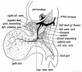
Peritoneal Cavity
Embryo Stage 13 Peritoneal Cavity

Week 8
Pleural

Peritoneal

Mesentery and Retroperitoneal

Stages in the development of the bursa omentalis, the greater omentum, and the fusion of the latter with the transverse mesocolon.

Mesothelium
The epithelial covering of coelomic organs and also line their cavities.
- Contribute to the vasculature of the heart and the intestinal tract.
- Undergo epithelial-mesenchymal transition (EMT), migration, and differentiation into endothelial cells, vascular smooth muscle cells, and pericytes.
- contribute most of the vascular smooth muscle to the respiratory and gastrointestinal tract. (Remainder derived from endothelium?)
References
- ↑ Carmona R, Ariza L, Cano E, Jiménez-Navarro M & Muñoz-Chápuli R. (2018). Mesothelial-mesenchymal transitions in embryogenesis. Semin. Cell Dev. Biol. , , . PMID: 30243860 DOI.
- ↑ Aiello D, Giambona A, Leto F, Passarello C, Damiani G, Maggio A, Siciliano C & Napoli A. (2018). Human coelomic fluid investigation: A MS-based analytical approach to prenatal screening. Sci Rep , 8, 10973. PMID: 30030477 DOI.
- ↑ Ariza L, Carmona R, Cañete A, Cano E & Muñoz-Chápuli R. (2016). Coelomic epithelium-derived cells in visceral morphogenesis. Dev. Dyn. , 245, 307-22. PMID: 26638186 DOI.
- ↑ Renda MC, Giambona A, Fecarotta E, Leto F, Makrydimas G, Renda D, Damiani G, Jakil MC, Picciotto F, Piazza A, Valtieri M & Maggio A. (2010). Embryo-fetal erythroid megaloblasts in the human coelomic cavity. J. Cell. Physiol. , 225, 385-9. PMID: 20533375 DOI.
- ↑ Menzies BR, Pask AJ & Renfree MB. (2011). Placental expression of pituitary hormones is an ancestral feature of therian mammals. Evodevo , 2, 16. PMID: 21854600 DOI.
Reviews
Hall NJ, Drewett M & Burge D. (2019). Nutritional role of amniotic fluid: clues from infants with congenital obstruction of the digestive tract. Arch. Dis. Child. Fetal Neonatal Ed. , 104, F199-F201. PMID: 29666202 DOI.
Ariza L, Carmona R, Cañete A, Cano E & Muñoz-Chápuli R. (2016). Coelomic epithelium-derived cells in visceral morphogenesis. Dev. Dyn. , 245, 307-22. PMID: 26638186 DOI.
Articles
Cindrova-Davies T, Jauniaux E, Elliot MG, Gong S, Burton GJ & Charnock-Jones DS. (2017). RNA-seq reveals conservation of function among the yolk sacs of human, mouse, and chicken. Proc. Natl. Acad. Sci. U.S.A. , 114, E4753-E4761. PMID: 28559354 DOI.
Funayama N, Sato Y, Matsumoto K, Ogura T & Takahashi Y. (1999). Coelom formation: binary decision of the lateral plate mesoderm is controlled by the ectoderm. Development , 126, 4129-38. PMID: 10457021
Search PubMed
Search Pubmed: Coelomic Cavity Development | pericardial cavity development | pleural cavity development | peritoneal cavity development
Additional Images
Historic
| Historic Disclaimer - information about historic embryology pages |
|---|
|

Yolk sac


Terms
- atresia - obstruction.
- eppiglottis - develops from hypobrachial eminence.
- fistula - abnormal comunication.
- hypopharyngeal eminence - fusion of 3rd pharyngeal arches, precursor of root of tongue.
- laryngotracheal groove - forms on anterior (ventral) wall of pharynx, gives rise to larynx, trachea, respiratory tree.
- larynx - lining from endoderm, cartilage from pharyngeal arch 4 and 6.
- lung buds - primordia of lungs.
- parietal pleura - outer lining of pleural cavity derived from epithelia of pericardioperitoneal canals from intra-embryonic coelom.
- pleural cavity - walls derived from pericardioperitoneal canals -> intra-embryonic coelom ->coelomic spaces -> lateral mesoderm -> mesoderm.
- pleuropericardial fold - restricts the communication between pleural cavity and pericardiac cavity, contains cardinal vein and phrenic nerve.
- pleuroperitoneal membrane - forms inferiorly at transverse septum to separate peritoneum from pleural cavity.
- septum transversum- mesoderm separating thoracic cavity and yolk sac, forms central tendon of diaphragm (and some of liver?).
- stenosis - narrowing
- surfactant - a detergent secreted by Type 2 alveolar cells between alveolar epithelium. Functions to lower surface tension, allowing lungs to remain inflated.
- visceral pleura - inner lining of pleural cavity derived from contact epithelia with lung bud of pericardioperitoneal canals from intra-embryonic coelom.
| System Links: Introduction | Cardiovascular | Coelomic Cavity | Endocrine | Gastrointestinal Tract | Genital | Head | Immune | Integumentary | Musculoskeletal | Neural | Neural Crest | Placenta | Renal | Respiratory | Sensory | Birth |
Glossary Links
- Glossary: A | B | C | D | E | F | G | H | I | J | K | L | M | N | O | P | Q | R | S | T | U | V | W | X | Y | Z | Numbers | Symbols | Term Link
Cite this page: Hill, M.A. (2026, July 28) Embryology Coelomic Cavity Development. Retrieved from https://embryology.med.unsw.edu.au/embryology/index.php/Coelomic_Cavity_Development
- © Dr Mark Hill 2026, UNSW Embryology ISBN: 978 0 7334 2609 4 - UNSW CRICOS Provider Code No. 00098G