Carnegie stage 14
| Embryology - 5 May 2026 |
|---|
| Google Translate - select your language from the list shown below (this will open a new external page) |
|
العربية | català | 中文 | 中國傳統的 | français | Deutsche | עִברִית | हिंदी | bahasa Indonesia | italiano | 日本語 | 한국어 | မြန်မာ | Pilipino | Polskie | português | ਪੰਜਾਬੀ ਦੇ | Română | русский | Español | Swahili | Svensk | ไทย | Türkçe | اردو | ייִדיש | Tiếng Việt These external translations are automated and may not be accurate. (More? About Translations) |
Introduction

Facts
Week 5, 31 - 35 days, 5 - 7 mm
Gestational Age GA - week 7
View: Lateral view. Amniotic membrane removed.
Summary
- Ectoderm: sensory placodes, lens pit, otocyst, nasal placode, primary/secondary vesicles, fourth ventricle of brain,
- Mesoderm: continued segmentation of paraxial mesoderm (more than 30 somite pairs), heart prominence
- Head: 1st, 2nd and 3rd pharyngeal arch, forebrain, site of lens placode, site of otic placode, stomodeum
- Body: heart, liver, umbilical cord, mesonephric ridge
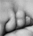
- Limb: upper and lower limb buds
See also Carnegie stage 14 Events
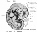
Features
- midbrain, nasal placode, lens pit, 1,2,3 pharyngeal arches, fourth ventricle of brain, 1st pharyngeal groove, heart prominence, cervical sinus, upper limb bud, mesonephric ridge, lower limb bud, umbilical cord.
- Identify: midbrain region, nasal placode, lens pit, 1st, 2nd and 3rd pharyngeal arches, 1st pharyngeal groove, maxillary and mandibular components of 1st pharyngeal arch, fourth ventricle of brain, heart prominence, cervical sinus, upper limb bud, mesonephric ridge, lower limb bud, umbilical cord.
| Week: | 1 | 2 | 3 | 4 | 5 | 6 | 7 | 8 |
| Carnegie stage: | 1 2 3 4 | 5 6 | 7 8 9 | 10 11 12 13 | 14 15 | 16 17 | 18 19 | 20 21 22 23 |
- Carnegie Stages: 1 | 2 | 3 | 4 | 5 | 6 | 7 | 8 | 9 | 10 | 11 | 12 | 13 | 14 | 15 | 16 | 17 | 18 | 19 | 20 | 21 | 22 | 23 | About Stages | Timeline
Bright Field
| Lateral view | |
|---|---|

|

|
| Ventral view | |

|

|
Embryo Virtual Slide
|
|


Scanning EM


Dorsolateral view
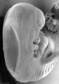
Cranial end
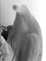
Cloacal region
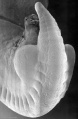
Caudal end


Image Source: Scanning electron micrographs of the Carnegie stages of the early human embryos are reproduced with the permission of Prof Kathy Sulik, from embryos collected by Dr. Vekemans and Tania Attié-Bitach. Images are for educational purposes only and cannot be reproduced electronically or in writing without permission.
Kyoto Collection

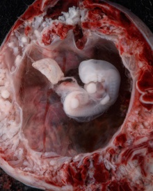
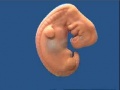
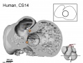
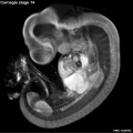
Lateral view of embryo surface at Carnegie stage 14.


|

|
| ||||||||||||||||||||||||||||||||||||||||||||||||||||||||||||||||||||||
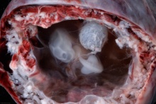
| Lateral view of the central nervous system of embryo at Carnegie stage 14 (Scale bar is 1 mm). | ||||||||||||||||||||||||||||||||||||||||||||||||||||||||||||||||||||||||
Carnegie Collection


- Carnegie stage 14: 6830 left | 1380 left | 7333 left | 6502 left | 5654 left | 4154 left | 4629 right | 7394 right | 6848 left | 7400 right | 7394 left | 1620 left | 7400 left
| Carnegie Collection Embryos - Stage 14 | ||||||||||
|---|---|---|---|---|---|---|---|---|---|---|
| Serial No. | Size (mm) | Grade | Fixative | Embedding Medium | Plane | Thinness (µm) | Stain | Year | Notes | |
| 4 | E.,7 | Poor | p | P | Transverse | 10 | Al. coch. | 1892 | ||
| 18 | E.,7 Ch., 18x18 | Poor | p | P | Transverse | 20 | Al. coch. | 1895 | ||
| 80 | E., 5.0 Ch., 24x18x8 | Good | Alc. | P | Transverse | 20 | Al. coch. | 1897 | ||
| 187 | E.,7 Ch., 35x30x25 | Poor | ? | P | Sagittal | 20 | Al. coch. | 1902 | ||
| 1208 | E.,7 Ch., 22x11Xx1 | Poor | ? | P | Sagittal | 20 | Al. coch. | 1902 | ||
| 245 | E.,6 Ch., 13x12x10 | Poor | Formol, Zenker | ? | Transverse | 5 | (Stain - Haematoxylin Eosin) | 1904 | ||
| 372 | E.,7 | Fair | p | P | Transverse | 10 | H.-Congo red | 1902 | ||
| 380 | E,6 Ch., 20x20x14 | Poor | p | P | Sagittal | 20 | (Stain - Haematoxylin Eosin) | 1906 | ||
| 387 | E.,7 Ch., 45x40x50 | Good | Formalin | P | Transverse | 20 | (Stain - Haematoxylin Eosin) | 1907 | ||
| 442 | E.,6 Ch., 25x20 | Poor | Formalin | P | Coronal? | 50 | Al. coch. | 1908 | ||
| 552 | E.,6 Ch, 40x28Xx8 | Poor | p | P | Sagittal | 40 | Al. coch. | 1911 | Possible anencephaly | |
| 560 | E., 7.0 Ch, 24x24 | Poor | Formalin | P | Coronal | 40 | Al. coch. | 1912 | ||
| 676 | E., 6.0 Ch, 35x20x17 | Good | Carnoy | P | Tr.-Coronal | 20 | (Stain - Haematoxylin Eosin) | 1913 | Possible spina bifida | |
| 873 | E,6.0 Ch., 35x28x16 | Poor | Formalin | P | Sagittal | 20 | Al. coch. | 1914 | ||
| 988 | E,6.0 Ch., 38x30x23 | Good | Formol-corros. acetic | P | Transverse | 20 | Al. coch. | 1914 | ||
| 1380 | E , 5.7 Ch, 36x24x24 | Exc. | Formalin | P | Coronal | 20 | Al. coch. | 1916 | ||
| 1620 | E, 6.6 Ch, 35x30x8 | Good | Formalin | P | Sagittal | 20 | Al. coch. | 1916 | ||
| ?? | E, 6.68 | Fair | ? | ? | Transverse | 6 | Al. coch., or. G | 1919 | ||
| 2841 | E , sy Ch. 35x21 | Good | Alc. | P | Transverse | 20 | (Stain - Haematoxylin Eosin) or.G | 1920 | ||
| 3360 | E.6.0 | Good | Formalin | C | Transverse | 20 | (Stain - Haematoxylin Eosin) or.G | 1920 | In myomatous uterus. Advanced. | |
| 3805 | E., 5.9 | Exc. | Bouin | P | Transverse | 15 | (Stain - Haematoxylin Eosin) | 1921 | Evans embryo No. 168. Serial bromides only | |
| 3960 | E., 5.5 | Good | Formalin | C-P | Coronal | 20 | Al. coch. | 1922 | Blood vessels naturally injected | |
| 4154 | E, 6.8 Ch., 33x31x20 | Poor | Alc. | C-P | Transverse | 8 | (Stain - Haematoxylin Eosin) | 1923 | ||
| 4245-6 | E., 7.0 | Good | Formalin | P | Transverse | 15 | Al. coch. | 1923 | Univ. Pennsylvania No. 40. Ag added | |
| 4692 | E., 6.5 Ch., 32x23 | Good | Formalin | C-P | Sagittal | 10 | (Stain - Haematoxylin Eosin) | 1924 | ||
| 4672 | E,8.2 Ch., 40X34X25 | Good | Formalin | P | Transverse | 20 | Al. coch. | 1924 | Advanced | |
| 4805 | E., 7.3 Ch., 15X8X9 | Good | Formalin | C-P | Transverse | 10 | (Stain - Haematoxylin Eosin) | 1924 | Tubal | |
| 5437 | E., 7.0 | Good | Formalin | C-P | Transverse | 8 | (Stain - Haematoxylin Eosin) | 1927 | Advanced | |
| 5654 | E., 5.0 Ch., 30x23x17 | Good | Formalin | P | Transverse | 10 | Al. coch., eosin | 1928 | Less advanced | |
| 5787 | E., 6.8 Ch., 32x30x23 | Good | Formalin | P | Sagittal | 10 | Al. coch., eosin | 1928 | ||
| 6428 | E., 7.0 Ch., 30X28X25 | Good | Formalin | C-P | Coronal | 6, 10 | Al. coch. | 1931 | Advanced | |
| 6500 | E, 4.9* | Good | Souza? | C-P | Sagittal | 10 | Al. coch. | 1931 | E. Leitz, Berlin | |
| 6502 | E., 6.7* | Exc. | Souza? | C-P | Transverse | 5, 10 | (Stain - Haematoxylin Eosin) | 1931 | E. Leitz, Berlin. Ag added to slides 1-25 | |
| 6503 | E., 6.3* | Exc. | Souza? | C-P | Coronal | 10 | Al. coch. | 1931 | E. Leitz, Berlin | |
| 6739 | E.,8 | Poor | Formalin | C-P | Sagittal | 20 | (Stain - Haematoxylin Eosin) | 1933 | ||
| 6830 | E,5.5 Ch., 47x23x15 | Exc. | Formalin | C-P | Coronal | 8 | (Stain - Haematoxylin Eosin) | 1933 | ||
| 6848 | E., 7.8 | Good | Formalin | C-P | Coronal | 10 | (Stain - Haematoxylin Eosin) | 1934 | Tubal | |
| 7324 | E, 6.6 Ch., 17x13x10 | Good | Formalin | C-P | Transverse | 8 | (Stain - Haematoxylin Eosin) | 1936 | Low implantation | |
| 7333 | E, 6.3 | Good | Formalin | C-P | Transverse | 8 | (Stain - Haematoxylin Eosin) | 1936 | ||
| 7394 | E, 7.2 Ch., 45x20x20 | Exc. | Formalin | C-P | Transverse | 8 | (Stain - Haematoxylin Eosin) | 1937 | ||
| 7400 | E, 6.3 Ch., 35x25x20 | Good | Formalin | C-P | Coronal | 10 | (Stain - Haematoxylin Eosin) | 1937 | ||
| 7522 | E., 7.7 Ch., 33x16x16 | Good | Formalin | C-P | Transverse | 8 | (Stain - Haematoxylin Eosin) | 1938 | Natural blood injection | |
| 7598 | E., 7.0 Ch., 30x30x25 | Poor | Alc. | C-P | Transverse | 10 | (Stain - Haematoxylin Eosin) | 1938 | Macerated | |
| 7667 | E., 5 Ch., 16x14x12 | Fair | Formalin | P | Transverse | 8 | (Stain - Haematoxylin Eosin), phlox. | 1939 | ||
| 7829 | E., 7.0 | Exc. | Bouin | C-P | Transverse | 8 | (Stain - Haematoxylin Eosin) | 1940 | Advanced | |
| 7870 | E,7.2 Ch., 25x20x13 | Exc. | Bouin | C-P | Transverse | 8 | (Stain - Haematoxylin Eosin) | 1941 | On borderline of next stage. Ag added | |
| 8141 | E,7.3 Ch.,33x28 | Exc. | C-P | Coronal | 8 | (Stain - Haematoxylin Eosin) | 1943 | Shrinkage cracks in brain | ||
| 8306 | E.5.3 Ch., 27 | Exc. | Bouin | C-P | Transverse | 10, 20 | (Stain - Haematoxylin Eosin), phlox. | 1945 | ||
| 8308 | E, 5.85 Ch., 27x18x18 | Exc. | Formol & Bouin | C-P | Sagittal | 8 | Azan | 1945 | ||
| 8314 | E,8 Ch.,23x22 | Exc. | Formol | C-P | Transverse | 8 | Azan | 1945 | ||
| 8357 | E., 6.5 | Good | Formol | C-P | Sagittal | 8 | Azan | 1946 | ||
| 8552 | E,6.5 | Exc. | Alc. & Bouin | C-P | Transverse | 8 | Azan | 1947 | ||
| 8999 | E,6 Ch.,16x15 | Exc. | Alc. & Bouin | C-P | Sagittal | 8 | Azan | 1952 | ||
| 9695 | E,8.5 | ? | 1955 | Not cut | ||||||
Abbreviations
| ||||||||||
| iBook - Carnegie Embryos | |
|---|---|

|
|
Hinrichsen Collection
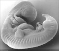
| Hinrichsen collection Human Embryo ME52 (stage14) CRL 6.6 mm. Embryo right lateral view.
|
|


Image source: The Hinrichsen Collection images are reproduced with the permission of Prof. Beate Brand-Saberi, Head, Department of Anatomy and Molecular Embryology, Ruhr-Universität Bochum. Images are for educational purposes only and cannot be reproduced electronically or in writing without permission.
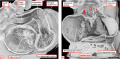
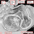
Embryo ME65 Carnegie stage 14, CRL 6.2 mm, Transverse Heart, (Semi-thin sections, Stain toluidine blue, total slides 37).
Digital Embryology Consortium - Hinrichsen Collection
Image source: The Hinrichsen Collection images are reproduced with the permission of Prof. Beate Brand-Saberi, Head, Department of Anatomy and Molecular Embryology, Ruhr-Universität Bochum. Images are for educational purposes only and cannot be reproduced electronically or in writing without permission.
Stage 14 Embryo Movie
|
| Stage 14 Model |
| Page | Play |
Events
- hearing - otic vesicle ventral portion elongates to form the cochlear duct and endolymphatic appendage becomes tapered.[1]
- vision - (about 32 days) the lens placode is indented by the lens pit, cup-shaped and still communicates with the surface by a narrowing pore.[2]
- Cardiovascular
- Coronary circulation plexus of blind epicardial capillaries appears on the heart.[3]
- Cerebral artery - basilar artery forms from the consolidation of the neural arteries.[4]
- endocrine[5]
- pituitary - craniopharyngeal pouch is prominent[1]) and the notochord appears to be inserted into its dorsal wall. The craniopharyngeal pouch has become elongated and blood vessels are beginning to grow in between the basement membranes of the pouch and brain (O'Rahilly 1973a).
- Epiphysis - a slight irregularity in the surface outline of the intact head corresponds to the future pineal body (O'Rahilly et al. 1982).
- thymus - Weller's (1933) "thymus" (the third pharyngeal pouch) becomes elongated.[6]
- parathyroid - "Parathyrogenic zones" (Politzer and Hann 1935) are recognizable (Streeter 1945). The parathyroid 4 primordium has been illustrated at this stage by Weller (Fig. 16)[6].
- thyroid - thyroid pedicle shows further elongation but is still connected to the epithelium of the pharynx.[6] Right and left lobes and an isthmus may perhaps be presaged (ibid.).
- adrenal cortex - A change in the characteristics of the cells of the coelomic epithelium appears between the mesogastrium and the lateral end of the mesonephros.[7]
- adrenal medulla - paravertebral sympathetic ganglia increase in size as a result of cell division and the addition of nerve fibres from the rami communicantes. The ganglia contain three types of cells: MI, M2, and M3. The M3 cells are the" parasympathetic cells" of Zuckerkandl.[7]
- pancreas - ventral pancreas (which may perhaps be distinguishable as early as stage 13) appears as an evagination from the bile duct at stages 14[8]( and 15.[9]. It is generally described as unpaired but, at least in some cases, may perhaps be bilobed[10] or even multiple.[11]
- meninges (spinal cord) - xiii and xiv (embryos of 4 to 8 mm.), advances are particularly evident in the vertebral rudiments and in the spinal ganglia, with the latter starting their migration ventrad. The vascularization of the tissues directly adjacent to the neural tube has continued, and endothelium-lined channels are beginning to form in the older embryos.[12]
- genital
- primordial germ cells are migrating into the genital ridges on the medial surface of mesonephros.[13]
References
- ↑ 1.0 1.1 Streeter GL. Developmental horizons in human embryos. Description of age group XIII, embryos about 4 or 5 millimeters long, and age group XIV, period of indentation of the lens vesicle. (1945) Carnegie Instn. Wash. Publ. 557, Contrib. Embryol., Carnegie Inst. Wash., 31: 27-63.
- ↑ Pearson AA. (1980). The development of the eyelids. Part I. External features. J. Anat. , 130, 33-42. PMID: 7364662
- ↑ Hutchins GM, Kessler-Hanna A & Moore GW. (1988). Development of the coronary arteries in the embryonic human heart. Circulation , 77, 1250-7. PMID: 3286038
- ↑ Menshawi K, Mohr JP & Gutierrez J. (2015). A Functional Perspective on the Embryology and Anatomy of the Cerebral Blood Supply. J Stroke , 17, 144-58. PMID: 26060802 DOI.
- ↑ O'Rahilly R. The timing and sequence of events in the development of the human endocrine system during the embryonic period proper. (1983) Anat. Embryol., 166: 439-451. PMID 6869855
- ↑ 6.0 6.1 6.2 Weller GL. Development of the thyroid, parathyroid and thymus glands in man. (1933) Contrib. Embryol., Carnegie Inst. Wash. 24: 93-139.
- ↑ 7.0 7.1 Crowder RE. The development of the adrenal gland in man, with special reference to origin and ultimate location of cell types and evidence in favor of the "cell migration" theory. (1957) Contrib. Embryol., Carnegie Inst. Wash. 36, 193-210.
- ↑ Blechschmidt E. Die prdnatalen Organsysteme des Menschen. (1973) Hippokrates, Stuttgart.
- ↑ Streeter GL. Developmental horizons in human embryos. Description of age groups XV, XVI, XVII, and XVIII, being the third issue of a survey of the Carnegie collection. (1948) Contrib. Embryol., Carnegie Inst. Wash. 575, 32: 133-203.
- ↑ Odgers PN. Some observations on the development of the ventral pancreas in man. (1930) J. Anat., 65(1): 1-7. PMID 17104298
- ↑ Delmas A. Les ebauches pancre'atiques dorsales et ventrales. Leurs rapports dans la constitution du pancreas definitif (Dorsal and ventral pancreatic outlines. Their relations in the constitution of the final pancreas). (1939) Ann. Anat. pathol, 16: 253-266.
- ↑ Sensenig EC. The early development of the meninges of the spinal cord in human embryos. (1951) Contrib. Embryol., Carnegie Inst. Wash. Publ. 611.
- ↑ Witschi E. Migration of the germ cells of human embryos from the yolk sac to the primitive gonadal folds. (1948) Carnegie Instn. Wash. Publ. 575, Contrib. Embryol., 32: 67-80.
- ↑ Shinohara H. Naora H. Hashimoto R. Hatta T. and Tanaka O. Development of the innervation pattern in the upper limb of staged human embryos. (1990) Acta Anat (Basel) 138: 265-269. PMID 2389673
Additional Images
Heart
- Heart
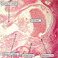
fig 7a Human CS 14 heart
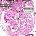
fig 7b Human CS 14 heart
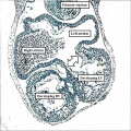
fig 13b Human CS 14 heart septum primum
EFIC outflow tract PMID 27587491
EFIC - OFT RA RV PMID 27587491
EFIC - OFT RA LA PMID 27587491
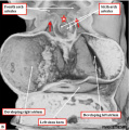
MRI Heart PMID 23755108


Various
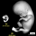
Stage 14 compare size to Stage 23
Stage 14 Optical Projection Tomography

External ear Stages 14-23 and adult


Historic
| Historic Disclaimer - information about historic embryology pages |
|---|
 |
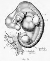
1907 Kollmann 37 somites
1921 Bailey 7mm embryo 27 somite
1922 Streeter Embryo No. 1380


- Carnegie Stages: 1 | 2 | 3 | 4 | 5 | 6 | 7 | 8 | 9 | 10 | 11 | 12 | 13 | 14 | 15 | 16 | 17 | 18 | 19 | 20 | 21 | 22 | 23 | About Stages | Timeline
Image Source: Scanning electron micrographs of the Carnegie stages of the early human embryos are reproduced with the permission of Prof Kathy Sulik, from embryos collected by Dr. Vekemans and Tania Attié-Bitach. Images are for educational purposes only and cannot be reproduced electronically or in writing without permission.
Image source: The Kyoto Collection images are reproduced with the permission of Prof. Kohei Shiota and Prof. Shigehito Yamada, Anatomy and Developmental Biology, Kyoto University Graduate School of Medicine, Kyoto, Japan for educational purposes only and cannot be reproduced electronically or in writing without permission.
Cite this page: Hill, M.A. (2026, Mayıs 5) Embryology Carnegie stage 14. Retrieved from https://embryology.med.unsw.edu.au/embryology/index.php/Carnegie_stage_14
- © Dr Mark Hill 2026, UNSW Embryology ISBN: 978 0 7334 2609 4 - UNSW CRICOS Provider Code No. 00098G