BGDA Lecture - Development of the Nervous System: Difference between revisions
mNo edit summary |
mNo edit summary |
||
| Line 52: | Line 52: | ||
* Chapter 10 [https://ebookcentral-proquest-com.wwwproxy1.library.unsw.edu.au/lib/unsw/reader.action?docID=2074524&ppg=252 Development of the Peripheral Nervous System] | * Chapter 10 [https://ebookcentral-proquest-com.wwwproxy1.library.unsw.edu.au/lib/unsw/reader.action?docID=2074524&ppg=252 Development of the Peripheral Nervous System] | ||
|} | |} | ||
See also: Purves D, Augustine GJ, Fitzpatrick D, et al., editors. [https://www.ncbi.nlm.nih.gov/books/NBK10799/ Neuroscience]. 2nd edition. Sunderland (MA): Sinauer Associates; 2001. Available from: https://www.ncbi.nlm.nih.gov/books/NBK10799/ | |||
* [https://www.ncbi.nlm.nih.gov/books/NBK11113/ Chapter 22 Early Brain Development] | |||
** [https://www.ncbi.nlm.nih.gov/books/NBK10954/ Formation of the Major Brain Subdivisions] | |||
** [https://www.ncbi.nlm.nih.gov/books/NBK11045/ Genetic Abnormalities and Altered Human Brain Development] | |||
[[Embryology_Textbooks|More Textbooks?]] | [[Embryology_Textbooks|More Textbooks?]] | ||
|} | |||
==Week 3== | ==Week 3== | ||
===Ectoderm=== | ===Ectoderm=== | ||
Revision as of 10:36, 28 May 2017
| Embryology - 26 Apr 2024 |
|---|
| Google Translate - select your language from the list shown below (this will open a new external page) |
|
العربية | català | 中文 | 中國傳統的 | français | Deutsche | עִברִית | हिंदी | bahasa Indonesia | italiano | 日本語 | 한국어 | မြန်မာ | Pilipino | Polskie | português | ਪੰਜਾਬੀ ਦੇ | Română | русский | Español | Swahili | Svensk | ไทย | Türkçe | اردو | ייִדיש | Tiếng Việt These external translations are automated and may not be accurate. (More? About Translations) |
Introduction

|
Neural development is a complex and ongoing process that commences in week 3 and continues through into the postnatal period. This lecture will introduce concepts about the timing, origin and abnormalities of the nervous system. Final lecture content will be added to this current page, the linked online textbook chapters are available as pre-reading for this lecture. |
Aim
To develop an understanding of the development of the nervous system and the consequences of abnormal development.
Draft Lecture Timetable - Monday 29 May 2017 09:00 AM - 10:00 AM Development of the nervous system Kensington - Rex Vowels Theatre
Textbooks


Week 3
Ectoderm
- neural plate - midline (columnar cells)
- neural crest - outside lateral edges of neural plate
- surface ectoderm - lateral (cuboidal cells)
- head - sensory and anterior pituitary (placodes)
- integument - epidermis of skin, hair, glands, teeth enamel
Neural Plate
- extends from buccopharyngeal membrane (oral membrane) to primitive node (Hensen's node)
- forms above notochord and paraxial mesoderm
- neuroectodermal cells - neural plate, neural crest
- rostrocaudal width
- brain plate (broad)
- spinal cord (narrow)

|
<html5media height="520" width="320">File:Neuralplate_001.mp4</html5media> |
| Week 3 Movies | |||
|---|---|---|---|
|
Week 4
Neural Tube
| neural groove | neural tube and neural crest |
|---|---|

|

|
| Stage 10 - Dorsal View
|
| Stage 10 | Embryo Slides |
| <html5media height="480" width="480">File:Neuraltube_001.mp4</html5media> | <html5media height="440" width="380">File:Mouse neural tube 01.mp4</html5media> |
| Week 4 Movies | |||||||
|---|---|---|---|---|---|---|---|
|
|
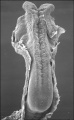
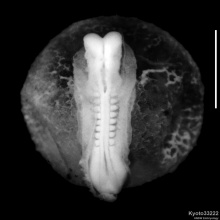
Stage 10 (22 - 23 days)
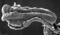
Stage 10 (22 - 23 days)
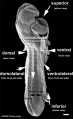
Stage 11 (23 - 26 days)
Stage 12 (26 - 30 days)
Stage 12 (26 - 30 days)


Neural Crest

|
<html5media height="380" width="410">File:Chicken-neural crest migration 01.mp4</html5media>
Chicken neural crest cell migration into pharyngeal arches. |
| System | Cell Type |
|---|---|
| Peripheral Nervous System (PNS) | Neurons - sensory ganglia, sympathetic and parasympathetic ganglia, enteric nervous system, and plexuses
Glia (neuroglial cells) - Schwann cells[1], satellite cells, olfactory ensheathing cells[2] |
| endocrine | Adrenal medulla Calcitonin-secreting cells Carotid body type I cells |
| integumentary | Epidermal pigment cells melanocyte |
| Facial cartilage and bone | Facial and anterior ventral skull cartilage and bones |
| Sensory | inner ear, cornea endothelium and stroma |
| Connective tissue | tooth odontoblast
smooth muscle, and adipose tissue of skin in head and neck Connective tissue of meninges, salivary, lachrymal, thymus, thyroid, and pituitary glands Connective tissue and smooth muscle in arteries of aortic arch origin |
| Links: neural crest | Category:Neural Crest | Neural Crest collapsible table | |
Primary Brain Vesicles
Traditional vesicle description (simplified name and alternate neuromere description in brackets)

Brain
- Prosencephalon (forebrain, prosomeres)
- Mesencephalon (midbrain, mesomeres)
- Rhombencephalon (hindbrain, rhombomeres)

|

|
Spinal Cord


| Neural Tube Regions | |||||||||||||||||||||
|---|---|---|---|---|---|---|---|---|---|---|---|---|---|---|---|---|---|---|---|---|---|
Table above shows the future transient regions that develop from the early neural tube. | |||||||||||||||||||||

Links: Spinal Cord
Week 5
Secondary Brain Vesicles

Brain Flexures

|
Rapid growth folds the neural tube forming 3 brain flexures (cranial to caudal)
|
Ventricles
|

CSF-filled spaces in adult brain. |

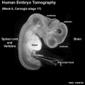
Week 6
| <html5media height="600" width="520">File:Human embryo tomography Carnegie stage 17.mp4</html5media> | 
Note the shape and size of the different regions of the brain and spinal cord.
|
Week 8
The human MRI movie below (head, sagittal plane, left to right) shows the central nervous system (CNS) development at the end of the embryonic period (week 8; GA week 10).
<html5media height="500" width="550">File:Stage23 MRI S01.mp4</html5media>
|
|
|
|
|
Cortex

| Week 8 Developing Cortex |
|---|
| Human embryo, Week 8, (GA week 10) Carnegie stage 22 section from the neural tube at the level of the developing cortex. Inset (upper right) shows whole section overview and approximate level of section (red line). Grey box shows detailed image region of developing cerebrum layer thicknesses are shown in microns.
Developing Cortex will form from the thin outer layer called the cortical plate. The underlying layers transient structures that continue to supply cells to the cortex through fetal period, most of these layers will eventually be lost, except for a thin ventricular layer. Cells migrate out along radial glia that establish the initial columnar and layered structure of the cortex. Layers are named according to the nervous system revised terminology (1970)[3] Developing Vascular blood vessels can also be seen spanning the developing layers. In the adult, these vessels will be lined with non-fenestrated endothelial cells that together with other vascular cells (pericytes and vascular smooth muscle cells), glial cells (astrocytes and microglia) and neurons will form the "blood-brain barrier". Developing Ventricular Space is cerebrospinal fluid (CSF) filled and the lateral ventricles form within the cortical region. The inset image shows lying within the lateral ventricles, the choroid plexus the modified vascular structure that forms and secretes the CSF. Developing Meninges layers lie outside the neural tube. The thin pia mater that closely covers the entire brain. The mesh-like arachnoid mater and the sub-arachnoid space that will also be CSF filled. The dense dura mater lies outside these 2 layers and under the skull, it cannot be seen in the enlarged image. |
Spinal Cord

Fetal
Second Trimester

Human week 10 fetus

|

|
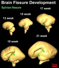
| Brain and Ventricular Development[4] | Brain Fissure Development[4] |
Sylvian Fissure Development
<html5media height="480" width="400">File:Neural_-_Sylvian_fissure.mp4</html5media>
Third Trimester

The brain goes from a smooth surface to begin to fold.
|

Human Fetus (CRL 240mm) Brain |
| Human Brain Growth | ||||||||||||||||||||||||||
|---|---|---|---|---|---|---|---|---|---|---|---|---|---|---|---|---|---|---|---|---|---|---|---|---|---|---|
| Embryonic | ||||||||||||||||||||||||||
Table below shows a direct comparison of brain growth in size between week 4 to 8 (GA 6-10)
| ||||||||||||||||||||||||||
| Fetal | ||||||||||||||||||||||||||

| ||||||||||||||||||||||||||
| Adult | ||||||||||||||||||||||||||

| ||||||||||||||||||||||||||
| Adult CNS Structures | ||||||||||||||||||||||||||
| ||||||||||||||||||||||||||


Fetal Timeline

Postnatal

Movies
| Neural Development | |||||||||||||||||||||||
|---|---|---|---|---|---|---|---|---|---|---|---|---|---|---|---|---|---|---|---|---|---|---|---|
|
|
|
|
|
| ||||||||||||||||||
|
|
|
|
|
| ||||||||||||||||||
| Abnormalities Ultrasound | |||||||||||||||||||||||
|
|
|
|
| |||||||||||||||||||
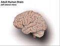
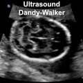
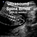
Abnormalities
| There are a large number of different neural abnormalities associated with genetic, environmental and unknown causes. These can also involve several different systems including: neural tube, neural crest, sensory development, ventricular and vascular system development.
It would be difficult to cover all in this current lecture so a few examples are given and students should explore the topic more widely themselves. |

|
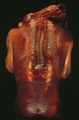
spina bifida and anencephaly
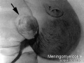
meningomyelocele
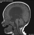
Congenital hydrocephalus (MRI)
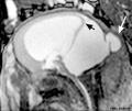
Dandy Walker malformation (MRI)
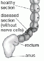
Intestinal aganglionosis


.jpg)


Environmental

|
The long time course of neural development (week 3 through to postnatal) also means that a large number of different environmental factors, including dietary deficiency, can impact upon its development and also have a range of different effects. |
Postnatal Neural Assessment - there are several basic clinical motor assessments that can identify normal and abnormal development.
| Abnormality Links | ||||
|---|---|---|---|---|
| ||||
| ||||
| ||||
| ||||
| ||||
| ||||
|
Terms
| Neural Terms |
|---|
Neural Development
|
| Other Terms Lists |
|---|
| Terms Lists: ART | Birth | Bone | Cardiovascular | Cell Division | Endocrine | Gastrointestinal | Genital | Genetic | Head | Hearing | Heart | Immune | Integumentary | Neonatal | Neural | Oocyte | Palate | Placenta | Radiation | Renal | Respiratory | Spermatozoa | Statistics | Tooth | Ultrasound | Vision | Historic | Drugs | Glossary |
BGDA: Lecture 1 | Lecture 2 | Practical 3 | Practical 6 | Practical 12 | Lecture Neural | Practical 14 | Histology Support - Female | Male | Tutorial
Glossary Links
- Glossary: A | B | C | D | E | F | G | H | I | J | K | L | M | N | O | P | Q | R | S | T | U | V | W | X | Y | Z | Numbers | Symbols | Term Link
Cite this page: Hill, M.A. (2024, April 26) Embryology BGDA Lecture - Development of the Nervous System. Retrieved from https://embryology.med.unsw.edu.au/embryology/index.php/BGDA_Lecture_-_Development_of_the_Nervous_System
- © Dr Mark Hill 2024, UNSW Embryology ISBN: 978 0 7334 2609 4 - UNSW CRICOS Provider Code No. 00098G
- ↑ Woodhoo A & Sommer L. (2008). Development of the Schwann cell lineage: from the neural crest to the myelinated nerve. Glia , 56, 1481-90. PMID: 18803317 DOI.
- ↑ Barraud P, Seferiadis AA, Tyson LD, Zwart MF, Szabo-Rogers HL, Ruhrberg C, Liu KJ & Baker CV. (2010). Neural crest origin of olfactory ensheathing glia. Proc. Natl. Acad. Sci. U.S.A. , 107, 21040-5. PMID: 21078992 DOI.
- ↑ <pubmed>5414696</pubmed>
- ↑ 4.0 4.1 <pubmed>19339620</pubmed>| PMC2721010 | J Neurosci.