RPAH Cardiac Embryology 2014
| Embryology - 20 Jul 2026 |
|---|
| Google Translate - select your language from the list shown below (this will open a new external page) |
|
العربية | català | 中文 | 中國傳統的 | français | Deutsche | עִברִית | हिंदी | bahasa Indonesia | italiano | 日本語 | 한국어 | မြန်မာ | Pilipino | Polskie | português | ਪੰਜਾਬੀ ਦੇ | Română | русский | Español | Swahili | Svensk | ไทย | Türkçe | اردو | ייִדיש | Tiếng Việt These external translations are automated and may not be accurate. (More? About Translations) |
Embryology of the Heart

22nd October, 2014 ROYAL PRINCE ALFRED HOSPITAL, SYDNEY, AUSTRALIA
This brief presentation will introduce development of the heart from its earliest origins through to the end of the embryonic period. The main morphological changes in the human heart will be shown in the context of the normal human timeline of development. Ultrasound scans that occur at the end of the first trimester should show a heart that has completed all aspects of septation (atrial, ventricular and outflow tract). Heart differences from the adult circulation will be present within the heart the atrial shunt (foramen ovale, or historically foramen Botalli) and just outside the heart the pulmonary-aortic arch shunt (ductus arteriosus). Please feel free to provide the author Feedback, as I continue to improve the content even after the presentation.
Note - Please take the opportunity now in your own time to look at the various animations on this page that help your understanding of cardiac development. This page also contains "collapsed tables" that can be opened to show more detailed information.
Developmental Timings
- Clinical timings refer to Gestational Age GA from the last day of the Last Menstrual Period (LMP).
- Embryonic timings refer to Ovulation age (OA) from day 14 of the cycle when fertilization occurs.
Other Cardiovascular Pages
Teach yourself about heart development with these online tutorials designed at different 3 levels.
| Cardiac Embryology | Begin Basic | Begin Intermediate | Begin Advanced |
The pages below are more detailed descriptions of cardiovascular development, most outside the scope of this presentation. Probably the most relevant page is Abnormalities.
Human Embryo Development

Weeks shown are ovulation age (OA) for gestational age (GA) add 2 weeks.
- Carnegie Stages: 1 | 2 | 3 | 4 | 5 | 6 | 7 | 8 | 9 | 10 | 11 | 12 | 13 | 14 | 15 | 16 | 17 | 18 | 19 | 20 | 21 | 22 | 23 | About Stages | Timeline

The above images of human embryonic development to week 8 (gestational age week 10) show the changing growth in size (all to scale) and external appearance of the embryo. Note that by the end of this period, covering most of the first trimester, the embryo is only about 3 cm long.
Heart Embryonic Timeline
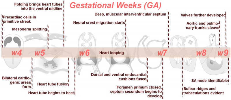
Weeks shown above are for clinical Gestational Age (GA). Ages shown below are Ovulation Age (OA), subtract 2 weeks from GA. If the linked Advanced pages below are too difficult, try starting with the Basic or Intermediate level descriptions of the same events.
| Begin Advanced | Heart Fields | Heart Tubes | Cardiac Looping | Cardiac Septation | Outflow Tract | Valve Development | Cardiac Conduction | Cardiac Abnormalities | Molecular Development |
Cardiovascular Movies
|
These simple cartoons show, almost in developmental sequence, aspects of cardiac development. Each linked page is supported by simple descriptions of events shown in the movie.
|

|
| Additional Movies | |||||||||||||||||||||||||||||||||||||
|---|---|---|---|---|---|---|---|---|---|---|---|---|---|---|---|---|---|---|---|---|---|---|---|---|---|---|---|---|---|---|---|---|---|---|---|---|---|
| |||||||||||||||||||||||||||||||||||||
The historic movies below include an audio commentary of cardiac development events (note the audio is unfortunately quite poor).
| |||||||||||||||||||||||||||||||||||||
Heart Field
Gastrulation
Week 5 (GA) |

|
.jpg)
Dorsal view of 18 day embryo |
.jpg)
Lateral view of 18 day embryo | ||||||
|
|
- The heart primordium arises predominantly from the primary heart field in splanchnic mesoderm forming in the cardiogenic region of the trilaminar embryo.
- The cardiogenic region can be thought of as bilateral fields that merge cranially to form a horseshoe-shaped field.
- During the third week (PO) of development (approximately day 18) angioblastic cords develop in this cardiogenic mesoderm and canalise to form bilateral endocardial heart tubes.
- The secondary heart field has been described as pharyngeal mesenchyme that contributes myocardium and smooth muscle to the arterial pole.
Heart Tube

Week 5 (GA) to beginning Week 6 (GA) |

|
Cardiac Looping

Image day 21 to 25 (GA Week 6)
|

|
.jpg)
- First stage occurs from late in the sixth week (GA) to early in the seventh week (GA)
- studied in chick embryos whose initial, straight heart tube is representative of that developing in humans.
- Second stage (creating S-shape) occurs as the ventricular bend moves caudally and the distance between the outflow and inflow tracts diminishes.
- dorsal mesocardium degenerates forming the transverse pericardial sinus (a point of communication across the pericardial coelom).
- atrial and outflow poles converge and myocardial cells are added, forming the truncus arteriosus.
- Final stage is the wedging of the aorta between the atrioventricular (AV) valves.
- occurs during septation and is dependent on the retraction and rotation of the myocardium by 45°.
Left-Right Asymmetry
- Heart is the first organ in the body to express left-right asymmetry in the form of looping.
- The axis of the heart is established earlier during gastrulation (week 3, GA week 5).
In the first images below the cardiac sac (pericardial sac) has been opened to show the developing heart tube.

|

|

|
| Stage 11 | Stage 12 | Stage 13 |


Septation
Atrioventricular Canal
- Partitioning of the primitive heart occurs between the middle of the week 6 (GA) and the end of week 7 (GA).
- Division of the atrioventricular canal is described below while
Two endocardial cushions form on the dorsal and ventral surfaces of the AV canal. Following expansion of the cardiac jelly, epithelial to mesenchymal transformation (EMT) of the endocardial cells in the canal occurs forming the cushions. Synergistic signalling between BMP and TGFβ facilitates EMT. The cushions grow as they are invaded by mesenchymal cells from the endocardium during the fifth week, eventually fusing to create the right and left AV canals, hence partially separating the primitive atrium and ventricle.
Atrial and Ventricular Septation
| Development of the Atria | ||||||||
|---|---|---|---|---|---|---|---|---|
|
Membranous tissue forming the septum primum grows from the roof of the atrium, dividing it into left and right halves. The septum primum originates from myocardium that differentiates from splanchnic mesoderm near the venous pole and approaches the endocardial cushions. The foramen primum refers to the decreasing communication between the septum primum and endocardial cushions. The junction of the septum primum and endocardial cushions becomes myocardialised by ingrowth of myocardial cells, although the centre is maintained as dense connective tissue and is referred to as Todaro’s tendon. Apoptosis-induced perforations appear in the centre of the septum primum to produce the foramen secundum. At this time, the strong, muscular septum secundum grows immediately to the right of the septum primum and gradually overlaps the foramen secundum during the fifth and sixth weeks of development. Part of the atrial septum and dorsal right atrium, as well as the septum secundum develop from left-sided mesenchyme. The incomplete partition of the atrium by the septum secundum forms the foramen ovale. Blood flows through the foramen ovale and foramen secundum to the left atrium. The remaining portion of the septum primum acts as the valve of the foramen ovale. Blood cannot flow in the opposite direction as the muscular strength of the septum secundum prevents prolapse of the septum primum.
Remodelling of the venous pole, including the further induction of myocardial cells, contributes to the development of the atria. In the mouse, myocardial differentiation occurs in the dorsal mesocardium and cells are then recruited to the venous pole. The development of two left to right shunts in the venous system leads to an increase in the right horn of the sinus venosus and consequentially a decrease in left horn by the end of the fourth week (GA 6 week). The sinuatrial orifice correspondingly shifts to the right thus becomes located in the right atrium. Hence the right atrium receives the superior vena cava (SVC) and inferior vena cava (IVC) in the adult. In mice and chicks the left sinus horn develops as the left SVC, however this regresses to form the coronary sinus in humans. Thus the sinus venosus gradually becomes incorporated into the right atrium. It contributes to the smooth walled part of the adult right atrium, referred to as the sinus venarum. The trabeculated right atrium corresponds to the primordial atrium; the division between these structures is indicated by the inner crista terminalis and outer sulcus terminalis. The primordial pulmonary vein develops in the dorsal wall of the left atrium. As the atrium increases in size it incorporates more of the branches of the pulmonary vein, culminating in its receiving the four pulmonary veins. The smooth wall of the adult left atrium originated from the primordial pulmonary vein, while the trabeculated wall represents the primordial atrium. |
|
| Development of the Ventricles | ||||
|---|---|---|---|---|
|
Minor trabeculations appear during early development of the primordial ventricle. Following growth of the ventricles further trabeculations appear and grow as larger, muscular structures. Some authors tout the idea that as trabeculations grow they coalesce resulting in the formation of the ventricular septum. However, the more commonly described theory of septation begins with the appearance of a primordial muscular interventricular (IV) ridge developing in the floor of the ventricle near the apex. As either side of the ventricle grows and dilates, their medial walls fuse forming the prominent IV septum. The foramen located between the cranial portion of the IV septum and the endocardial cushions, the IV foramen, closes by the end of the seventh week (GA 9 week) as the bulbar ridges fuse with the endocardial cushions. |
Outflow Tract Septation
- Cardiac neural crest - levels associated with somites 1 and 3 of the neural tube migrate through the pharyngeal arches to contribute to the conotruncal septum.
- Week 7 (GA) - proliferation of pharyngeal mesenchyme in the bulbus cordis forms bulbar ridges, continuous in the truncus arteriosus. Cardiac neural crest migrates into these ridges, condensing as cellular columns to support the outflow tract septum. The ridges form a 180° spiral to create the helical aorticopulmonary septum. Myocardialisation of the ridges gives a zippering effect resulting in fusion.
- Week 8 (GA) - fusion occurs distal to proximal direction, allowing for cleavage of the aorta and pulmonary trunk. The spiralling nature of the ridges causes the pulmonary trunk to twist around the aorta. The bulbus cordis accounts for the smooth conus arteriosus (or infundibulum) in the right ventricle and the aortic vestibule in the left ventricle.

.jpg)
- Links: Movie - Outflow tract
Heart Week 7 and 10 (GA)
| The images below and the associated collapsed tables show detailed histological images of heart development at about the midpoint (week 5, GA week 7) and at the end (week 8, GA week 10) of human embryonic development. (Note - These are in more detail than you currently require, but clearly show the position, size and shape of the heart in the embryo).
|


|
|


Valve Development
|
|
Development of the mitral and tricuspid valves
|
.jpg)


Development of the semilunar valves |
Development of the semilunar cusps |

Aortic and pulmonary valves (semilunar valves)
- formed from the bulbar ridges and subendocardial valve tissue.
- primordial semilunar valve consists of a mesenchymal core covered by endocardium.
- mechanisms of valve remodelling may involve apoptotic pathways.
Cardiac Conduction
|

Diagram of the adult cardiac conduction system |
Embryonic Heart Rate
The heart rate data shown below is from a 1996 ultrasound study of normal successful human gestations (34-56 days GA) developmental stages.[1]
- Week 5-6 (GA) Stage 9 to 10 - 2 mm embryo (gestational sac diameter of 20 mm) EHR at least 75 beats / minute
- Week 6 (GA) Stage 11 to 12 5 mm embryo (gestational sac diameter of 30 mm) EHR at least 100 beats / minute
- Week 8 (GA) Stage 16 - 10 mm embryo EHR at least 120 beats / minute
- Week 9 (GA) Stage 18 - 15 mm embryo EHR at least 130 beats / minute
Fetal Heart
Beyond the scope of this current talk.
Study using 3 and 4-dimensional fetal echocardiography between 12 and 41 weeks gestational age measured outflow tract angles.[2]
- ductal arch and thoracic aorta angle decreased with age.
- between the ductal arch and the aortic arch angle increased with age.
- between the left outflow tract (LOT) and right outflow tract (ROT) angle increased with age.
Abnormalities
Shown below are images showing the major Congenital Heart Disease (CHD). More details about the abnormality and the associated epidemiology and disease classification (ICD) can be seen by opening the table beside the image. Within the tables for the major CHDs, there are linked pages just about that abnormality.
| About - The International Classification of Diseases (ICD) |
|---|
| The International Classification of Diseases (ICD) World Health Organization's classification used worldwide as the standard diagnostic tool for epidemiology, health management and clinical purposes. This includes the analysis of the general health situation of population groups. It is used to monitor the incidence and prevalence of diseases and other health problems. Within this classification "congenital malformations, deformations and chromosomal abnormalities" are (Q00-Q99) but excludes "inborn errors of metabolism" (E70-E90). |
CHD is a feature of many genetic abnormalities, the most relevant statistically would be Trisomy 21, but there are others that can be identified from the genetic links below.
| Genetic Links: genetic abnormalities | maternal age | Trisomy 21 | Trisomy 18 | Trisomy 13 | Trisomy X | trisomy mosaicism | Monosomy | Fragile X | Williams | Alagille | Philadelphia chromosome | mitochondria | VACTERL | hydatidiform mole | epigenetics | Prenatal Diagnosis | Neonatal Diagnosis | meiosis | mitosis | International Classification of Diseases | genetics |
Ventricular Septal Defect

|
|
Atrial Septal Defects

|
|
Patent Ductus Arteriosus

|
|


Tetralogy of Fallot

|
|
Hypoplastic Left Heart

|
|
Double Outlet Right Ventricle

|
|
Tricuspid Atresia

|
|
Dextrocardia

Dextrocardia anatomical heart position[4] |
Dextrocardia (postnatal 1 year old)[4] |

| About - Dextrocardia |
|---|
|
Abnormalities of Conducting System
Also variously called the cardiac conduction system (CCS), cardiac pacemaking and conduction system (CPCS), or atrioventricular conduction system (AVCS). Recently animal models (CCS-lacZ transgenic mouse) have helped identify key processes in the development of this specialized conduction system.
"Known arrhythmogenic areas including Bachmann's bundle, the pulmonary veins, and sinus venosus derived internodal structures, demonstrate lacZ expression." (Jongbloed et al, 2004)
Long QT Syndrome
Congenital long QT syndrome (LQTS) is a group of rare genetic disorders with prolonged ventricular repolarization and a risk of ventricular tachyarrhythmias. Cause is mutations in genes encoding either cardiac ion channels or channel interacting proteins.
Search NCBI Bookshelf: Congenital long-QT syndrome
- Links: Search PubMed
Magnetic Resonance Imaging
The following movies show aspects of heart development from mid-embryonic to the end of embryonic development.[5] Original image resolution of the scans ranges from 30-150 microns/pixel.
Gestational Age
- Week 6 to 7 - Stage 13 Heart (atrioventricular junction)
- Week 8 - Stage 16 Heart (outflow tract)
- Week 9 - Stage 18 Heart AV valves
- Week 10 - Stage 23 Heart septation (ventricular+inlet)
Image source: The Kyoto Collection embryo MRIs are reproduced with the permission of Prof. Shige Yamada, Cecilia Lo and Kohei Shiota. This material is provided for educational purposes only and cannot be reproduced electronically or in writing without permission. MRI Atlas of Human Embryo Ref:<pubmed>20503356</pubmed>| PMC3401072
Further Online Reading
| Cardiovascular Links: cardiovascular | Heart Tutorial | Lecture - Early Vascular | Lecture - Heart | Movies | 2016 Cardiac Review | heart | coronary circulation | heart valve | heart rate | Circulation | blood | blood vessel | blood vessel histology | heart histology | Lymphatic | ductus venosus | spleen | Stage 22 | cardiovascular abnormalities | OMIM | 2012 ECHO Meeting | Category:Cardiovascular | ||
|
References
- ↑ <pubmed>8921130</pubmed>
- ↑ <pubmed>17384040</pubmed>| MC2190734 J Ultrasound Med.
- ↑ <pubmed>19876418</pubmed>
- ↑ 4.0 4.1 <pubmed>19142355</pubmed>| Arq Bras Cardiol.
- ↑ <pubmed>20503356</pubmed>| PMC3401072 | MRI Atlas of Human Embryo
Online Textbooks
- Developmental Biology (6th ed.) Gilbert, Scott F. Sunderland (MA): Sinauer Associates, Inc.; c2000. Figure 15.6 Cascade of heart development
Search Bookshelf heart development
Reviews
<pubmed>21940548</pubmed> <pubmed>17224285</pubmed>| PMC1858673 <pubmed></pubmed>
Articles
<pubmed></pubmed> <pubmed></pubmed> <pubmed></pubmed> <pubmed>9475206</pubmed>
Search Pubmed
Search Pubmed heart development
External Links
External Links Notice - The dynamic nature of the internet may mean that some of these listed links may no longer function. If the link no longer works search the web with the link text or name. Links to any external commercial sites are provided for information purposes only and should never be considered an endorsement. UNSW Embryology is provided as an educational resource with no clinical information or commercial affiliation.
- Cardiovascular Ultrasound is an open access, peer-reviewed, online journal covering clinical, technological, experimental, biological, and molecular aspects of ultrasound applications in cardiovascular physiology and disease. Search - Fetal
Glossary Links
- Glossary: A | B | C | D | E | F | G | H | I | J | K | L | M | N | O | P | Q | R | S | T | U | V | W | X | Y | Z | Numbers | Symbols | Term Link
Cite this page: Hill, M.A. (2026, July 20) Embryology RPAH Cardiac Embryology 2014. Retrieved from https://embryology.med.unsw.edu.au/embryology/index.php/RPAH_Cardiac_Embryology_2014
- © Dr Mark Hill 2026, UNSW Embryology ISBN: 978 0 7334 2609 4 - UNSW CRICOS Provider Code No. 00098G