Paper - Development of the human heart from its earliest appearance to the stage found in embryos of twenty paired somites (1927)
| Embryology - 27 Apr 2024 |
|---|
| Google Translate - select your language from the list shown below (this will open a new external page) |
|
العربية | català | 中文 | 中國傳統的 | français | Deutsche | עִברִית | हिंदी | bahasa Indonesia | italiano | 日本語 | 한국어 | မြန်မာ | Pilipino | Polskie | português | ਪੰਜਾਬੀ ਦੇ | Română | русский | Español | Swahili | Svensk | ไทย | Türkçe | اردو | ייִדיש | Tiếng Việt These external translations are automated and may not be accurate. (More? About Translations) |
Davis CL. Development of the human heart from its earliest appearance to the stage found in embryos of twenty paired somites. (1927) Carnegie Instn. Wash. Publ 380. 19: 245-284.
| Historic Disclaimer - information about historic embryology pages |
|---|
| Pages where the terms "Historic" (textbooks, papers, people, recommendations) appear on this site, and sections within pages where this disclaimer appears, indicate that the content and scientific understanding are specific to the time of publication. This means that while some scientific descriptions are still accurate, the terminology and interpretation of the developmental mechanisms reflect the understanding at the time of original publication and those of the preceding periods, these terms, interpretations and recommendations may not reflect our current scientific understanding. (More? Embryology History | Historic Embryology Papers) |
Development of the Human Heart from its Earliest Appearance to the Stage Found in Embryos of Twenty Paired Somites
By Carl L. Davis
with 8 plates and 10 text-figures.
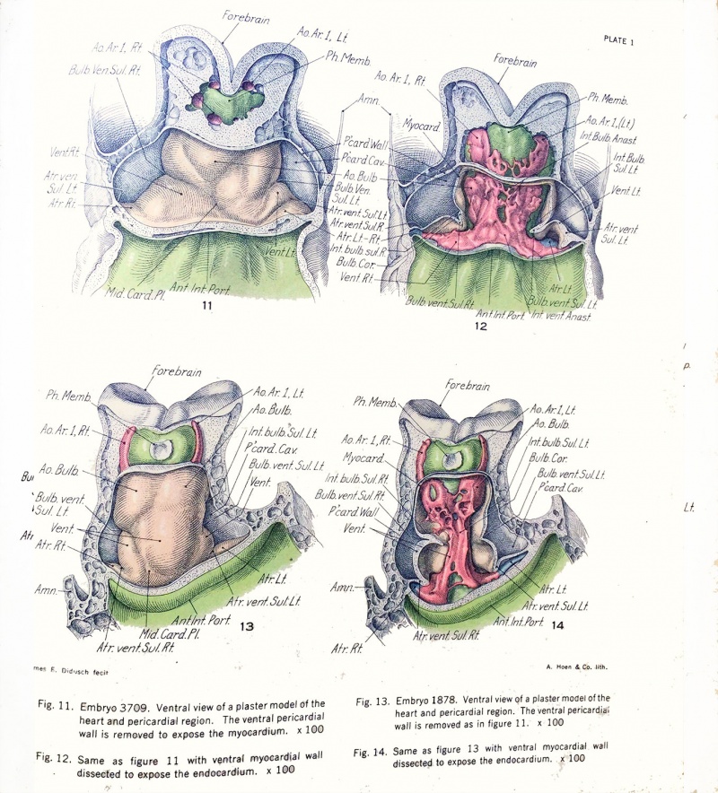
Plate 1
| Fig. 11. Embryo 3709. | Fig. 13. Embryo 1878. | |
|---|---|---|
| Ventral view of a plaster model of the heart and pericardial region. The ventral pericardial wall is removed to expose the myocardium. x100. | Ventral view of a plaster model of the heart and pericardial region. The ventral pericardial wall is removed as in figure 11. x100. | |
| Fig. 12. Embryo 3709. | Fig. 14. Embryo 1878. | |
| The ventral myocardial wall dissected to expose the endocardium. x100 | The ventral myocardial wall dissected to expose the endocardium. x100 |
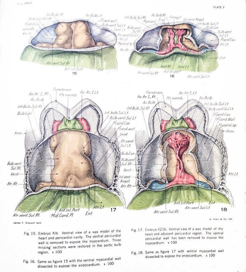
Plate 2
| Fig. 15. Embryo Klb. | Fig. 17. Embryo 4216. | |
|---|---|---|
| Ventral view of a wax model of the heart and pericardial cavity. The ventral pericardium wall is removed to expose the myocardium. Three missing sections were restored in the aortic bulb region. x100. | Ventral view of a wax model of the heart and pericardial region. The ventral pericardium wall is removed to expose the myocardium. x100 | |
| Fig. 16. Embryo Klb. | Fig. 18 Embryo 4216. | |
| The ventral myocardial wall dissected to expose the endocardium. x100 | The ventral myocardial wall dissected to expose the endocardium. x100 |
Reference
Davis CL. Development of the human heart from its earliest appearance to the stage found in embryos of twenty paired somites. (1927) Carnegie Instn. Wash. Publ 380. 19: 245-284.
Cite this page: Hill, M.A. (2024, April 27) Embryology Paper - Development of the human heart from its earliest appearance to the stage found in embryos of twenty paired somites (1927). Retrieved from https://embryology.med.unsw.edu.au/embryology/index.php/Paper_-_Development_of_the_human_heart_from_its_earliest_appearance_to_the_stage_found_in_embryos_of_twenty_paired_somites_(1927)
- © Dr Mark Hill 2024, UNSW Embryology ISBN: 978 0 7334 2609 4 - UNSW CRICOS Provider Code No. 00098G


