Embryology History - Ziegler Models
Introduction

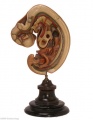
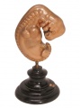
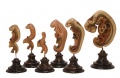
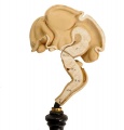
Not a collection as such, but a historic series of wax models made in the 1860-80s based upon the embryos of Prof. Wilhelm His, Leipzig. Wilhelm His had earlier prepared a series of freehand models. These Ziegler models are the basis of some of the teaching models that are still commercially available and used today.
Adolf Ziegler (1820 - 1889) a German modeller developed these models at his Studio for Scientific Teaching Models (Atelier für wissenschaftliche Unterrichtsmodelle) from the 1860's onwards of both human and animal embryos. Ziegler's models were first used in Germany, but were also later exhibited in Paris (1867) and Vienna (1873).
His son, Friedrich Ziegler (- 1936), continued the modelling company.
The Carnegie Institute later in the early 1900's also developed many additional models based upon their own collection. (More? Carnegie Models)
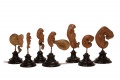
Embryo Model Sets


Embryo Models
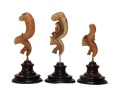
4-5 Somite Embryo


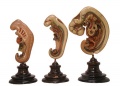
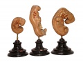
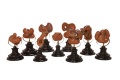
Month 1 Embryos


Neural Models

|

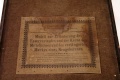
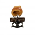
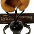
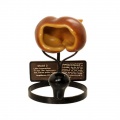
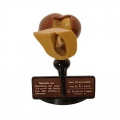
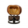
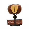
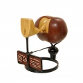
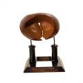
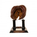
Image of information attached to underside of Medulla model base. Model for explaining the course and the fiber cores of the midbrain and the medulla oblongata of the newborn. (Modell zur Erläuterung des Faserverlaufes und der Kerne des Mittelhirnes und des verlängerten Markes eines Neugeborenen.) |

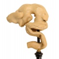
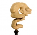
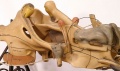
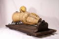
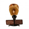
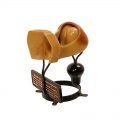
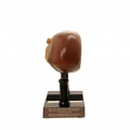
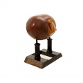
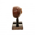
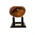
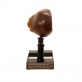
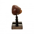
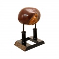
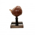
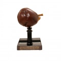
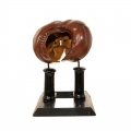
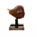
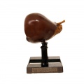
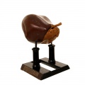
Medulla Model |

Medulla Model |

|

|

|

|
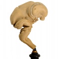
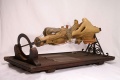
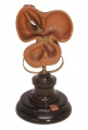
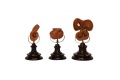
Eye Models
Development history of the eye series 8b consists of 3 models while series 8c consists of 5 models of early eye development.
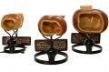
Series 8b Models 1-3
Series 8b, three wax models, submitted by F. Ziegler, c 1930. Models are made to example of Prof. von Szily. Showing the development of the papilla nervi optici primitiva s. lamina typus "sauger". max. height 18 cm.

Series 8c Models 1-5
Models of the right eye of the developing rabbit embryo.
Series 8c, five wax models, submitted by F. Ziegler, c 1930. Models are made to example of Prof. von Szily. it is a view of the ontogenesis of the idiotype gap formation of the eye of microphthalmia and the orbital cistern. it concerns a rabbit eye. maximum height 32 cm.

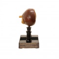
Series 8b Model 1
Left primordial eye of 12 days old embryo.
Images show model viewed laterally and from the external and internal view.
|  |-
|
|-
|  |
|  |-
|
|-
|  |
|  |}
|}
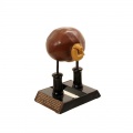
Series 8b Model 2
Left primordial eye of 13 days old embryo, 9.6 mm greatest length.
Images show model viewed laterally and from the external and internal view.


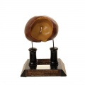
Series 8b Model 3
Left primordial eye of 13 days old embryo, 10.5 mm greatest length.
Images show model viewed laterally and from the external and internal view.


Series 8c Model 1


Series 8c Model 2


Series 8c Model 3


Series 8c Model 4


Series 8c Model 5


- Links: Vision Development
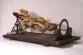
Heart Models

|

|

Gallery
Eye - Series 8b Model 1
Eye - Series 8b Model 1 legend

Eye - Series 8c Model 1
Eye - Series 8c Model 2 Right eye of a 18-day rabbit embryo.
Eye - Series 8c Model 2 Right eye of a 18-day rabbit embryo.
Eye - Series 8c Model 2 Right eye of a 18-day rabbit embryo.
Eye - Series 8c Model 2 Right eye of a 18-day rabbit embryo.
Eye - Series 8c Model 2 Right eye of a 18-day rabbit embryo.
Eye - Series 8c Model 3
Eye - Series 8c Model 4 Right eye of a 24-day rabbit embryo.
Eye - Series 8c Model 4

Eye - Series 8c Model 4
Eye - Series 8c Model 4 Right eye of a 24-day rabbit embryo, ventral view.
Eye - Series 8c Model 5 Right eye of a 26-day rabbit embryo.
Eye - Series 8c Model 5 Right eye of a 26-day rabbit embryo.
Eye - Series 8c Model 5 Right eye of a 26-day rabbit embryo.
Eye - Series 8c Model 5 Right eye of a 26-day rabbit embryo.

External Links
External Links Notice - The dynamic nature of the internet may mean that some of these listed links may no longer function. If the link no longer works search the web with the link text or name. Links to any external commercial sites are provided for information purposes only and should never be considered an endorsement. UNSW Embryology is provided as an educational resource with no clinical information or commercial affiliation.
Glossary Links
- Glossary: A | B | C | D | E | F | G | H | I | J | K | L | M | N | O | P | Q | R | S | T | U | V | W | X | Y | Z | Numbers | Symbols | Term Link
Cite this page: Hill, M.A. (2024, April 27) Embryology Embryology History - Ziegler Models. Retrieved from https://embryology.med.unsw.edu.au/embryology/index.php/Embryology_History_-_Ziegler_Models
- © Dr Mark Hill 2024, UNSW Embryology ISBN: 978 0 7334 2609 4 - UNSW CRICOS Provider Code No. 00098G