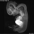
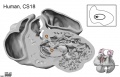
Carnegie stage 18
| Embryology - 5 May 2026 |
|---|
| Google Translate - select your language from the list shown below (this will open a new external page) |
|
العربية | català | 中文 | 中國傳統的 | français | Deutsche | עִברִית | हिंदी | bahasa Indonesia | italiano | 日本語 | 한국어 | မြန်မာ | Pilipino | Polskie | português | ਪੰਜਾਬੀ ਦੇ | Română | русский | Español | Swahili | Svensk | ไทย | Türkçe | اردو | ייִדיש | Tiếng Việt These external translations are automated and may not be accurate. (More? About Translations) |
Introduction

Facts
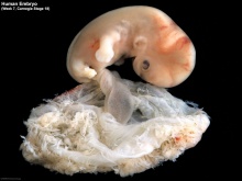
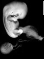
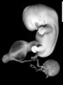
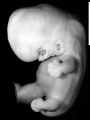
Week 7, 44 - 48 days, 13 - 17 mm
Gestational Age GA - week 9
Summary
- Ectoderm: sensory placodes, lens pit, otocyst,nasal pits moved ventrally, fourth ventricle of brain
- Mesoderm: heart prominence
- Head: 1st, 2nd and 3rd pharyngeal arch, forebrain, eye, auricular hillocks
- Body: heart, liver, umbilical cord
- Limb: upper and lower limb buds, foot plate, wrist, hand plate with digital rays
See also Carnegie stage 18 Events
Features
- Development indices: number of semicircular ducts (1-3) and length of the paramesonephric duct.
- Identify: pigmented eye, eyelid, nasolacrimal groove, external acoustic meatus, heart, digital rays, liver prominance, thigh, ankle, foot plate, umbilical cord
| Week: | 1 | 2 | 3 | 4 | 5 | 6 | 7 | 8 |
| Carnegie stage: | 1 2 3 4 | 5 6 | 7 8 9 | 10 11 12 13 | 14 15 | 16 17 | 18 19 | 20 21 22 23 |
- Carnegie Stages: 1 | 2 | 3 | 4 | 5 | 6 | 7 | 8 | 9 | 10 | 11 | 12 | 13 | 14 | 15 | 16 | 17 | 18 | 19 | 20 | 21 | 22 | 23 | About Stages | Timeline
Bright Field

|

|
| Embryo in gestational sac | Embryo open sac |

|

|
| Embryo with placentation (ectopic) | Embryo in amniotic sac |
- Stage 18 Links: Embryo in gestational sac | Embryo open sac | Embryo 2 and gestational sac | Embryo 2 | Carnegie stage 18
Embryo Virtual Slides
|
|

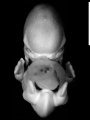
Scanning EM


Ventral view of head showing upper lip, maxilla and nasal region.
Image Source: Prof Virginia Diewert
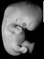
Kyoto Collection

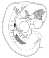
View: This is a dorsolateral view of embryo. Amniotic membrane removed.
Image source: Embryology page Created: 19.03.1999

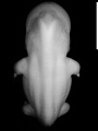
Ventral view of head region (1 mm scale).
- Kyoto Embryo 25783
right+yolk
left+yolk
left
ventral
right
dorsal


Image source: The Kyoto Collection images are reproduced with the permission of Prof. Kohei Shiota and Prof. Shigehito Yamada, Anatomy and Developmental Biology, Kyoto University Graduate School of Medicine, Kyoto, Japan for educational purposes only and cannot be reproduced electronically or in writing without permission.
Carnegie Collection


| Carnegie Collection Embryos - Stage 18 | |||||||||||
|---|---|---|---|---|---|---|---|---|---|---|---|
| Serial No. | Size (mm) | Grade | Fixative | Embedding Medium | Plane | Thinness (µm) | Stain | Semi. ducts | P.-M. duct (mm) | Year | Notes |
| 109 | E., 12.0* Ch.,30 | Poor | Alc. | P | Transverse | 20 | Al. coch. | 1 | 0.4 | 1897 | Tubal Least—advanced third |
| 144 | E., 16.0* Ch, 40x30x30 | Good | Formalin | P | Sagittal | 40 | Al. eoch. | 3 | 0.85 | 1899 | Most—advanced third |
| 175 | E., 13.0 Ch, 30x25x25 | Poor | Alc. | P | Transverse | 20 | Al. coch. | 2 | 0.6 | 1900 | Tubal Partly macerated |
| 296 | E., 17.0 | Poor | Ale. | P | Coronal | 20 | Various | 3 | 0.85 | 1905 | Most—advanced third |
| 317 | E., 16.0 | Good | Formalin | P | Coronal | 20 | (Stain - Haematoxylin Eosin) or. G. | 2 | 0.7 | 1905 | Middle third |
| 351 | E.,14.0* | Good | Formalin | P | Coronal | 250 | Slightly carmine— | 2 | 038 | 1904 | Injected (Berlin blue) |
| 406 | E., 16.0 Ch., 40x40x40 | Good | Formalin | P | Sagittal | 20 | (Stain - Haematoxylin Eosin) | 3 | 0.7 | 1907 | Operative. Most—advanced third |
| 423 | E., 15.2 | Good | Formol—Zenker | P | Transverse | 50 | Carmine | 3 | 0.85 | 1904 | |
| 424 | E., 172 | Good | Formalin | P | Transverse | 50 | Carmine | 3 | 10 | 1904 | Double infection. Advanced |
| 492 | E, 16.8 Ch, 40 x 40 | Exc. | Zenker | P | Coronal | 40 | Al. coch. v | 3 | 0.7 | 1911 | Injected (India ink) |
| 511 | E., 160* Ch., 3?* 32x32 | Good | Ale. | P | Sagittal | 40 | Al. coch. | 3 | 1.1 | 1911 | Head injured. Most advanced in group |
| 670 | E, 12.5 | Poor | Ale. | P | Sagittal | 50 | (Stain - Haematoxylin Eosin) | 3 | 10 | 1913 | Tubal Advanced |
| 719 | E, 15.0 Ch, 50x50x50 | Good | Formalin | P | Trans | 40 | Al. coch. | 2 | 0.6 | 1913 | Median in group |
| 733 | E., Ch., 4Sx40x2S | ISO Poor | Formalin | P | Sagittal | 50 | Al. coch. | 2 | 0.6 | 1913 | Median in group |
| 841 | E. 15.0 Ch., 18 x 16x9 | Good | Formalin | P | Coronal & Trans, 20 | 10 | (Stain - Haematoxylin Eosin), carmine | 2 | 0.32 | 1914 | Operative. Head cut separately |
| 899 | E, 160* Ch. 50 x 18 x IS | Good | Bouin | P | Sagittal | S0 | Al. coch. | 3 | 0,65 | 1914 | Tubal Head injured |
| 991 | E. l?.0 | Good | Formalin | P | Sag | so | R, V, Gieson | 3 | 0.9 | 1914 | Advanced |
| 1909 | E., 14.6 | Good | Formalin | P | Coronal | 20 | Al. coch,or. G. | 1 | 0.3 | 1917 | Less advanced |
| 2673 | E.,15.5 | Good | Formalin | P | Transverse | 40 | Al. coch. | 2 | 0.52 | 1919 | Median in group |
| 4430 | E., 14.0 Ch, 51 x40x21 | Exc. | Corros. acetic | P | Transverse | 15 | Al. coch,or. G. | 3 | 0.9 | 1923 | Most—advanced third |
| 5542B | E., 16.0 Ch, 37x32x25 | Good | Formalin | P | Transverse | 40 | Al. coch. | 2 | 0.7 | 1927 | Other twin abnormal |
| 5747 | E, 15.2 Ch, 32x27x25 | Poor | Alc.—formol | P | Sagittal | 25 | Al. coch. | 2 | 0.25 | 1928 | Least—advanced or middle third |
| 5935A | E, 13.5 Ch, 40x30x30 | Good | Formalin | P | Coronal | 40 | Al. coch. | 1 | 0.38 | 1929 | Other twin stunted |
| 6522 | E, 13.2* | Good | Corros. acetic | C—P | Coronal | 10 | Al. coch. | 3 | 0.8 | 7 | Middle or most—advanced third |
| 6524 | E, 11.7* | Exc. | Corros. acetic | C—P | Transverse | 10 | Al. coch. | 1 | 0.4 | ? | Least—advanced third |
| 6525 | E, 13.8* | Exc. | Corros. acetic | C—P | Sagittal | 8 | Al. coch. | 2 | 0.42 | ? | Weak staining |
| 6527 | E, 14.4* | Exc. | Corros. acetic | C—P | Transverse | 15 | Al. coch. | 2 | 0.67 | ? | Mechanical damage |
| 6528 | E, 13.4* | Exc. | Corros. acetic | C—P | Coronal | 8 | Al. coch. | 1 | 0.33 | ? | Least—advanced third |
| 6529 | E, 15.6* | Good | Corros. acetic | C—P | Coronal | 10 | Al. coch. | 2 | 0.4 .5 | Middle third | |
| 6533 | E, 12.5* | Good | Corros. acetic | C—P | Sagittal | 6, 8, 10 | Al. coch. | 2 | 0.45 | ? | Middle third |
| 6551 | E, 18.0 | Poor | Formalin | p | Coronal | 40 | (Stain - Haematoxylin Eosin) | 3 | 0.8 | 1932 | Tubal |
| 7707 | E, 14.5 Ch ,37x32 | Exc. | Bouin | C—P | Transverse | 10 | (Stain - Haematoxylin Eosin), phlox. | 2 | 0.54 | 1939 | Operative. Middle third |
| 8097 | E, 15.5 Ch, 37x25x21 | Good | Formalin | C—P | Transverse | 10 | (Stain - Haematoxylin Eosin) | 1 | 0.19 | 1942 | Least advanced in group |
| 8172 | E, 16.5 | Exc. | Bouin | C—P | Transverse | 20 | (Stain - Haematoxylin Eosin) | 3 | 0.58 | 1943 | Operative. Very advanced |
| 8235 | E, 14.0* | Good | Bouin | C—P Sagittal | 10 | (Stain - Haematoxylin Eosin) | Mallory | 2 | 0.25 | 1944 | Tubal |
| 8355 | E, 15.0 Ch, 23 | Exc. | Formalin | C—P | Coronal | 10 | Azan | 1946 | Tubal. Duplicated spinal cord caudally | ||
| 8812 | E, 12_9 | Exc | Formalin | C—P | Transverse | 10 | (Stain - Haematoxylin Eosin) | 1950 | Rubella. Medical abortion. Midbrain punctured | ||
| 8945 | E, 13.9 | Good | Zenker | p | Transverse | 8 | Borax, carm. | 1952 | Univ. Chicago No. H 1254 | ||
| 9107 | E, 17.0 Ch, 38x28x22 | Good | Bouin | p | Transverse | 15 | Borax, carm. | 1918 | Univ. Chicago No. H 516 | ||
| 9247 | E, 15.0 | Exc. | Bouin | C—P | Sagittal | 8 | Azan | 1954 | Tubal | ||
Abbreviations
| |||||||||||
| iBook - Carnegie Embryos | |
|---|---|

|
|
Hill Collection

|

|

|

|
| right ventral | right ventrolateral | right ventrolateral | right lateral |

|

|

|
|
| right ventral (smaller) | left lateral (smaller) | right dorsolateral (smaller) |
- Links: Hill Collection
Madrid Collection
| Madrid Collection Embryos | ||||||
|---|---|---|---|---|---|---|
| Carnegie Stage |
Embryo | Days | CRL (mm) | Section thickness |
Staining | Section plane |
| 18 | GV-6 | 44 | 13 | 10 | (Stain - Haematoxylin Eosin)-Azan | transverse |
| 18 | Ve | 44 | 13.5 | 10 | (Stain - Haematoxylin Eosin) | sagittal |
| 18 | JF2 | 45 | 14.5 | 10 | (Stain - Haematoxylin Eosin)-trichrome | transverse |
| 18 | NO | 45 | 15 | 10 | (Stain - Haematoxylin Eosin) | transverse |
| 18 | J5 | 46 | 16.5 | 10 | (Stain - Haematoxylin Eosin) | frontal |
Hinrichsen Collection

Embryo 226 Carnegie stage 18, 16 mm slide 20 (Sagittal, quality good, Stain ((Stain - Haematoxylin Eosin), Tri, Azan), total slides 114).
Digital Embryology Consortium - Hinrichsen Collection
Embryo ME53 Carnegie stage 18, CRL 16 mm, (Frontal whole Embryo, Stain ((Stain - Haematoxylin Eosin)), total slides 132).
Image source: The Hinrichsen Collection images are reproduced with the permission of Prof. Beate Brand-Saberi, Head, Department of Anatomy and Molecular Embryology, Ruhr-Universität Bochum. Images are for educational purposes only and cannot be reproduced electronically or in writing without permission.
Events
- hearing - otic capsule precartilaginous. Semicircular ducts form in the order anterior, posterior, and lateral from thickened epithelial areas and adjacent epithelial layers fuse. Cochlear duct is now L-shaped. First pharyngeal arch bar begins to chondrify (Meckel’s cartilage), second arch may chondrify (Reichert’s cartilage), stapes and stapedius commence. Auricular hillocks merge to form auricle primordia.
- smell - the olfactory bulb begins to appear.[2] Stages Description | primitive olfactory bulb
- cardiovascular
- Coronary circulation connection of the proximal coronary arteries to the aorta.[4]
- endocrine[5]
- Epiphysis - cellular migration in the pineal body forms a distinct "anterior lobe" in which follicles appear (Stadium 3 of Turkewitsch 1933) (O'Rahilly 1973 a).
- thymus - thymus makes contact with the thyroid gland and contains a series of canals internally[6].
- thyroid - median thyroid is in contact with "lateral thyroid components"[6] but others have maintained that the telopharyngeal body should not be regarded as a thyroid component (Bejdl and Politzer 1953). The lobes of the thyroid are "composed of series of continuously communicating solid annectent bars" (Weller 1933). This is "the earliest stage of the definitive thyroid" (ibid.). First differentiation occurs in Weller's (1933) "lateral thyroid component," which is beginning to "blend into uniformly constituted thyroid tissue". Weller (1933)[6] illustrated (Fig. 11) a thyroid gland that still showed continuity between its pedicle and the epithelium of the pharynx.
- adrenal cortex - gland becomes reorganized. The C1, 2, and 3 cells form cords as sinusoids develop. Cells divide at or near the surface, where new cells are added.[7]
- meninges (spinal cord) - vertebral canal clearly distinguishable from its wall, within which chondrification is rapidly advancing. Ventral migration of ganglia has continued, ventral extremities are entering the intervertebral foramina, dorsal extremities lie at about spinal cord mid-level. Meninx primitiva is loosening and spaces are becoming evident within it, particularly in the region ventral to the cord. Over the dorsal surfaces of the centra and the intervertebral disks the cells of the meninx primitiva are, for the first time, showing indications of stratification. This stratification is more evident over the intervertebral disks, where it extends laterally and then sweeps dorsad over the medial surface of the ganglia. Similar stratification appears in the pia mater, and this is particularly evident adjacent to the anterior longitudinal sulcus. These conditions are most pronounced in the cervical region and become less distinct in a caudal direction. The rudiments of the dentate processes now form denser and more restricted concentrations and can be identified in the interganglionic transverse sections, where they extend in a dorsoventral direction from the pia, slightly above the emergence of the ventral root, to the mid-point of the rudimentary pedicle.[8]
- Renal - Collecting tubules develop from the calices at stages 17 and 18. They are surrounded by sharply outlined condensed primordia in the process of forming secretory tubules. Renal corpuscules are not yet present. By stage 18 the mesonephric duct and the ureter open almost independently into the vesico-urethral canal[9] : i.e., the common excretory duct is disappearing. The cloacal membrane is ready to rupture.[10]
- genital[11]
- Primordial Germ Cells, extragonadal germ cells were identified in gut epithelium from this stage.[12]rimordial germ cells were identified in the testis.
- gonad forms a pronounced elevation and extends to the rostral pole of the mesonephros and the rete is beginning.[13]
- testis gonadal blastema differentiates into testicular cords.[12]
- ovary gonadal cords identifiable in the ovary.[14]
- ventral body wall - separate intercostal and abdominal wall muscles differentiated, and ribs, sterna, and muscles began to expand ventromedially and caudally.[15]
References
- ↑ Pearson AA. (1980). The development of the eyelids. Part I. External features. J. Anat. , 130, 33-42. PMID: 7364662
- ↑ Bossy J. Development of olfactory and related structures in staged human embryos. (1980) Anat. Embryol., 161(2);225-36 PMID 7469043
- ↑ Mérida-Velasco JA, Sánchez-Montesinos I, Espín-Ferra J, Mérida-Velasco JR, Rodríguez-Vázquez JF & Jiménez-Collado J. (1997). Development of the human knee joint ligaments. Anat. Rec. , 248, 259-68. PMID: 9185992
- ↑ Hutchins GM, Kessler-Hanna A & Moore GW. (1988). Development of the coronary arteries in the embryonic human heart. Circulation , 77, 1250-7. PMID: 3286038
- ↑ O'Rahilly R. The timing and sequence of events in the development of the human endocrine system during the embryonic period proper. (1983) Anat. Embryol., 166: 439-451. PMID 6869855
- ↑ 6.0 6.1 6.2 Weller GL. Development of the thyroid, parathyroid and thymus glands in man. (1933) Contrib. Embryol., Carnegie Inst. Wash. 24: 93-139.
- ↑ Crowder RE. The development of the adrenal gland in man, with special reference to origin and ultimate location of cell types and evidence in favor of the "cell migration" theory. (1957) Contrib. Embryol., Carnegie Inst. Wash. 36, 193-210.
- ↑ Sensenig EC. The early development of the meninges of the spinal cord in human embryos. (1951) Contrib. Embryol., Carnegie Inst. Wash. Publ. 611.
- ↑ Shikinami J. Detailed form of the Wolffian body in human embryos of the first eight weeks. (1926) Contrib. Embryol., Carnegie Inst. Wash. Publ. 363, 18: 46-61.
- ↑ O'Rahilly R. and Müller F. Developmental Stages in Human Embryos. Contrib. Embryol., Carnegie Inst. Wash. 637 (1987).
- ↑ O'Rahilly R. (1983). The timing and sequence of events in the development of the human reproductive system during the embryonic period proper. Anat. Embryol. , 166, 247-61. PMID: 6846859
- ↑ 12.0 12.1 Jirásek JE. Development of the Genital System and Male Pseudohermaphroditism. (1971) Johns Hopkins Press, Baltimore.
- ↑ Wilson KM. Origin and development of the rete ovarii and the rete testis in the human embryo. (1926) Carnegie Instn. Wash. Publ. 362, Contrib. Embryol., Carnegie Inst. Wash., 17:69-88.
- ↑ Gillman J. The development of the gonads in man, with a consideration of the role of fetal endocrines and the histogenesis of ovarian tumors. (1948) Contr Embryol Carneg Instn. 32: 81-131.
- ↑ Mekonen HK, Hikspoors JP, Mommen G, Köhler SE & Lamers WH. (2015). Development of the ventral body wall in the human embryo. J. Anat. , 227, 673-85. PMID: 26467243 DOI.
Additional Images
Stage 18 Optical Projection Tomography
Stage 18 Heart MRI

External ear Stages 14-23 and adult


Historic Images
Human embryonic shoulder girdle
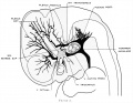
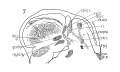
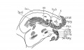
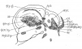
1921. Dural venous system human embryo 15 mm long
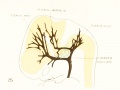
1921 Vascular system of the brain of the human embryo
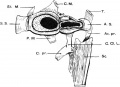
Embryo 109 Sabin 1909 Fig. 4
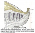
Fig. 273. Sagittal cut caudal end of embryo of 14mm

17 mm Embryo


Lisser H. Studies on the development of the human larynx. (1911) Amer. J Anat. 12: 27-66.
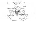
1911 Cross section to show cricoid cartilage and M. criooarytaenoideus posterior.
1911 Cross section to show thyreoid cartilage, M. criooarytaenoideus lateralis,
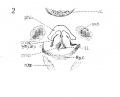
1911 Sagittal section to show, especially, M. interarytaenoideus.
1911 Sagittal section to show hyoid bone and thyreoid cartilage, M. cricothyreoideus, and tongue region
1911 Sagittal section


- Carnegie Stages: 1 | 2 | 3 | 4 | 5 | 6 | 7 | 8 | 9 | 10 | 11 | 12 | 13 | 14 | 15 | 16 | 17 | 18 | 19 | 20 | 21 | 22 | 23 | About Stages | Timeline
Cite this page: Hill, M.A. (2026, Mayıs 5) Embryology Carnegie stage 18. Retrieved from https://embryology.med.unsw.edu.au/embryology/index.php/Carnegie_stage_18
- © Dr Mark Hill 2026, UNSW Embryology ISBN: 978 0 7334 2609 4 - UNSW CRICOS Provider Code No. 00098G