Cardiovascular System - Heart Development
| Embryology - 9 Jul 2026 |
|---|
| Google Translate - select your language from the list shown below (this will open a new external page) |
|
العربية | català | 中文 | 中國傳統的 | français | Deutsche | עִברִית | हिंदी | bahasa Indonesia | italiano | 日本語 | 한국어 | မြန်မာ | Pilipino | Polskie | português | ਪੰਜਾਬੀ ਦੇ | Română | русский | Español | Swahili | Svensk | ไทย | Türkçe | اردو | ייִדיש | Tiếng Việt These external translations are automated and may not be accurate. (More? About Translations) |
Introduction

In human embryos the heart begins to beat at about 22-23 days, with blood flow beginning in the 4th week. The heart is therefore one of the earliest differentiating and functioning organs.
The heart begins very early in mesoderm within the trilaminar embryonic disc. The heart forms initially in the embryonic disc as a simple paired tube inside the forming pericardial cavity, which when the disc folds, gets carried into the correct anatomical position in the chest cavity.
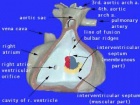
A key aspect of heart development is the septation of the heart into separate chambers. As the embryonic/fetal circulation is different to the neonatal circulation (lung/pulmonary activation), several defects of heart septation may only become apparent on this transition. One septal "defect" occurs in us all, the foramen ovale (between the 2 atria) which in general closes in the neonate over time.
Embryonic Heart Rate (EHR), early in development the heart starts to spontaneously beat and a recent study by Wisser and Dirschedl in dated human embryos showed an increase up to 63 postmenstrual days or 22 mm greatest length. Thereafter a steady decrease of EHR was noted. Maximal EHR is reached when morphological development of the embryonic heart is completed.
| Heart Links: Heart Tutorial | Detailed Cardiac Development | Heart Histology | Neural Crest | 2016 Review |
Basic Heart
Advanced Heart
| Cardiovascular Links: cardiovascular | Heart Tutorial | Lecture - Early Vascular | Lecture - Heart | Movies | 2016 Cardiac Review | heart | coronary circulation | heart valve | heart rate | Circulation | blood | blood vessel | blood vessel histology | heart histology | Lymphatic | ductus venosus | spleen | Stage 22 | cardiovascular abnormalities | OMIM | 2012 ECHO Meeting | Category:Cardiovascular | ||
|
Some Recent Findings
|
| More recent papers |
|---|
 This table allows an automated computer search of the external PubMed database using the listed "Search term" text link.
More? References | Discussion Page | Journal Searches | 2019 References | 2020 References Search term: Heart Development | Heart Embryology | Cardiac Embryology | Cardiac Development | Cardiomyocyte Development |
| Older papers |
|---|
| These papers originally appeared in the Some Recent Findings table, but as that list grew in length have now been shuffled down to this collapsible table.
See also the Discussion Page for other references listed by year and References on this current page. |
Textbooks
UNSW Embryology

Hill, M.A. (2020). UNSW Embryology (20th ed.) Retrieved July 9, 2026, from https://embryology.med.unsw.edu.au
| Cardiovascular Links: cardiovascular | Heart Tutorial | Lecture - Early Vascular | Lecture - Heart | Movies | 2016 Cardiac Review | heart | coronary circulation | heart valve | heart rate | Circulation | blood | blood vessel | blood vessel histology | heart histology | Lymphatic | ductus venosus | spleen | Stage 22 | cardiovascular abnormalities | OMIM | 2012 ECHO Meeting | Category:Cardiovascular | ||
|
Heart Tutorial | 2013 Lecture Slides
The Developing Human: Clinically Oriented Embryology

Moore, K.L., Persaud, T.V.N. & Torchia, M.G. (2015). The developing human: clinically oriented embryology (10th ed.). Philadelphia: Saunders.
(links available to UNSW students)
Larsen's Human Embryology

Schoenwolf, G.C., Bleyl, S.B., Brauer, P.R., Francis-West, P.H. & Philippa H. (2015). Larsen's human embryology (5th ed.). New York; Edinburgh: Churchill Livingstone.
(links available to UNSW students)
- Links: Embryology Textbooks
- Human Embryology (2nd ed.) Larson Ch7 p151-188 Heart
- The Developing Human: Clinically Oriented Embryology (6th ed.) Moore and Persaud Ch14: p304-349
- Before we Are Born (5th ed.) Moore and Persaud Ch12; p241-254
- Essentials of Human Embryology Larson Ch7 p97-122 Heart
- Human Embryology Fitzgerald and Fitzgerald Ch13-17: p77-111
- Links: Embryology Textbooks
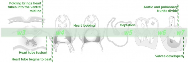
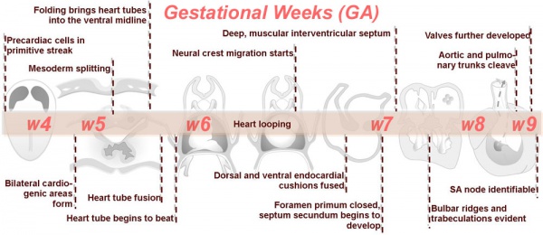
Timeline
.jpg)
- Cardiogenic region - in splanchnic mesenchyme of prechordal plate region
- Week 2 pair of thin-walled tubes
- Week 3 tubes fused, truncus arteriosus outflow, heart contracting
- Week 4 heart tube continues to elongate, curving to form S shape
- Week 5 Septation starts, atrial and ventricular
- Septation continues, atrial septa remains open, foramen ovale
- Week 40 At birth pressure difference closes foramen ovale leaving a fossa ovalis
| Characteristic | Carnegie stage: | 13 | 14 | 15 | 16 | 17 | 18 | 19 | 20 | 21 | 22 | 23 |
|---|---|---|---|---|---|---|---|---|---|---|---|---|
| Septum primum | ||||||||||||
| Foramen primum | ||||||||||||
| Atrioventricular bundle | ||||||||||||
| Atrioventricular cushions | ||||||||||||
| Conotruncal ridges | ||||||||||||
| Foramen secundum | ||||||||||||
| Semilunar cusps | ||||||||||||
| Conotruncal septum; atria | ||||||||||||
| Closure primum foramen | ||||||||||||
| Fusion atrioventricular cushions | ||||||||||||
| Septum secundum and foramen ovale | ||||||||||||
| Closure secondary interventricular foramen | ||||||||||||
| Chordae tendineae | ||||||||||||
| Colour Coding: | beginning to appear | present | Table data[5] Links: heart | Madrid Collection | |||||||||
Link: timeline
Comparison Human and Mouse
Table from Anderson RH. Teratogenecity in the setting of cardiac development and maldevelopment. (2016)
| Table 1. Comparison of Human and Murine Development | |||||
|---|---|---|---|---|---|
| Human | Mouse | ||||
| Carnegie Stage |
Post-ovulatory Days |
Size (mm) Greatest Length |
Theiler Stage |
Post-conception Days |
Size (mm) Crown-rump |
| 11 | 24 | 2.5 - 4.5 | 14 | 9 - 9.5 | 2.1 |
| 12 | 26 | 3 - 5 | 15 | 9.5 - 10.25 | 2.5 |
| 13 | 28 | 4 - 6 | 16 | 10.25 - 10.5 | 3.6 |
| 14 | 32 | 5 - 7 | 17 | 10.5 | 4.1 |
| 15 | 33 | 7 - 9 | 18 | 11 | 4.6 |
| 16 | 37 | 8 - 11 | 19 | 11.5 | 6 - 7 |
| 17 | 41 | 11 - 14 | 20 | 12 | 7 |
| 18 | 44 | 13 - 17 | 21 | 12.5 - 13 | 8.2 |
| 19 | 47.5 | 16 - 18 | 21 | 12.5 - 13 | 8.2 |
| 20 | 50.5 | 18 - 22 | 22 | 13.5 - 14 | 9.1 |
| 21 | 52 | 22 - 24 | 22 | 13.5 - 14 | 9.1 |
| 22 | 54 | 23 - 28 | 22 | 13.5 - 14 | 9.1 |
| 23 | 56.5 | 27 - 31 | 22 | 13.5 - 14 | 9.1 |
| 26 | 17.5 - 18 | 17.8 | |||
|
Notes: Human Embryonic Development | Carnegie Stages | Mouse Development | Theiler Stage | Post-conception Days | Carnegie Stage Comparison Reference: Anderson RH. Teratogenecity in the setting of cardiac development and maldevelopment. (2016) | |||||
Heart Innervation
Comparison of Human, Mouse and Chicken heart innervation timeline.[6]

|
|
Week 9
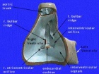
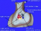
Images from human heart (week 9)[7]
- Heart (week 9)
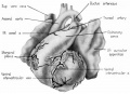
fig 1 Ventrocephalic heart 31.5 mm embryo
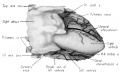
fig 2 Dextral heart 31.5 mm embryo superficial cardiac vessels
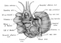
fig 3 Dorsal heart 31.5 mm embryo
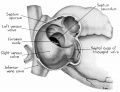
fig 4 Dextral heart 25 mm embryo atrium opened
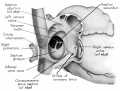
fig 5 Dextral heart 25 mm embryo
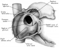
fig 6 Dextral heart 31.5 mm embryo
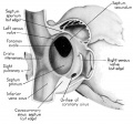
fig 7 Dextral heart 31.5 mm embryo
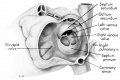
fig 8 Sinistral heart 25 mm embryo
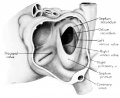
fig 9 Sinistral heart 31.5 mm embryo interatrial septal complex
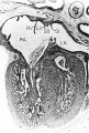
fig 10 Tricuspid valve 31.5 mm embryo
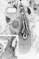
fig 11 Pulmonary semilunar valves 31.5 mm embryo
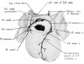
fig 12 Branches coronary arteries supply sino-atrial node
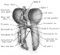
fig 13 Branches coronary arteries supply bundle of His and its main branches
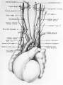
fig 14 Ventral heart and great vessels 31.5 mm embryo
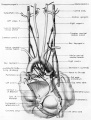
fig 15 Dorsal heart 31.5 mm embryo
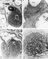
fig 16 Sinoventricular conduction system heart 31.5 mm embryo
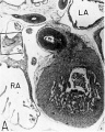
fig 16a Right atrium near level of entrance of superior vena cava
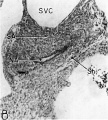
fig 16b Detail from A box
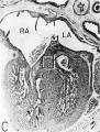
fig 16c Interventricular septum
fig 16d Detail from C box


Movies
Cardiovascular System Development | Heart Tutorial
|
|
|
|
|
| Heart Cartoons | |||||||||||||||||||||||||||
|---|---|---|---|---|---|---|---|---|---|---|---|---|---|---|---|---|---|---|---|---|---|---|---|---|---|---|---|
|
|
|
|
|
|
|
|
|
|
|
| Heart (1951) |
| Page | Play |
| Historic Animations | |||||||||||||||
|---|---|---|---|---|---|---|---|---|---|---|---|---|---|---|---|
|
|
|
| ||||||||||||
|
|
|
|
| About Historic Animations | ||||
|---|---|---|---|---|
The sound quality is quite poor and some of the information is now out of date, most general concepts are still correct. Please note the relatively large size (Mb) of each excerpt will effect download and viewing. March 2013
|
Molecular

Human heart developmental functional networks[8]
Heart Layers
- pericardium - covers the heart, formed by 3 layers consisting of a fibrous pericardium and a double layered serous pericardium (parietal layer and visceral epicardium layer).
- myocardium - muscular wall of the heart, thickest layer formed by spirally arranged cardiac muscle cells.
- endocardium - lines the heart, epithelial tissue lining the inner surface of heart chambers and valves.
Heart Volume
| Week | Heart Volume (ml) | Lung Volume (ml) | |
|---|---|---|---|
| 10 | 0.6 | 1.6 | |
| 18 | 4.3 | 10.9 | |
| 30 | 26.6 | 49.3 | |
| Table data is "embryonic age" while original reference used gestational age GA (from LMP)[9] | |||
| Week | Left Stroke Volume (ml) | Right Stroke Volume (ml) | Cardiac Output L/R (ml/min) |
|---|---|---|---|
| 10 | 0.02 | 0.01 | 2.39 / 1.8 |
| 18 | 0.32 | 0.30 | 43.46 / 46.72 |
| 32 | 2.07 | 2.67 | 284.71 / 365.99 |
| Table data is "embryonic age" while original reference used gestational age GA (from LMP) The stroke volume (SV) can be calculated from ultrasound measurement of end diastole volume (EDV) minus end systole volume (ESV); (SV = EDV - ESV).[10] | |||
Additional Images
Historic Images
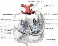
Fig. 528. Heart of a Rabbit Embryo seen from behind at 3.4 mm head length
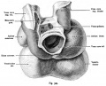
Fig. 529. The heart of a 24 mm Embryo
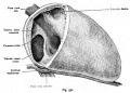
Fig. 530. Fetal heart (6 months) in normal situation
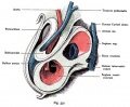
Fig. 531. Heart included in the pericardium of a human embryo of 7.5 mm body length
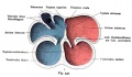
Fig. 532. Development of the heart chambers and septa
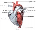
Fig. 533. Heart of a newborn viewed from the front and placed in the vertical direction
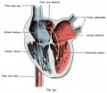
Fig. 534. Fetal heart, dorsal half with the afferent paths, open and colored according to the physiological condition of the blood
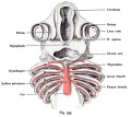
Fig. 535. The aortic arch in the shark embryo (Pristiurus)
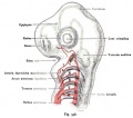
Fig. 536. The arteries of the gill arch region of a shark embryo (Pristiurus)
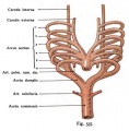
Fig. 537. Aortic arch of mammals and man
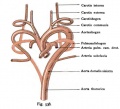
Fig. 538. Arteries in mammals and humans from the Aortic Arch


References
- ↑ Morris GM & Ariyaratnam JP. (2019). Embryology of the Cardiac Conduction System Relevant to Arrhythmias. Card Electrophysiol Clin , 11, 409-420. PMID: 31400866 DOI.
- ↑ Miao L, Li J, Li J, Lu Y, Shieh D, Mazurkiewicz JE, Barroso M, Schwarz JJ, Xin HB, Singer HA, Vincent PA, Zhong W, Radice GL, Wan LQ, Fan ZC, Huang G & Wu M. (2019). Cardiomyocyte orientation modulated by the Numb family proteins-N-cadherin axis is essential for ventricular wall morphogenesis. Proc. Natl. Acad. Sci. U.S.A. , 116, 15560-15569. PMID: 31300538 DOI.
- ↑ Garcia-Canadilla P, Cook AC, Mohun TJ, Oji O, Schlossarek S, Carrier L, McKenna WJ, Moon JC & Captur G. (2019). Myoarchitectural disarray of hypertrophic cardiomyopathy begins pre-birth. J. Anat. , , . PMID: 31347708 DOI.
- ↑ Odelin G, Faure E, Coulpier F, Di Bonito M, Bajolle F, Studer M, Avierinos JF, Charnay P, Topilko P & Zaffran S. (2018). Krox20 defines a subpopulation of cardiac neural crest cells contributing to arterial valves and bicuspid aortic valve. Development , 145, . PMID: 29158447 DOI.
- ↑ Arráez-Aybar LA, Turrero-Nogués A & Marantos-Gamarra DG. (2008). Embryonic cardiac morphometry in Carnegie stages 15-23, from the Complutense University of Madrid Institute of Embryology Human Embryo Collection. Cells Tissues Organs (Print) , 187, 211-20. PMID: 18057862 DOI.
- ↑ Végh AMD, Duim SN, Smits AM, Poelmann RE, Ten Harkel ADJ, DeRuiter MC, Goumans MJ & Jongbloed MRM. (2016). Part and Parcel of the Cardiac Autonomic Nerve System: Unravelling Its Cellular Building Blocks during Development. J Cardiovasc Dev Dis , 3, . PMID: 29367572 DOI.
- ↑ Licata RH. The human embryonic heart in the ninth week. (1954) Amer. J Anat., 94: 73-125. PMID 13124266
- ↑ Lage K, Møllgård K, Greenway S, Wakimoto H, Gorham JM, Workman CT, Bendsen E, Hansen NT, Rigina O, Roque FS, Wiese C, Christoffels VM, Roberts AE, Smoot LB, Pu WT, Donahoe PK, Tommerup N, Brunak S, Seidman CE, Seidman JG & Larsen LA. (2010). Dissecting spatio-temporal protein networks driving human heart development and related disorders. Mol. Syst. Biol. , 6, 381. PMID: 20571530 DOI.
- ↑ Peralta CF, Cavoretto P, Csapo B, Falcon O & Nicolaides KH. (2006). Lung and heart volumes by three-dimensional ultrasound in normal fetuses at 12-32 weeks' gestation. Ultrasound Obstet Gynecol , 27, 128-33. PMID: 16388511 DOI.
- ↑ Molina FS, Faro C, Sotiriadis A, Dagklis T & Nicolaides KH. (2008). Heart stroke volume and cardiac output by four-dimensional ultrasound in normal fetuses. Ultrasound Obstet Gynecol , 32, 181-7. PMID: 18634132 DOI.
Reviews
Végh AMD, Duim SN, Smits AM, Poelmann RE, Ten Harkel ADJ, DeRuiter MC, Goumans MJ & Jongbloed MRM. (2016). Part and Parcel of the Cardiac Autonomic Nerve System: Unravelling Its Cellular Building Blocks during Development. J Cardiovasc Dev Dis , 3, . PMID: 29367572 DOI.
Articles
Cho KH, Kim JH, Murakami G, Abe H, Rodríguez-Vázquez JF & Chai OH. (2019). Nerve distribution in myocardium including the atrial and ventricular septa in late stage human fetuses. Anat Cell Biol , 52, 48-56. PMID: 30984452 DOI.
Wu SM, Chien KR & Mummery C. (2008). Origins and fates of cardiovascular progenitor cells. Cell , 132, 537-43. PMID: 18295570 DOI.
Person AD, Klewer SE & Runyan RB. (2005). Cell biology of cardiac cushion development. Int. Rev. Cytol. , 243, 287-335. PMID: 15797462 DOI.
Matsui H, Ikeda K, Nakatani K, Sakabe M, Yamagishi T, Nakanishi T & Nakajima Y. (2005). Induction of initial cardiomyocyte alpha-actin--smooth muscle alpha-actin--in cultured avian pregastrula epiblast: a role for nodal and BMP antagonist. Dev. Dyn. , 233, 1419-29. PMID: 15977172 DOI.
Moorman A, Webb S, Brown NA, Lamers W & Anderson RH. (2003). Development of the heart: (1) formation of the cardiac chambers and arterial trunks. Heart , 89, 806-14. PMID: 12807866
Anderson RH, Webb S, Brown NA, Lamers W & Moorman A. (2003). Development of the heart: (2) Septation of the atriums and ventricles. Heart , 89, 949-58. PMID: 12860885
Brand T. (2003). Heart development: molecular insights into cardiac specification and early morphogenesis. Dev. Biol. , 258, 1-19. PMID: 12781678
Yutzey KE & Kirby ML. (2002). Wherefore heart thou? Embryonic origins of cardiogenic mesoderm. Dev. Dyn. , 223, 307-20. PMID: 11891982 DOI.
Eisenberg LM. (2002). Belief vs. scientific observation: the curious story of the precardiac mesoderm. Anat. Rec. , 266, 194-7. PMID: 11920381
Davidson AJ & Zon LI. (2000). Turning mesoderm into blood: the formation of hematopoietic stem cells during embryogenesis. Curr. Top. Dev. Biol. , 50, 45-60. PMID: 10948449
Heart - Historic References
| Heart - Historic References | ||
|---|---|---|
Tandler J. The Development of the Heart. (1912) Sect. II, chapt. 18, vol. 2, in Keibel F. and Mall FP. Manual of Human Embryology II. (1912) J. B. Lippincott Company, Philadelphia., pp. 534-570. Mall FP. On the development of the human heart. (1912) Amer. J Anat. 13: 249-298. Abbott ME. Congenital Cardiac Disease (1915) Osler & Mccrae's Modern Medicine 6, 2nd Edition. Frazer JE. The formation of the pars membranacea septi. (1916) J Anat. 51(1): 19-29. PMID 17103800 Waterston D. The development of the heart in man. (1917) Trans. Roy. Soc. Edin., 7(2): 258-302. McClure CFW. and Butler EG. The development of the vena cava inferior in man. (1925) Amer. J Anat. 35(3): 331-383. Odgers PNB. The formation of the venous valves, the foramen secundum and the septum secundum in the human heart. (1935) J. Anat., 69: 412-422. PMID 17104548 Odgers PN. The development of the pars membranacea septi in the human heart. (1938) J Anat. 72(2): 247-59. https://www.ncbi.nlm.nih.gov/pubmed/17104688 PMID 17104688] Patten BM. Developmental defects at the foramen ovale. (1938) Am J Pathol. 14(2):135-162. PMID 19970381 Odgers PNB. The development of the atrio-ventricular valves in man. (1939) J Anat. 73: 643-57. PMID 17104787 Kramer TC. The partitioning of the truncus and conus and the formation of the membranous portion of the interventricular septum in the human heart. (1942) Amer. J Anat. 71(3): 343-370. |
Terms
- angioblastic cords - Groups or ‘columns’ of embryonic precursor cells which will form the walls of both arteries and veins.
- apex - Anatomical term referring to the most inferior, left, downwards pointing part of the heart.
- aorta - The largest artery in the human body originating in the left ventricle. The aorta ascends, arches over the heart and then descends through the abdomen.
- aortic arch arteries - (pharyngeal arch arteries) Each early developing pharyngeal arch contains a lateral pair of arteries arising from the aortic sac, above the heart, and running into the dorsal aorta. Later in development these arch arteries are extensively remodelled to form specific components of the vascular system.
- aortic valve - Three-leaflet valve located at the junction between the left ventricle and aortic entrance.
- aortic vestibule - Smooth-walled portion of the left ventricle directly below the aortic valve.
- aorticopulmonary septum - Division between the aorta and the pulmonary trunk formed from the bulbar ridges.
- atresia - Abnormal closure or absence of a body vessel or orifice.
- atrioventricular canal - Junction between the primitive atrium and primitive ventricle in the embryo. This canal splits to later form two atrioventricular canals which consequently form the valves of the adult heart.
- bulbar ridges - Endocardial cushion tissue located in the bulbus cordis extending into the truncus arteriosus thus forming ridges. These fuse together to form the aorticopulmonary septum.
- bulbus cordis - A region of the early developing heart tube forming the common outflow tract, will differentiate to form three regions of the heart.
- cardiac jelly - Term used in early heart development to describe the initial gelatinous or sponge-like connective tissue separating the myocardium and heart tube endothelium.
- cardiogenic region - The area in the embryo where the precursor cells for heart development lie.
- chordae tendineae - Cord-like tendons connecting the papillary muscles to the leaflets of the mitral and tricuspid valves.
- congenital heart disease - Abnormal structure or function of the heart due to a developmental defect arising prior to birth.
- connective tissue - Fibrous tissue that acts to support body structures or bind other forms of tissue.
- conus arteriosus - An embryological heart outflow structure, that forms in early cardiac development and will later divides into the pulmonary artery and aorta. Term is also used clinically to describe the malformation of the cardiac outflow pattern, where only one artery arises from the heart and forms the aorta and pulmonary artery.
- coronary sinus - a venous sinus emptying into the right atrium that collects blood from the myocardium of the heart. The coronary sinus is the largest cardiac vein.
- cyanosis - Blue colouration of the skin and mucous membrane due to poor oxygenation of the blood.
- dorsal aortae - Two largest arteries either side of the midline which later fuse to form the descending portion of the aorta.
- dorsal mesocardium - The mesentery attaching the heart to the dorsal wall of the pericardial coelom. This breaks down to form a space known as the transverse pericardial sinus.
- dyspnoea - Shortness of breath.
- endocardial cushions - Swellings of migrated cells on the inner lining of the heart located in the atrioventricular canal.
- endocardial heart tubes - Two tubes formed from the cardiogenic plate in the developing embryo. These form the primordium of the truncus arteriosus, the atrium and the ventricles; later invested with myocardium.
- endocardium - The epithelial membrane lining the inside surface of heart, which along with the endothelial layer forms a continuous lining of the entire cardiovascular system. The endocardium, like the majority of the heart is mesoderm in origin.
- endothelium - A simple squamous epithelium lining blood vessels.
- epicardium - The outer layer of heart tissue.
- fibrous trigone - (trigonum fibrosum) term describing the dense connective tissue between the aortic ring and the atrioventricular ring and has a left and right component. The right fibrous trigone (trigonum fibrosum dextrum) lies between the aortic ring and the right atrioventricular ring. The left fibrous trigone (trigonum fibrosum sinistrum) lies between the aortic ring and the left atrioventricular ring.
- foramen ovale - Shunt allowing blood to enter the left atrium from the right atrium. It is located in the septum secundum.
- foramen primum - Original space between the septum primum and the fused endocardial cushions as the septum primum grows towards the cushions.
- foramen secundum - Refers to the coalesced perforations in the septum primum after it has fused with the endocardial cushions.
- fossa ovalis - (oval fossa, annulus ovalis) Region in the postnatal atrial septum where the foramen ovale was located during embryonic development. Appears as an indentation in the right atrium septum.
- heart murmur - Extra heart sounds appearing upon auscultation due to turbulent blood flow.
- hypertrophy - Increase in size of an organ or tissue due to enlargement of component cells.
- inferior vena cava - (IVC) Large vein which carries deoxygenated blood from the lower half of the body to the right atrium.
- inflow tract - Entrance of blood into the heart tube; the sinus venosus portion of the tube.
- infundibulum - Smooth-walled portion of the right ventricle directly below the pulmonary valve.
- interventricular septum - Wall of muscular tissue growing from the base of the heart dividing the primitive ventricle into the left and right ventricles.
- interventricular foramen - Space between the interventricular septum and the fused endocardial cushions. The foramen closes when the septum fuses with the endocardial cushions and bulbar ridges.
- intraembryonic coelom - Initial embryonic space in lateral plate mesoderm that will be separated to form the three major body cavities: pericardial, pleural and peritoneal cavity. The lateral plate mesoderm is divided by this space into splanchnic and somatic mesoderm.
- left horn of sinus venosus - The left side of the sinus venosus (initially symmetrical with the right) collecting blood from half of the paired veins: common cardinal veins, umbilical veins and vitelline veins. Later the left horn diminishes and becomes the small coronary sinus.
- mitral valve - (Bicuspid valve) two leaflet valve located on the left side of the heart, that is between the left atrium and ventricle.
- myocardium - The middle layer of the heart wall composed of cardiac muscle.
- neural crest mesenchyme - Connective tissue arising from critical cells in the cranial region of the embryo. These paired dorsal lateral streaks of cells migrate throughout the embryo and can differentiate into many different cell types (= pluripotential). Those that remain on the dorsal neural tube form the sensory spinal ganglia (DRG), those that migrate ventrally form the sympatheitic ganglia. Neural crest cells also migrate into the somites and regions through the entire embryo.
- outflow tract - Exit of blood from the heart tube formed by the truncus arterioles.
- oval fossa - (fossa ovalis, annulus ovalis) Region in the postnatal atrial septum where the foramen ovale was located during development. Appears as an indentation in the right atrium septum.
- papillary muscles - Small muscles found on the inner myocardium of the left and right ventricles. They are attached to the mitral and tricuspid valves via the chordae tendineae and serve to limit the movements of the valves.
- patent foramen ovale - Abnormality in atrial septum due to failure of the atrial septum to close at the foramen ovale. (More? Atrial Septal Defects)
- pericardial coelom - (pericardial cavity) The anatomical body cavity in which the heart lies. The pericardial cavity forms in the lateral plate mesoderm above the buccopharyngeal membrane, as part of the early intraembryonic coelom. This cavity is initially continuous with the two early pleural cavities. Note the single intraembryonic coelom forms all three major body cavities: pericardial cavity, pleural cavity, peritoneal cavity.
- primordial atrium - Common cavity in the upper portion of the developing heart. Later divides to form the left and right atria.
- primordial ventricle - Common cavity in the lower portion of the developing heart. Later divides to form the left and right ventricles.
- pulmonary circulation - Carries blood between the heart and lungs.
- pulmonary trunk - A vessel that arises from the right ventricle of the heart, extends upward, and divides into the right and left pulmonary arteries that transport deoxygenated blood to the lungs.
- pulmonary valve - Three-leaflet valve located at the junction between the right ventricle and the pulmonary trunk.
- pulmonary veins - Four veins that allow oxygenated blood from the lungs to empty into the left atrium.
- right horn of sinus venosus - The right side of the sinus venosus (initially symmetrical with the left) collecting blood from half of the paired veins: common cardinal veins, umbilical veins and vitelline veins. Later the right horn dilates, receiving all the veins, and becomes the sinus venarum of the right atrium.
- second heart field - (SHF) splanchnic mesoderm that forms progenitors important for heart formation.
- semilunar valves - Flaps of endocardium and connective tissue reinforced by fibres which prevent the valves from turning inside out. They are shaped like a half moon, hence the name semilunar. The semilunar valves are located between the aorta and the left ventricle and between the pulmonary artery and the right ventricle.
- septum primum - Original structure growing from the roof of the heart towards the endocardial cushions dividing the primitive atrium.
- septum secundum - Second structure growing to the right of the septum primum dividing the primitive atrium.
- sinoatrial node - Specialised cardiomyocyte pacemaking region of the heart located at the entry of the right superior caval vein (SVC) into the right atrium. (More? Detailed Cardiac - Sinus Node)
- sinus venosus - (systemic venous sinus) An early developmental cardiovascular structure, thin walled cavity, forming the input to developing heart which has 3 venous inputs (vitelline vein, umbilical vein, common cardinal vein). Later in heart development this structure gets incorporated into the wall of the future right atrium. (More? Systemic Venous Sinus)
- sinuatrial orifice - The opening between the sinus venosus and right atrium which has two valve leaflets to prevent backflow of blood.
- sinus venarum - Smooth-walled portion of the adult right atrium; originally the left horn of the sinus venous.
- splanchnic mesoderm - Gastrointestinal tract (endoderm) associated mesoderm formed by the separation of the lateral plate mesoderm into two separate components by a cavity, the intraembryonic coelom. Splanchnic mesoderm is the embryonic origin of the second heart field, gastrointestinal tract connective tissue, smooth muscle, blood vessels and contribute to organ development (heart, pancreas, spleen, liver).
- stenosis - Abnormal narrowing of a blood vessel or orifice.
- superior vena cava - (SVC) Short vein which carries deoxygenated blood from the upper half of the body to the right atrium.
- systemic circulation - Carries oxygenated blood away from the heart to the other body organs and returns to the heart deoxygenated.
- tachypnoea - Abnormally rapid breathing rate.
- trabeculae - (trabeculations)Muscular beams located within the ventricles and parts of the atria of the heart.
- transverse pericardial sinus - Dorsal area within the pericardial coelom, initially occupied by the dorsal mesocardium.
- tricuspid valve - Three leaflet valve located in the right atrioventricular canal i.e. between the right atrium and ventricle.
- tricuspid atresia - Abnormality of the tricuspid valve between the right atrium and right ventricle.
- truncus arteriosus - An embryological heart outflow structure, that forms in early cardiac development and will later divides into the pulmonary artery and aorta. Term is also used clinically to describe the malformation of the cardiac outflow pattern, where only one artery arises from the heart and forms the aorta and pulmonary artery.
| Other Terms Lists |
|---|
| Terms Lists: ART | Birth | Bone | Cardiovascular | Cell Division | Endocrine | Gastrointestinal | Genital | Genetic | Head | Hearing | Heart | Immune | Integumentary | Neonatal | Neural | Oocyte | Palate | Placenta | Radiation | Renal | Respiratory | Spermatozoa | Statistics | Tooth | Ultrasound | Vision | Historic | Drugs | Glossary |
External Links
External Links Notice - The dynamic nature of the internet may mean that some of these listed links may no longer function. If the link no longer works search the web with the link text or name. Links to any external commercial sites are provided for information purposes only and should never be considered an endorsement. UNSW Embryology is provided as an educational resource with no clinical information or commercial affiliation.
- Embryo Images - Embryo Images Online Early Cell Populations (cardiogenic section) | Cardiovascular Development | Week 3 Development | Week 4 Development | Heart Chambers and Outflow Tract | Atrioventricular Septation | Outflow Tract Septation | Ventricular Septation | Atrial Septation | Atrial Walls Aortic Arch Vessels | Changes at Birth
Glossary Links
- Glossary: A | B | C | D | E | F | G | H | I | J | K | L | M | N | O | P | Q | R | S | T | U | V | W | X | Y | Z | Numbers | Symbols | Term Link
Cite this page: Hill, M.A. (2026, July 9) Embryology Cardiovascular System - Heart Development. Retrieved from https://embryology.med.unsw.edu.au/embryology/index.php/Cardiovascular_System_-_Heart_Development
- © Dr Mark Hill 2026, UNSW Embryology ISBN: 978 0 7334 2609 4 - UNSW CRICOS Provider Code No. 00098G