Carnegie Stages
| Embryology - 19 Jul 2026 |
|---|
| Google Translate - select your language from the list shown below (this will open a new external page) |
|
العربية | català | 中文 | 中國傳統的 | français | Deutsche | עִברִית | हिंदी | bahasa Indonesia | italiano | 日本語 | 한국어 | မြန်မာ | Pilipino | Polskie | português | ਪੰਜਾਬੀ ਦੇ | Română | русский | Español | Swahili | Svensk | ไทย | Türkçe | اردو | ייִדיש | Tiếng Việt These external translations are automated and may not be accurate. (More? About Translations) |
Introduction

Carnegie stages are named after the famous US Institute which began collecting and classifying embryos in the early 1900's. Stages are based on the external and/or internal morphological development of the embryo, and are not directly dependent on either age or size. The human embryonic period proper is divided into 23 Carnegie stages covering the first 8 weeks post-ovulation (GA week 10). Criteria beyond morphological features include ranges of age in days, number of somites present, and embryonic crown rump lengths (CRL). See also the timeline tabulation of both whole embryo and systematic development. The table below also has detailed descriptions of each Carnegie stage as well as identifying embryo examples from different collections and the published literature.
| Week: | 1 | 2 | 3 | 4 | 5 | 6 | 7 | 8 |
| Carnegie stage: | 1 2 3 4 | 5 6 | 7 8 9 | 10 11 12 13 | 14 15 | 16 17 | 18 19 | 20 21 22 23 |
Note that many photographs of staged (using Carnegie criteria) human embryos on this current site are from the Kyoto collection in collaboration with Prof Kohei Shiota and Prof Shigehito Yamada. Scanning electron micrographs are published in collaboration with Prof Kathy Sulik.
- Links: Embryonic Development | Carnegie Stage Comparison | Carnegie Institution - Contributions to Embryology | Human Embryo Collections
- Carnegie Stages: 1 | 2 | 3 | 4 | 5 | 6 | 7 | 8 | 9 | 10 | 11 | 12 | 13 | 14 | 15 | 16 | 17 | 18 | 19 | 20 | 21 | 22 | 23 | About Stages | Timeline
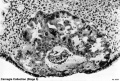
Carnegie Embryos

Embryo/Fetal Definition
The division of human development into an embryonic (embryo) and fetal (fetus) periods was an historically based arbitrary system. Defined by the probability that more than 90 percent of the identifiable structures of the adult body have appeared by Carnegie stage 23.
Streeter also defined the fetal period as beginning when the humerus cartilage was replaced by bone marrow.
There have been several other human systems of embryo categorisation developed, sometimes to establish a standard between species. To prevent confusion and consistency with the historic literature the Carnegie stages are used for human development on this site.
History
The following text and information about the collection is modifed from the original Carnegie Institute website.

Franklin Mall
Franklin P. Mall (1862-1917) is most remembered for his work done at the Department of Embryology at the Carnegie Institute of Washington. Mall began collecting human embryos while a postgraduate student in Lepzig with Wilhelm His, but didn't receive the first Carnegie specimen until his position at Johns Hopkins University. (More? Franklin Mall)
- Franklin Mall Links: Franklin Mall | 1891 26 Day Human Embryo | 1905 Blood-Vessels of the Brain | 1906 Human Ossification | 1910 Manual of Human Embryology 1 | 1912 Manual of Human Embryology 2 | 1911 Mall Human Embryo Collection | 1912 Heart Development | 1915 Tubal Pregnancy | 1916 Human Magma in Normal and Pathological Development | 1917 Frequency Human Abnormalities | 1917 Human Embryo Cyclopia | 1918 Embryo Age | 1918 Appreciation | 1934 Franklin Mall biography PDF | Mall photograph | Mall painting | Mall painting | Carnegie Stages | Carnegie Embryos | Carnegie Collection | Category:Franklin Mall
Surprizingly age and size proves a poor way to organize embryos. It is very difficult to accurately age an embryo, and it could shrink a full 50% in the preserving fluids. Mall took it upon himself to find a better way. He had more success basing his "staging" scheme on morphological characteristics. To that end, Mall and his colleagues not only prepared and preserved serial sections of the embryos, they also made hundreds of three-dimensional models at different stages of growth. According to Adrianne Noe, who managed the collection at the National Museum of Health and Medicine, Mall gathered the most renowned scientists, scholars, artists, photographers, and craftspeople ever to apply their interests and skills to embryology.
- Links: Franklin Mall
George Streeter

Began as an assistant professor at the Wistar Institute of Anatomy and Biology in Philadelphia, then went to the University of Michigan as professor of gross anatomy. In 1914, he became research professor in the department of embryology of the Carnegie Institution, at the Johns Hopkins Medical School under Franklin Mall and succeeded him as director of the Carnegie Institution.
- Links: George Streeter
Collection Location
The embryo collection is now held at the National Museum of Health and Medicine, located at the Walter Reed Army Medical Center in Washington, D.C. the Carnegie collection is still available for use by researchers. The raw data, which will be copyright free, may be made available to all legitimate researchers and students. HDAC - Agreement Policies
Contributions to Embryology
| The Contributions to Embryology are a historic series of papers published by the Carnegie Institution of Washington early in the 20th Century. Many of the collection embryos were first described and characterised in these papers and from serial sectioning of these embryos.
The 1920 volume (Volume IX) was prepared as a memorial by present and former members of the staff of the Institute to the late Professor Franklin Paine Mall. |

Carnegie Institute Staff (1935)
James F. Didusch

James F. Didusch (1890 - 1955) was a medical illustrator in the Department of Art as Applied to Medicine, Johns Hopkins. He was the main illustrator for the Carnegie Institute of Embryology (1913-1955) with his drawings and plates forming the main visual component of many Carnegie publications. There has been a 1992 article on his artistic contribution to embryology[1] and his papers are held in the Alan Mason Chesney Medical Archives at Johns Hopkins.
- Links: James Didusch
Osborne O. Heard
| One of the first to be hired, in 1913, was modeler Osborne O. Heard, who spent 42 years at the department and made over 700 wax-based reconstructions.
The results of this team effort still stand as the international standard by which human embryos are described and classified. The models were mainly made by the lost-wax casting process and his models were also more detailed than the earlier (1880's) Ziegler embryo models. |

- Links: Osborne Heard
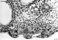
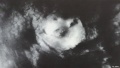
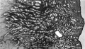
Carnegie Collection Embryos
| iBook - Carnegie Embryos | |
|---|---|

|
|
| Week: | 1 | 2 | 3 | 4 | 5 | 6 | 7 | 8 |
| Carnegie stage: | 1 2 3 4 | 5 6 | 7 8 9 | 10 11 12 13 | 14 15 | 16 17 | 18 19 | 20 21 22 23 |
Stage 3
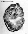
Embryo No.8794
Embryo No.8663


| Stage 3 Links: Week 1 | zona pellucida | blastocyst | trophoblast | mitosis | Lecture | Medicine Practical | Science Practical | Blastocyst Day 3-6 Movie | Next Stage 4 |
| Historic Papers: 1954 | 1956 |
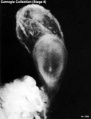
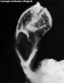
Stage 4


| Stage 4 Links: Week 2 | Implantation | | Trophoblast | Human Chorionic Gonadotropin | Lecture | Practical | Category:Carnegie Stage 4 | Next Stage 5 |
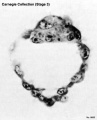
Stage 5
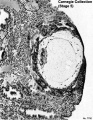
Embryo No.8155
Embryo No.8155
Embryo No.8155
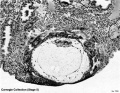
Embryo No.8004
Embryo No.8004
Embryo No.8004
Embryo No.7700
Embryo No.7700
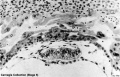
Embryo No.7700
Embryo No.7700


| Stage 5 Links: Week 2 | Implantation | Lecture | Practical | Carnegie Embryos | Category:Carnegie Stage 5 | Next Stage 6 | ||
| Historic Papers: 1941 | 1944 day 9-10 | 1945 day 7.5 | 1945 day 9-10 | ||
|
Stage 6


| Stage 6 Links: Week 2 | Implantation | Lecture | Practical | Carnegie Embryos | Category:Carnegie Stage 6 | Next Stage 7 |
| Historic Papers: 1909 | 1925 | 1937 |
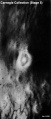
Stage 7


| Stage 7 Links: Week 3 | Gastrulation | Lecture | Practical | Carnegie Embryos | Category:Carnegie Stage 7 | Next Stage 8 |
| Historic Papers: 1923 head-process | 1933 tubal | 1940 | 1949 |
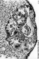
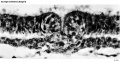
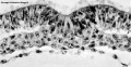
Stage 8
Embryo 5960
Embryo 5960
primitive pit
Embryo 7545 primitive groove and primitive streak
Embryo 7545 notochordal process and notochordal canal
Embryo 7545 section series
Embryo 7545 notochordal process and notochordal canal


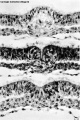
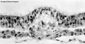
Stage 9


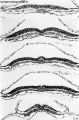
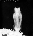
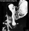
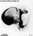
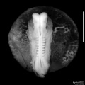
Stage 10
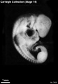
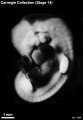
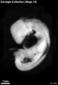
6330 dorsal
6330 lateral
7251 lateral
3710 lateral


| Stage 10 Links: Week 4 | Gastrulation | Lecture | Practical | Image Gallery | Carnegie Embryos | Embryos | Category:Carnegie Stage 10 | Next Stage 11 |
| Historic Papers: 1910 | 1917 | 1926 | 1939 | 1943 | 1957 | 1985 |
Stage 11
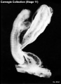
6344 dorsolateral
6344 dorsal
6784 ventral
6050 right


Stage 12


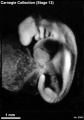
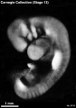
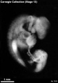
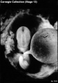
Stage 13
6473 left
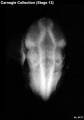
6473 dorsal
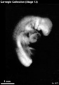
6473 right
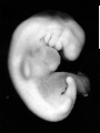
6469 right
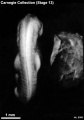
8066 dorsal
8066 left
8119 left
7433 right
7433 ventral


Stage 14
6830 left
1380 left
7333 left
6502 left
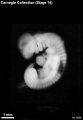
left

6502 left
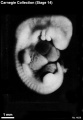
4629 right
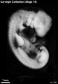
7394 right
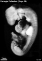
6848 left
7400 right
7394 left

1620 left
7400 left


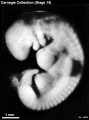
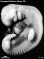
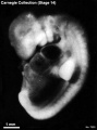
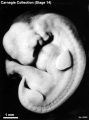
Stage 15


Stage 16


Stage 17


Stage 18


Stage 19


Stage 20


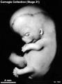
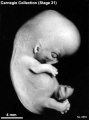
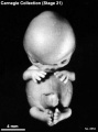
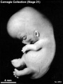
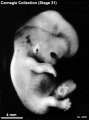
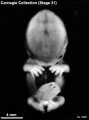
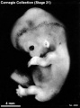
Stage 21
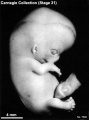
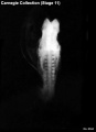
Embryo No.7392 (right)
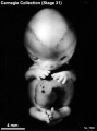
Embryo No.7392 (front)
Embryo No.7392 (left)
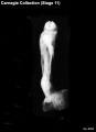
Embryo No.8553 (right)
Embryo No.8553 (front)
Embryo No.8553 (left)
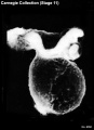
Embryo No.4090 (right)
Embryo No.4090 (front)
Embryo No.4090 (left)


Stage 22


| Stage 22 Links: Week 8 | System Development | Lecture - Limb | Lecture - Head Development | Lecture - Sensory | Science Practical - Head | Science Practical - Sensory | Science Practical - Urogenital | Historic - Skull Development | Carnegie Embryos | Madrid Embryos | Category:Carnegie Stage 22 | Next Stage 23 |
| Week 8, GA week 10, 54 - 56 days, CRL 23 - 28 mm, Carnegie Embryos |
| Historic Papers: 1914 | 1954 Stage 19-23 |
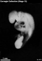
Stage 23


Fetal
Stage 23 is the final embryonic stage, after this development is described as "fetal" through the entire second and third trimester.
Carnegie Human Embryo Glossary
Age
Postovulatory age is one criterion for the overall staging of embryos.
It is the length of time since the last ovulation before fertilization took place and is estimated by assigning an embryo to a developmental stage and then referring to a standard table of norms. A range of +/- 1 day is expected. Postovulatory age is stated in days or weeks.
Case Number
The basis of the Carnegie Collection of Embryos was Franklin P. Mall's personal collection of 813 embryos, which he began in 1887 with the first specimen. Carnegie Embryo No. 1. was acquired by Mall while he was an assistant in Pathology at the Johns Hopkins Hospital. All subsequent specimens in the Collection were numbered sequentially at the time of acquisition, i.e. case number.
Crown-Rump (C-R)

One criterion for the overall classification of human embryonic development.
A measurement of prenatal length, from the vertex of the skull (crown), along the curvature of the spine to the midpoint between the apices of the buttocks (rump), of the developing embryo. This measurement was developed for smaller specimens, 35mm or less, so that their natural curved posture is not disturbed. C-R length is stated in millimeters.
An alternative measurement of prenatal length is the greatest length (G.L.), which some researchers find to be more useful in the assessment of length of an embryo. The G.L. is determined by measuring the embryo in a straight line, (i.e. caliper length) without any attempt to straighten the natural curvature of the specimen. G.L. is stated in millimeters.
Grade
Specimens in the Carnegie Collection have been graded Excellent, Good, Fair or Poor.
This reference is based on the total grade of the specimen, including both its original quality and the condition of the specimen.
Normal/Abnormal
Of the approximately 600 sectioned embryos in the Carnegie Collection assigned to the 23 stages, a majority have been classified as normal. However, variations in, and anomolies of, individual organs are known to occur.
Section Information
Plane of Section
The surface formed by extension through an axis of the embryo.
There are three primary descriptive terms referring to the planes of the embryo:
- Coronal A vertical plane dividing the body into anterior and posterior portions.
- Sagittal Any plane parallel to the median.
- Transverse A plane horizontal to the median.
Thinness of Section
The specified thinness of the cut embryonic section for mounting on a glass slide in serial order. Thinness is measured in micrometers. Some regions of a few of the specimens in the Collection were cut at various thinnesses; these instances are represented in the search results.
Slides
Total number of glass slides containing serial histologic sections of each specimen in the Collection.
Sections
Total number of serial histologic sections on any number of glass slides for each specimen in the Collection.
Sex
Gender identification, i.e. male or female, is noted where apparent. In the embryo, the gonads do not acquire male or female morphological characteristics until the 7th or 8th week of development (stages 18-23). Therefore, many specimens in the embryonic period are not identified by gender. This assignment applies mostly to very late embryonic period specimens in the Collection.
Somites
One criterion for the overall classification of human embryonic development.
Somites are paired segments of paraxial mesoderm appearing in longitudinal rows along the left and right side of the neural groove and notochord. They commence in the third or early fourth week of development (approximately the 20th day), appearing first in the cervical region of the embryo. Their formation proceeds in a craniocaudal direction. New somites appear approximately three per day, until at the end of the 5th week when 42 to 44 pairs are present. Differentiation of the somites leads to formation of the axial skeleton.
Stage of Development
The definitive classification of human embryos into developmental groups termed stages.
Stages are based on the external and/or internal morphological development of the embryo, and are not directly dependent on either age or size. The human embryonic period proper is divided into 23 Carnegie stages.
Criteria beyond morphological features include age in days, number of somites present, and embryonic length. This chart shows the relationship between Stage, Age and embryonic length.
Stain Type
The type of individual dye or staining substance, or combination of dyes and reagents, used in histologic technique to color the constituents of cells and tissues.
Carnegie Stage Table
Weeks shown in the table below are embryonic post ovulation age, for clinical Gestational Age (GA) measured from last menstrual period, add 2 weeks.
(not to scale) |
||||
 |
fertilized oocyte, zygote, pronuclei | |||
 |
morula cell division with reduction in cytoplasmic volume, blastocyst formation of inner and outer cell mass | |||
 |
loss of zona pellucida, free blastocyst | |||
| attaching blastocyst | ||||
(week 2) |
 |
implantation | ||
 |
extraembryonic mesoderm, primitive streak, gastrulation | |||
| gastrulation, notochordal process | ||||
| primitive pit, notochordal canal | ||||
 |
Somitogenesis Somite Number 1 - 3 neural folds, cardiac primordium, head fold | |||
| Somite Number 4 - 12 neural fold fuses | ||||
| Somite Number 13 - 20 rostral neuropore closes | ||||
| Somite Number 21 - 29 caudal neuropore closes | ||||
| Somite Number 30 leg buds, lens placode, pharyngeal arches | ||||
| lens pit, optic cup | ||||
| lens vesicle, nasal pit, hand plate | ||||
| nasal pits moved ventrally, auricular hillocks, foot plate | ||||
| finger rays | ||||
| ossification commences | ||||
| straightening of trunk | ||||
| upper limbs longer and bent at elbow | ||||
| hands and feet turned inward | ||||
| eyelids, external ears | ||||
| rounded head, body and limbs | ||||
The embryos shown in the table are from the Kyoto and Carnegie collection and other sources.
References
- ↑ <pubmed>1624478</pubmed>
- Carnegie Stages: 1 | 2 | 3 | 4 | 5 | 6 | 7 | 8 | 9 | 10 | 11 | 12 | 13 | 14 | 15 | 16 | 17 | 18 | 19 | 20 | 21 | 22 | 23 | About Stages | Timeline
Glossary Links
- Glossary: A | B | C | D | E | F | G | H | I | J | K | L | M | N | O | P | Q | R | S | T | U | V | W | X | Y | Z | Numbers | Symbols | Term Link
Cite this page: Hill, M.A. (2026, July 19) Embryology Carnegie Stages. Retrieved from https://embryology.med.unsw.edu.au/embryology/index.php/Carnegie_Stages
- © Dr Mark Hill 2026, UNSW Embryology ISBN: 978 0 7334 2609 4 - UNSW CRICOS Provider Code No. 00098G