Human Embryo Collections
| Embryology - 26 Apr 2024 |
|---|
| Google Translate - select your language from the list shown below (this will open a new external page) |
|
العربية | català | 中文 | 中國傳統的 | français | Deutsche | עִברִית | हिंदी | bahasa Indonesia | italiano | 日本語 | 한국어 | မြန်မာ | Pilipino | Polskie | português | ਪੰਜਾਬੀ ਦੇ | Română | русский | Español | Swahili | Svensk | ไทย | Türkçe | اردو | ייִדיש | Tiếng Việt These external translations are automated and may not be accurate. (More? About Translations) |
Introduction
While many universities hold collections of embryos from many species, very few have well-characterised collections of embryos showing human development. Many of those that are available are historic in nature, consist of histological sections, some with limited information about the embryo history.
There are groups now taking advantage of new imaging techniques to either re-evaluate these historic collections, or analysing new embryonic material. Some of these new databases are being made available online for research purposes.
See also Digital Embryology Consortium information page about the project to digitise and preserve these extremely valuable human collections.[1]
Remember this current site is for educational use only.
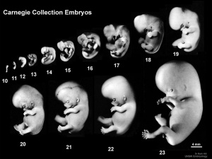
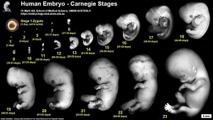
- Carnegie Stages: 1 | 2 | 3 | 4 | 5 | 6 | 7 | 8 | 9 | 10 | 11 | 12 | 13 | 14 | 15 | 16 | 17 | 18 | 19 | 20 | 21 | 22 | 23 | About Stages | Timeline
Carnegie Collection
(Carnegie Institution, USA)
- Begun by Franklin Mall in the early 1900's.
- Many of these embryos were used in the historic papers in the Contributions to Embryology series - Carnegie Institution of Washington Series.
- Franklin Mall Links: Franklin Mall | 1891 26 Day Human Embryo | 1905 Blood-Vessels of the Brain | 1906 Human Ossification | 1910 Manual of Human Embryology 1 | 1912 Manual of Human Embryology 2 | 1911 Mall Human Embryo Collection | 1912 Heart Development | 1915 Tubal Pregnancy | 1916 Human Magma in Normal and Pathological Development | 1917 Frequency Human Abnormalities | 1917 Human Embryo Cyclopia | 1918 Embryo Age | 1918 Appreciation | 1934 Franklin Mall biography PDF | Mall photograph | Mall painting | Mall painting | Carnegie Stages | Carnegie Embryos | Carnegie Collection | Category:Franklin Mall | Carnegie Embryos | Contributions to Embryology Series
Harvard Collection
This historic collection of human and other embryos was originally collected by Charles Minot (1852–1914), and sometimes referred to as the Minot Collection, now forms part of the larger Carnegie Collection.
- Links; Carnegie Collection | Harvard Collection
Kyoto Collection
(Kyoto University, Japan)
- Begun by Dr. Hideo Nishimura in 1961 and has over 44,000 human embryo specimens.
- Developed and managed by Kohei Shiota.
- Currently managed by Shigehito Yamada.
- Polydactyly in human embryos[2]
- 129 embryos with polydactyly in 36,380 human conceptuses obtained through induced abortion during the period from 1962 to 1974.
- Human embryo imaging with a super-parallel magnetic resonance (MR) microscope[3]
- Links: Kyoto Collection | Japan - Kyoto Collection
Blechschmidt Collection
(University of Goettingen, Germany)
- Erich Blechschmidt (1904–92) independently developed new methods of embryo reconstruction.
- Director of Göttingen University’s Anatomical Institute from 1942 until 1973.
- 200,000 serial sections of embryos and 64 models.
- Some of the embryological collection were assigned Carnegie Nos. 10315-10434 in 1972.
- Links: Blechschmidt Collection | The Human Embryology Collection | Collections of the University Medical Center
Hubrecht Collection
(Netherlands Institute for Developmental Biology in Utrecht, Hubrecht Laboratory)
In 2004 was relocated to the Museum fur Naturkunde, Berlin. Incorporating the Hill collection.
- The Hubrecht Laboratory[4] was founded in 1916 from Ambrosius Arnold Willem Hubrecht (1853-1915) personal collection.[5]
- The collection is for comparative embryology of vertebrates and includes a large number of animal embryos (also an associated historical library).
- 600 species of chordate, in 175 families and 10 classes.
- 2,000 wet specimens.
- histological sections (A. Dohrn - Pisces, Amphibia, Reptilia; L. Bolk - Vertebrates; and W. Kückenthal - Cetacea)
- material blocked out in paraffin but not sectioned.
Hill Embryological Collection
No relation to this Website's author/editor.
In 2004 this collection was relocated to the Museum fur Naturkunde, Berlin.
- James Peter Hill (1873-1954)
- University of Edinburgh, Royal College of Science in London, 1892 demonstrator in Sydney, Australia.
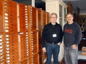
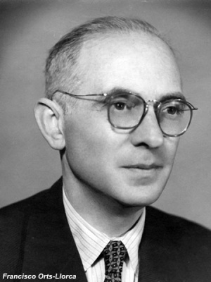
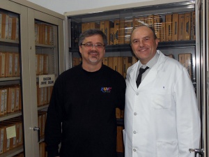
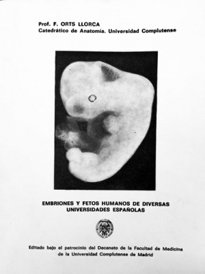
Madrid Institute of Embryology Human Embryo Collection
Complutense University of Madrid, Institute of Embryology.
- Links: Madrid Collection
Domenech-Mateu Collection
(Anatomy and Embryology Unit, Morphological Science Department, Universitat Autònoma de Barcelona)
- Links: Domenech-Mateu Collection
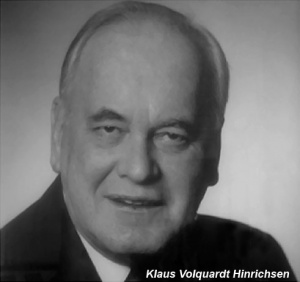
Hinrichsen Collection
| (Department of Anatomy and Molecular Embryology, Ruhr-Universität Bochum, Germany)
Human specimens were collected by Hinrichsen's team between 1969 and 1994. Some material from this collection was published in Hinrichsen's embryology textbook (Humanembryologie, 1990).
|
HDBR and HuDSeN Collection
Human Developmental Biology Resource
The HDBR is a MRC-Wellcome Trust funded resource to provide human embryological research materials (tissues, cells, slides, RNA, cDNA, DNA, protein) to registered users.
Human Developmental Studies Network
| HuDSeN is a new collection from optical projection tomography that links gene expression patterns to a specific set of previously scanned human embryos.
|
| |||
Hamilton-Boyd Collection
|
(Cambridge University, UK)
|
Radlanski Collection
22 human embryos and fetuses ranging from 18 to 270 mm CRL
Ralf J. Radlanski Telefax: +49–30–450562902 E-mail: ralfj.radlanski@charite.de
Charite – Campus Benjamin Franklin at Freie Universität Berlin, Center for Dental and Craniofacial Sciences, Department of Craniofacial Developmental Biology, Assmannshauser Str. 4-6, 14197 Berlin, Germany
An atlas of prenatal development of the human orofacial region.[9]
- "There are several atlases available showing prenatal human development. However, none is focused on prenatal orofacial development during maxillary and mandibular bone formation. These events, together with dental development and formation of the temporomandibular joint, take place during several fetal stages. While photographic atlases are limited to depicting the outer shape, and atlases based on histological sections only show a series of single sections, an atlas based on three-dimensional reconstructions from serial sections can show both the outer skin and the structures underneath, which can be electronically dissected layer by layer. In this Focus article, we present our atlas on prenatal human orofacial development, which is accessible online at the Journal's website."
Central Laboratory for Human Embryology
(University of Washington, USA)
- begun August 1963.
- after 18 years consists of 5,200 specimens.
- range from very young embryos to full-term fetus.
University of Wisconsin Otologic Collection
A historic collection of mainly fetal material described in the 1930's series of papers [10] and later in the 1940's by Anson.[11]
Normal Plates of the Development of the Human Embryo
A historic 1908 book by Franz Keibel (Normentafeln zur Entwicklungsgeschichte der Wirbeltiere - homo sapiens) documenting a range of available human embryonic materials.
Ziegler Models
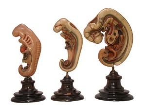
Not a collection as such, but a historic series of wax models made in the 1880s based upon the embryos of Prof. Wilhelm His, Leipzig. Wilhelm His had earlier prepared a series of freehand models. These Ziegler models are the basis of some teaching models still commercially available and used today.
The Carnegie Institute later in the early 1900's developed many additional models based upon their own collection. (More? Carnegie Models)
- Links: Ziegler Models | Wilhelm His
References
- ↑ Hill MA. (2019). Two Web Resources Linking Major Human Embryology Collections Worldwide. Cells Tissues Organs (Print) , , 1-10. PMID: 30673660 DOI.
- ↑ Shiota K & Matsunaga E. (1978). A genetic and epidemiologic study of polydactyly in human embryos in Japan. Jinrui Idengaku Zasshi , 23, 173-92. PMID: 691840 DOI.
- ↑ Matsuda Y, Ono S, Otake Y, Handa S, Kose K, Haishi T, Yamada S, Uwabe C & Shiota K. (2007). Imaging of a large collection of human embryo using a super-parallel MR microscope. Magn Reson Med Sci , 6, 139-46. PMID: 18037794
- ↑ Faasse P, Faber J & Narraway J. (1999). A brief history of the Hubrecht Laboratory. Int. J. Dev. Biol. , 43, 583-90. PMID: 10668967
- ↑ Richardson MK & Narraway J. (1999). A treasure house of comparative embryology. Int. J. Dev. Biol. , 43, 591-602. PMID: 10668968
- ↑ Kerwin J, Scott M, Sharpe J, Puelles L, Robson SC, Martínez-de-la-Torre M, Ferran JL, Feng G, Baldock R, Strachan T, Davidson D & Lindsay S. (2004). 3 dimensional modelling of early human brain development using optical projection tomography. BMC Neurosci , 5, 27. PMID: 15298700 DOI.
- ↑ Lindsay S & Copp AJ. (2005). MRC-Wellcome Trust Human Developmental Biology Resource: enabling studies of human developmental gene expression. Trends Genet. , 21, 586-90. PMID: 16154230 DOI.
- ↑ Kerwin J, Yang Y, Merchan P, Sarma S, Thompson J, Wang X, Sandoval J, Puelles L, Baldock R & Lindsay S. (2010). The HUDSEN Atlas: a three-dimensional (3D) spatial framework for studying gene expression in the developing human brain. J. Anat. , 217, 289-99. PMID: 20979583 DOI.
- ↑ Radlanski RJ & Renz H. (2010). An atlas of prenatal development of the human orofacial region. Eur. J. Oral Sci. , 118, 321-4. PMID: 20662903 DOI.
- ↑ Bast TH. Blood supply of the otic capsule of a 150 mm (C.R.) human fetus. (1931) Anat. Rec. 48: 141-151.
- ↑ Anson BJ. and Cauldwell EW. Stapes, fissula ante fenestram and associated structures in man: V . From the fetus of 160 mm to term. (1948) 48(3): 263-300.
Reviews
BLECHSCHMIDT E. (1964). [THE DEVELOPMENT OF ORGAN SYSTEMS IN MAN]. Med Monatsschr , 18, 99-103. PMID: 14193295
Articles
Werneburg I. (2009). A standard system to study vertebrate embryos. PLoS ONE , 4, e5887. PMID: 19521537 DOI.
Hopwood N. (2007). A history of normal plates, tables and stages in vertebrate embryology. Int. J. Dev. Biol. , 51, 1-26. PMID: 17183461 DOI.
Shepard TH & Hollingsworth RR. (1968). Teratologic monitoring through embryo and fetus collecting. Pediatrics , 42, 713-4. PMID: 5681297
Glossary Links
- Glossary: A | B | C | D | E | F | G | H | I | J | K | L | M | N | O | P | Q | R | S | T | U | V | W | X | Y | Z | Numbers | Symbols | Term Link
Cite this page: Hill, M.A. (2024, April 26) Embryology Human Embryo Collections. Retrieved from https://embryology.med.unsw.edu.au/embryology/index.php/Human_Embryo_Collections
- © Dr Mark Hill 2024, UNSW Embryology ISBN: 978 0 7334 2609 4 - UNSW CRICOS Provider Code No. 00098G
