Lecture - Limb Development
| Embryology - 10 Jul 2026 |
|---|
| Google Translate - select your language from the list shown below (this will open a new external page) |
|
العربية | català | 中文 | 中國傳統的 | français | Deutsche | עִברִית | हिंदी | bahasa Indonesia | italiano | 日本語 | 한국어 | မြန်မာ | Pilipino | Polskie | português | ਪੰਜਾਬੀ ਦੇ | Română | русский | Español | Swahili | Svensk | ไทย | Türkçe | اردو | ייִדיש | Tiếng Việt These external translations are automated and may not be accurate. (More? About Translations) |
Introduction

This lecture is an introduction to the events in limb development.
The limb has long been used as a model of how developmental patterning occurs by manipulation of the limb in animal models. This lecture will therefore also introduce some concepts and experiments that have identified patterning mechanisms within the limb.
The previous lecture covered the basics of bone, muscle and cartilage development, that can be applied to the same elements within the limb.
Cells of the ectoderm, cells derived from the dermatome and the hypaxial portion of the myotome mix with somatic component of the lateral plate mesoderm to give rise to the fore and hind limbs.
The appendicular skeleton consists of: Shoulder girdle, Upper limb (arm, hand), Pelvic girdle, Lower limb (leg, foot).
| Recent Reviews |
|---|
| 2017
<pubmed>28283405</pubmed> <pubmed>28131856</pubmed> 2015 <pubmed>28283405</pubmed> 2011 Developmental Dynamics - Special Issue: Special Issue on Limb Development May 2011 Volume 240, Issue 5 |
| 2016 Lecture Video Recording |
|---|
| This 2016 lecture video recording is similar in content to the current 2017 online lecture.
<html5media height="600" width="800">File:2016Lecture-Limb.mp4</html5media> Click to play new window - 2016 Lecture Video (50 MB) |
Lecture Objectives

- Review of the subdivisions of mesoderm development.
- Differentiation of somites
- Limb patterning (axes)
- Cartilage formation
- Bone formation
- Development of skeletal muscle
Lecture Resources
| Movies | |||
|---|---|---|---|
|
| References | ||
|---|---|---|
 |
| |

|
Moore, K.L., Persaud, T.V.N. & Torchia, M.G. (2015). The developing human: clinically oriented embryology (10th ed.). Philadelphia: Saunders. | |

|
Schoenwolf, G.C., Bleyl, S.B., Brauer, P.R., Francis-West, P.H. & Philippa H. (2015). Larsen's human embryology (5th ed.). New York; Edinburgh: Churchill Livingstone. |
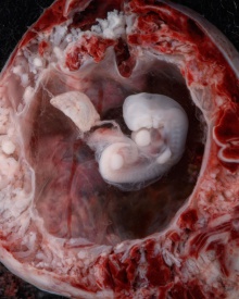
Limb Buds
Stage 13 |
|

Upper and Lower Limb
Human Limb Development during week 6

Human Limb Development during week 8


Limb development occurs at different times for forelimbs and hindlimbs. In the mid-4th week, human upper limb buds first form and lower limbs about 2 days later. The limbs form at vertebra segmental levels C5-C8 (upper limbs) L3-L5 (lower limbs).

Limb Axis Formation

Four Concepts - much of the work has been carried out using the chicken and more recently the mouse model of development.
- Limb Initiation
- Proximodistal Axis
- Dorsoventral Axis
- Anteroposterior Axis
Limb Initiation
- Fibroblast growth factor (FGF) coated beads can induce additional limb
- FGF10 is expressed in lateral plate mesoderm prior to bud formation induces expression of FGF8 in the overlying ectoderm. FGF8 induces continued growth in the underlying mesoderm - thus a positive feedback loop
- Anterior boundary of Hoxc6 expression coincides with the position of forelimb development

Limb induction-initiation signal[1]
Autoregulatory loop of induction between FGF10 and FGF8
Site of FGF10 expression in the chick embryo
Examples of Hox gene expression boundaries in the mouse Hoxb2 and Hoxb4
Limb Identity
Forelimb and hindlimb (mouse) identity appears to be regulated by T-box (Tbx) genes, which are a family of transcription factors.
- forelimb Tbx5 is expressed.
- hindlimb Tbx4 is expressed.
- Tbx2 and Tbx3 are expressed in both limbs.

Tbx3 and Tbx2 expression in E9.75 to 10.5 wild-type mouse embryonic forelimb.[2]
Related Research - PMID: 12490567 | Development 2003 Figures | Scanning electron micrographs of E9 Limb bud wild-type and Tbx5del/del A model for early stages of limb bud growth | PMID: 12736217 | Development 2003 Figures
Tbx4 expression can turn an experimentally induced forelimb into a hindlimb
Axes and Morphogens

- Anteroposterior - (Rostrocaudal, Craniocaudal, Cephalocaudal) from the head end to opposite end of body or tail.
- Dorsoventral - from the spinal column (back) to belly (front).
- Proximodistal - from the tip of an appendage (distal) to where it joins the body (proximal).
Model of patterning signals in the vertebrate limb
Diffusible morphogens create a concentration gradient accross an embryonic field
Proximodistal Axis
- Apical Ectodermal Ridge (AER) initially formed at the site of FGF10 induction
- then AER secretes FGF8 and FGF4 slightly later
- FGFs stimulate proliferation and outgrowth in the underlying mesenchyme
limb development at embryo images online
Morphogen production from the AER - The Fibroblast Growth Factors (FGFs)
- 22 FGF genes identified in humans
- bind membrane tyrosine kinase receptors
- Patterning switch with many different roles in different tissues
| FGF receptors |
|---|
FGF receptors are paired proteins on the cell surface with an internal tyrosine kinase domain |
Dorsoventral Axis
- Important for patterning muscles - ventral muscles - flexors; Dorsal muscles - extensors
- Early grafting experiments showed that the D/V signalling centre resided in the dorsal ectoderm
- Wnt7a is a diffusible morphogen that is secreted by dorsal ectoderm cells
- Wnt7a induces the expression of the homeobox gene Lmx1 in the underlying mesoderm adjacent to the dorsal surface
- The homeobox gene Engrailed (En1) is expressed in the opposite ventral ectoderm
Consequence of Wnt7a deficiency in the mouse forelimb
Morphogen production from the dorsal ectoderm - Wnt7a
- name was derived from 'wingless' and 'int’
- Wnt gene first defined as a protooncogene, int1
- Humans have 19 Wnt genes
- Wnt7a gene is at 3p25 encoding a 349aa secreted glycoprotein
- patterning switch with different roles in different tissues
- One WNT receptor is called Frizzled (FZD) - named after a drosophila phenotype
- Frizzled gene family encodes a G protein-coupled receptor with 7 transmembrane domains
Anteroposterior Axis
- Zone of polarizing activity (ZPA)
- a mesenchymal posterior region of limb
- secretes sonic hedgehog (SHH)
ZPA secretes SHH and determines the anteroposterior axis of the limb bud
Morphogen production from the ZPA - Sonic Hedgehog (SHH)

- Sonic hedgehog (SHH) is a diffusible morphogen secreted from cells, the protein product of the SHH gene
- The protein is processed by cleavage of the preprotein and addition of a palmitate molecule to the amino terminus and cholesterol to the carboxy terminus
- The SHH receptor is a cell surface protein called Patched which interacts with another cell surface protein Smoothened.
- Binding of SHH to Patched blocks its inhibitory effect on Smoothened and allows it to initiate an intracellular signaling cascade
The Time Axis - Dynamic development and temporal gene expression
- Different Hox genes are expressed at different times in the developing limb bud and pattern the fine structure of the limb.
- Structures are determined in a proximal>distal direction with time, i.e. proximal structures such as the humerus bone are laid down first.
Hox genes and dynamic patterning of the limb
Cellular origins of the limb
Limb cartilage and bone
- Derived from local proliferating mesenchyme derived from the somatic lateral plate mesoderm (somatopleure)
- BMP2 and BMP4 play crucial roles in the development of cartilage - sufficient BMP must be present to achieve chondrogenesis. However, the main role is in later bone formation. Loss of BMP2 and 4 leads to a severe impairment of osteogenesis

- Differentiation of somitic mesoderm in the chick embryo
Limb muscle and dermis
Origin of limb muscle cells - Migrations traced by grafting cells from a quail embryo into a chick embryo
|

|
Dorsal/Ventral Muscle Mass - sometimes referred to as the anterior and posterior muscle compartments.
Limb Muscle - Differentiation of Skeletal muscle is the same as in the myotome blocks but involves an extra migratory step
Muscle Development
- Muscle precursor cells migrate to the muscle location
- Form beds of proliferating myoblasts
- Myoblasts fuse together to form a syncitial structure called a myotube
- Myotubes begin to express contractile proteins, form sarcomeres
- mature into myofibers with tendon connections at each end, motor and sensory innervation.
Hand and Footplates

- 5th week- hand and footplates appear at the ends of limb buds and ridges form digital rays
- Cells between the digital rays are removed by programmed cell death (apoptosis)
- 3-5 day difference between hand and foot development

Interdigital apoptosis in the mous hindlimb.[4]
- Links: hand growth
Apoptosis
Cell Biology - Apoptosis Lecture
Fluorescent staining of cells undergoing apoptosis in the limb
Limb Rotation

- 8th week limbs rotate in different directions (Humans Stage 20-23)
- thumb and toe rostral
- knee and elbow face outward
- upper limb rotates dorsally
- lower limb rotates ventrally
Limb Innervation


- spinal cord segmental nerves form a plexus adjacent to each limb
- Brachial (upper) lumbar (lower)
- Plexus forms as nerves invade the limb bud mesechyme
- Fetal period - touch pads become visible on hands and feet

Mouse Limb Timeline<pubmed>22174793</pubmed>| PMC3235105 | PLoS One.
|
Embryonic period - the external appearance of both the upper and lower limb has been formed.
Play the associated animation to observe the relative change in limb dimensions.
|
Limb Abnormalities
Congenital Hip Dislocation

- Instability of the femoral head in the acetabulum - ligaments may stretch: 1:60 at birth
- congenital instability of hip, later dislocates by muscle pulls or gravity
- familial predisposition female predominance
- Growth of femoral head, acetabulum and innominate bone are delayed until the femoral head fits firmly into the acetabulum
Maternal
- thalidomide Phocomelia
- short ill-formed upper or lower limbs
- hyperthermia
- Links: Thalidomide
Genetic
- Trisomy 21 - Downs syndrome
- Human Gene Mutations - mutation of any of the patterning genes will result in limb abnormalities

Type II syndactyly- HoxD13
Syndactyly

Fusion of fingers or toes (Greek, syn = together, dactyly = digit) which may be single or multiple and may affect: skin only, skin and soft tissues or skin, soft tissues and bone. The condition is unimportant in toes but disabling in fingers and requires operative separation and is frequently inherited as an autosomal dominant. The presence of this additional "webbing" reflects preservation of the developmental tissues that in normal development are removed by programmed cell death (apotosis).
Talipes Equinovarus

(Latin, talipes = ankle bone, pes = foot, equinus = horse) Abnormality of the lower limb which begins in the embryonic period (first trimester of pregnancy) resulting in the foot is then turned inward and downward at birth, described as "club foot". Occurs in approximately 1 in 1,000 births, postnatally it affects how children walk on their toes with the foot pointed downward like a horse.
Muscle Development
Duchenne Muscular Dystrophy
- X-linked dystrophy
- large gene encoding cytoskeletal protein- Dystrophin
- progressive wasting of muscle, die late teens
Becker Muscular Dystrophy
- milder form, adult onset
References
- ↑ <pubmed>26212321</pubmed>
- ↑ <pubmed>20386744</pubmed>| PMC2851570 | PLoS Genet.
- ↑ <pubmed>17194222</pubmed>| PMC1713256 | PLoS Genet.
- ↑ <pubmed>17194222</pubmed>| PMC1713256
Online Textbooks
- Developmental Biology by Gilbert, Scott F. Sunderland (MA): Sinauer Associates, Inc.; c2000 Formation of the Limb Bud | Generating the Proximal-Distal Axis of the Limb
- Molecular Biology of the Cell Alberts, Bruce; Johnson, Alexander; Lewis, Julian; Raff, Martin; Roberts, Keith; Walter, Peter New York and London: Garland Science; c2002 Figure 21-13. Sonic hedgehog as a morphogen in chick limb development
Search
- Bookshelf limb development
- Pubmed limb development
Images
Stage13


Stage14


Historic Images
Keith, A. (1902) Human Embryology and Morphology. London: Edward Arnold. Chapter 20. The Limbs
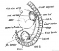
Fig. 233. Lateral view of a human embryo at the 28th day, showing the Limb Buds, Lateral Edges, and Primitive Segments.
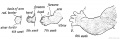
Fig. 234. Development of the Upper Limb.
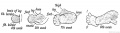
Fig. 235. Development of the Lower Limb.
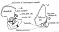
Fig. 236. The Corresponding Points {A, B, C, and D) in the Ilium and Scapula.
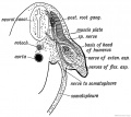
Fig. 237. Section of the Arm Bud of a human embryo at the end of the 4th week.
Fig. 238. Schematic Section showing the Origin and Arrangement of the Muscles and Nerves of the Limbs.
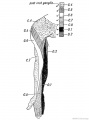
Fig. 239. The Distribution of the Posterior Roots of the Spinal Nerves on the Flexor Aspect of the Arm.
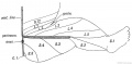
Fig. 240. Diagram to show the typical Manner in which the Posterior Nerve Roots are distributed in the Lower Limb.
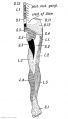
Fig. 241. Flexor Aspect of the Lower Limb showing the Sensory Distribution of the Segmental or Spinal nerves.
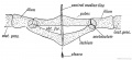
Fig. 242. Diagram of the Pelvic Girdle of a Lizard.
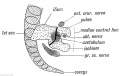
Fig. 243. The Pelvic Girdle of a Human Foetus at the 5th week.
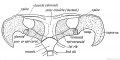
Fig. 244. The Shoulder Girdle of Ornithorynchus.
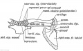
Fig. 245. The Parts in the Shoulder Girdle of a human foetus which correspond with those of Ornithorynehus.
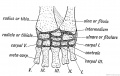
Fig. 246. The Carpal Bones of a Tortoise.
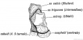
Fig. 247. The Os Trigenum and Bones of the Tarsus
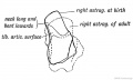
Fig. 248. The Foetal and Adult (in dotted outline) Forma of the Astralagus contrasted.
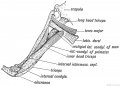
Fig. 249. Latissimo-condyloideus Muscle.
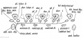
Fig. 250. The Morphology of the Short Muscles of the Digits.
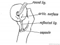
Fig. 251. Showing the Origin of the Ligamentum Teres and Reflected Bundle of the Capsular Ligament.
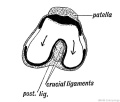
Fig. 252. Showing the Origin of the Crucial Ligaments of the Knee.


External Links
External Links Notice - The dynamic nature of the internet may mean that some of these listed links may no longer function. If the link no longer works search the web with the link text or name. Links to any external commercial sites are provided for information purposes only and should never be considered an endorsement. UNSW Embryology is provided as an educational resource with no clinical information or commercial affiliation.
- Embryo Images Limb Unit
- International J. Dev. Biology Vol 46 Special Issue- Limb Development 2002
- Research Labs - Rolf Zeller University of Basel Medical School