Hinrichsen Collection
| Embryology - 27 Apr 2024 |
|---|
| Google Translate - select your language from the list shown below (this will open a new external page) |
|
العربية | català | 中文 | 中國傳統的 | français | Deutsche | עִברִית | हिंदी | bahasa Indonesia | italiano | 日本語 | 한국어 | မြန်မာ | Pilipino | Polskie | português | ਪੰਜਾਬੀ ਦੇ | Română | русский | Español | Swahili | Svensk | ไทย | Türkçe | اردو | ייִדיש | Tiếng Việt These external translations are automated and may not be accurate. (More? About Translations) |
Introduction
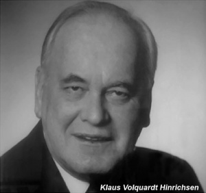
The human embryo histology collection specimens were added between 1969 and 1994 by Professor Klaus Volquardt Hinrichsen and is located at the Department of Anatomy and Molecular Embryology, Ruhr-Universität Bochum. Some material from this collection was published in Hinrichsen's embryology textbook (Humanembryologie, 1990).[1] [2]
Digital Embryology Consortium - Hinrichsen Collection
- Histological sections of approx. 100 specimens from 4 to 20 weeks
- Plastic sections of 20 specimens;
- Additional 70 unsectioned fetuses
- Animal sections
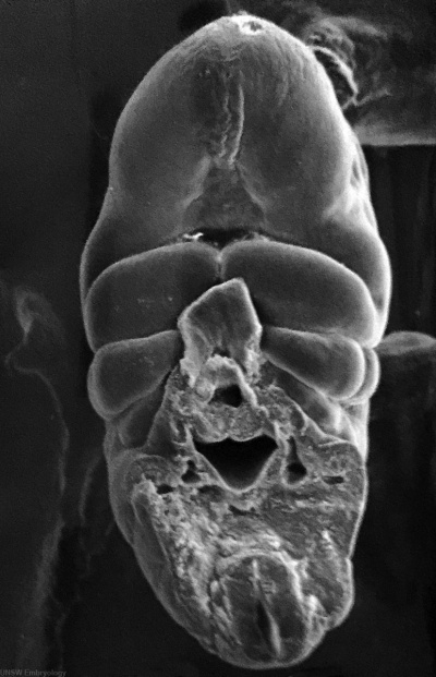
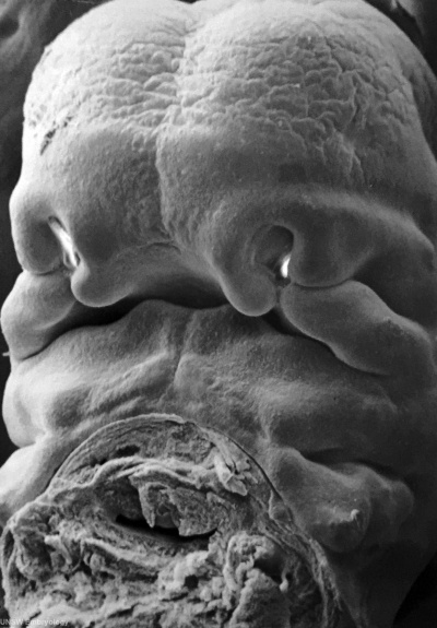
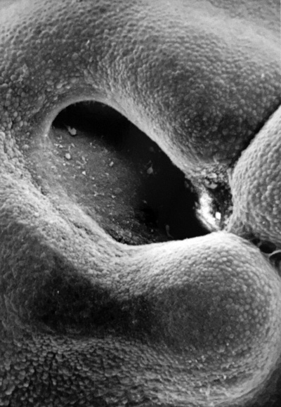
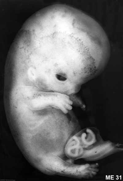
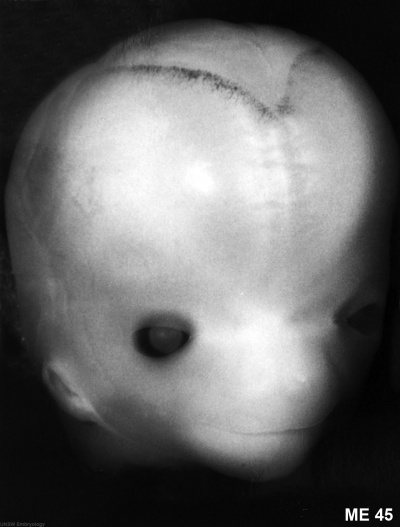
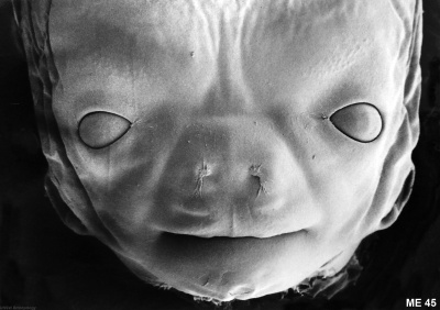
- Hinrichsen Images: ME50 stage 12 | ME18 stage 13 | ME52 stage 14 | ME52 stage 14 Head | ME34 stage 16 | ME34 stage 16 Arches | ME16 stage 17 Head | ME16 stage 17 Nasal | S226 stage 18 | ME28 stage 19 | ME28 stage 19 Hand | ME54 stage 21 | ME54 stage 22 | ME45 stage 23 Head | ME45 stage 23 Face | Hinrichsen Collection
Human Embryo Stage 12
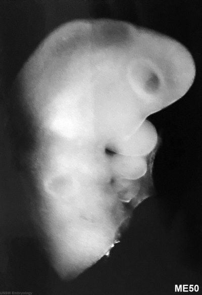
ME 50 Carnegie stage 12
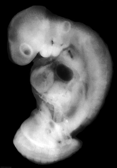
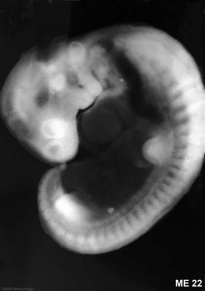
ME 22 Carnegie stage 12
Human Embryo Stage 13
ME 18 Carnegie stage 13
Human Embryo Stage 17
ME 16 Carnegie stage 17
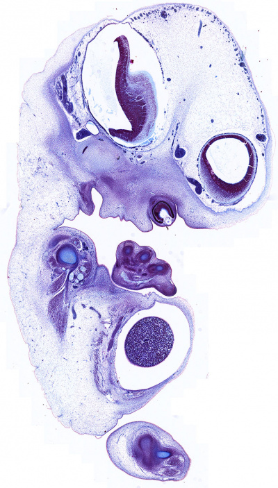
Human Embryo Stage 18
| Embryo S226 Carnegie stage 18 16 mm | |
|
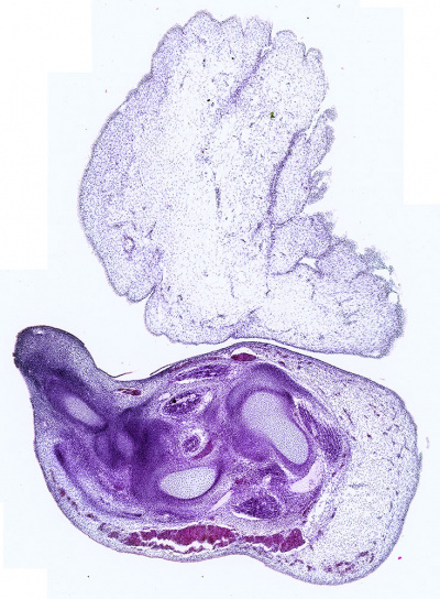
|
| slide 20 | slide 10 |
Human Embryo Stage 21
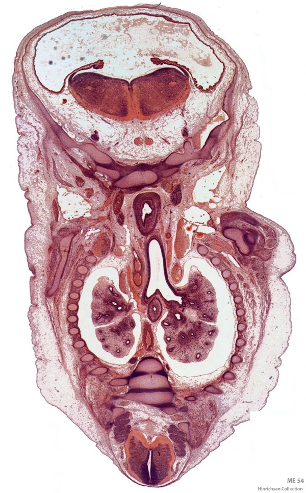
ME 54 Carnegie stage 21, 22.5 mm, 8 weeks, female, frontal, (Stain - Haematoxylin Eosin)
Human Embryo Stage 22
ME 31 Carnegie stage 22
Human Embryo Stage 23
ME 45 Carnegie stage 23
Image source: The Hinrichsen Collection images are reproduced with the permission of Prof. Beate Brand-Saberi, Head, Department of Anatomy and Molecular Embryology, Ruhr-Universität Bochum. Images are for educational purposes only and cannot be reproduced electronically or in writing without permission.
References
- ↑ Hinrichsen KV. Human Embryology (Humanembryologie). (1990) Springer-Verlag Berlin Heidelberg. ISBN 978-3-662-07815-0
- ↑ Maricic N, Khaveh N, Marheinecke C, Wald J, Helluy X, Liermann D, Zaehres H & Brand-Saberi B. (2019). The Hinrichsen Embryology Collection: Digitization of Historical Histological Human Embryonic Slides and MRI of Whole Fetuses. Cells Tissues Organs (Print) , 207, 1-14. PMID: 31189166 DOI.
External Links
External Links Notice - The dynamic nature of the internet may mean that some of these listed links may no longer function. If the link no longer works search the web with the link text or name. Links to any external commercial sites are provided for information purposes only and should never be considered an endorsement. UNSW Embryology is provided as an educational resource with no clinical information or commercial affiliation.
Glossary Links
- Glossary: A | B | C | D | E | F | G | H | I | J | K | L | M | N | O | P | Q | R | S | T | U | V | W | X | Y | Z | Numbers | Symbols | Term Link
Cite this page: Hill, M.A. (2024, April 27) Embryology Hinrichsen Collection. Retrieved from https://embryology.med.unsw.edu.au/embryology/index.php/Hinrichsen_Collection
- © Dr Mark Hill 2024, UNSW Embryology ISBN: 978 0 7334 2609 4 - UNSW CRICOS Provider Code No. 00098G
