Cardiovascular System - Abnormalities: Difference between revisions
mNo edit summary |
|||
| (167 intermediate revisions by 2 users not shown) | |||
| Line 1: | Line 1: | ||
{{Header}} | |||
==Introduction== | ==Introduction== | ||
[[File:Abnormal81-92-heart.png|thumb| | [[File:Abnormal81-92-heart.png|thumb|350px|{{AbnormalDataPie1981-1992}}]] | ||
Heart defects and preterm birth are the most common causes of neonatal and infant death. The long-term development of the heart combined with extensive remodelling and post-natal changes in circulation lead to an abundance of abnormalities associated with this system. | Heart defects and preterm birth are the most common causes of neonatal and infant death. The long-term development of the heart combined with extensive remodelling and post-natal changes in circulation lead to an abundance of abnormalities associated with this system. A good recent review of cardiac abnormalities is available online.<ref name=Anderson2016>{{Ref-Anderson2016}}</ref> | ||
"Baltimore-Washington Infant Study data on live-born cases and controls (1981-1989) was reanalyzed for potential environmental and genetic risk-factor associations in complete atrioventricular septal defects AVSD (n = 213), with separate comparisons to the atrial (n = 75) and the ventricular (n = 32) forms of partial AVSD. ...Maternal diabetes constituted a potentially preventable risk factor for the most severe, complete form of AVSD." | A UK study literature showed that preterm infants have more than twice as many cardiovascular malformations (5.1 / 1000 term infants and 12.5 / 1000 preterm infants) as do infants born at term and that 16% of all infants with cardiovascular malformations are preterm. (0.4% of live births occur at greater than 28 weeks of gestation, 0.9% at 28 to 31 weeks, and 6% at 32 to 36 weeks. Overall, 7.3% of live-born infants are preterm){{#pmid:16322141|PMID16322141}} | ||
"Baltimore-Washington Infant Study data on live-born cases and controls (1981-1989) was reanalyzed for potential environmental and genetic risk-factor associations in complete atrioventricular septal defects AVSD (n = 213), with separate comparisons to the atrial (n = 75) and the ventricular (n = 32) forms of partial AVSD. ...Maternal diabetes constituted a potentially preventable risk factor for the most severe, complete form of AVSD."{{#pmid:11241431|PMID11241431}} | |||
In addition, there are in several congenital abnormalities that exist in adults (bicuspid aortic valve, mitral valve prolapse, and partial anomalous pulmonary venous connection) which may not be clinically recognized. | In addition, there are in several congenital abnormalities that exist in adults (bicuspid aortic valve, mitral valve prolapse, and partial anomalous pulmonary venous connection) which may not be clinically recognized. | ||
Developmental abnormalities of the cardiovascular system are classified under {{ICD-11}} includes a coding based upon the International Pediatric Cardiac Code.{{#pmid:19063777|PMID19063777}}{{#pmid:23804886|PMID23804886}} | |||
<br> | |||
{{Heart abnormal cartoon gallery}} | |||
{{Heart Abnormal}} | |||
{{Heart Links}} | |||
{| class="wikitable mw-collapsible mw-collapsed" | |||
! System Abnormalities | |||
|- | |||
| {{Abnormality Links}} | |||
|} | |||
==Some Recent Findings== | ==Some Recent Findings== | ||
[[File:Australia Congenital heart disease 2016–17.png|thumb|alt=Australia Congenital heart disease 2016–17|Australia Congenital heart disease 2016–17]] | |||
{| | |||
|-bgcolor="F5FAFF" | |||
| | |||
* '''Congenital heart disease in {{Australia}}''' (Report 07 Nov 2019)<ref>Australian Institute of Health and Welfare 2019. Congenital heart disease in Australia. Cat. no. [https://www.aihw.gov.au/reports/heart-stroke-vascular-diseases/congenital-heart-disease-in-australia/contents/table-of-contents CDK 14]. Canberra: AIHW.</ref> "Globally, congenital heart disease affects around 9 in every 1,000 babies born. In {{Australia}}, a large number of children, young people and adults live with congenital heart disease. This report presents latest statistics and trends on the incidence of congenital heart disease, and on hospitalisation and mortality. There were almost 5,000 congenital heart disease hospitalisations in 2016–17. There were 70 infant deaths from congenital heart disease in 2017, comprising 6.9% of all infant deaths. Around 9 in every 1,000 live births are affected by congenital heart disease. Advances in paediatric cardiac care has led to more adults living with congenital heart disease." | |||
* '''First trimester combined screening biochemistry in detection of congenital heart defects'''{{#pmid:29683008|PMID29683008}} "To evaluate the performance of first trimester biochemical markers, pregnancy-associated plasma protein-A (PAPP-A), free beta human chorionic gonadotropin (fβ-hCG), and nuchal translucency (NT) in detection of severe congenital heart defects (CHDs). Methods: During the study period from 1 January 2008 to 31 December 2011, biochemical markers and NT were measured in 31,144 women as part of voluntary first trimester screening program for Down's syndrome in Northern Finland. Data for 71 severe CHD cases and 762 controls were obtained from the hospital records and from the National Medical Birth Register, which records the birth of all liveborn and stillborn infants, and from the National Register of Congenital Malformations that receives information about all the CHD cases diagnosed in Finland. Results: Both PAPP-A and fβ-hCG multiple of median (MoM) values were decreased in all severe CHDs: 0.71 and 0.69 in ventricular septal defects (VSDs), 0.58 and 0.88 in tetralogy of Fallot cases (TOFs), 0.82 and 0.89 in hypoplastic left heart syndromes (HLHSs), and 0.88 and 0.96 in multiple defects, respectively. NT was increased in all study groups except of VSD group. ROC AUC was 0.72 for VSD when combining prior risk with PAPP-A and fβ-hCG. Adding NT did not improve the detection rate. With normal NT but decreased (<0.5 MoM) PAPP-A and fβ-hCG odds ratios for VSD and HLHS were 19.5 and 25.6, respectively. Conclusions: Maternal serum biochemistry improves the detection of CHDs compared to NT measurement only. In cases with normal NT measurement but low concentrations of both PAPP-A and fβ-hCG, an alert for possible CHD, especially VSD, could be given with thorough examination of fetal heart in later ultrasound scans." | |||
* '''Association of NKX2-5, GATA4, and {{TBX}}5 polymorphisms with congenital heart disease in Egyptian children'''{{#pmid:30834692|PMID30834692}} "Venous blood samples from 150 congenital heart disease children (including a ventricular septal defect, atrial septal defect, tetralogy of Fallot, and patent ductus arteriosus) and 90 apparently healthy of matched age and sex were studied by polymerase chain reaction followed by direct sequencing in order to study two single-nucleotide variants of NKX2-5 (rs2277923, rs28936670), two single-nucleotide variants of GATA4 (rs368418329, rs56166237) and one single-nucleotide variant TBX5 (rs6489957). The distribution of genotype and allele frequency in the congenital heart diseases (CHD) group and control group were analyzed. Significantly higher frequency of different allelle of five variants was observed in cases when compared to the control group, with significant risky effect for the development of septal defect. In addition to two polymorphisms of NKX2-5 (rs2277923, rs28936670) variant in the cardiac septal defect, two variants in GATA4 (rs368418329, rs56166237) and one variant in TBX5 (rs6489957) seem to have a role in the pathogenesis of congenital heart disease." | |||
* '''Non-invasive fetal electrocardiography for the detection of fetal arrhythmias'''{{#pmid:30602066|PMID30602066}} "A total of 500 echocardiography and NI-FECG recordings were collected from pregnant women during a routine medical visit in this multicenter study. All the cases with fetal arrhythmias (n = 12) and a matching number of control (n = 14) were used. Two perinatal cardiologists analyzed the extracted NI-FECG while blinded to the echocardiography. The NI-FECG based diagnosis was compared to the reference fetal echocardiography diagnosis....It is possible to diagnose fetal arrhythmias using the NI-FECG technique. However, this study identifies that improvement in algorithms for reconstructing the P-wave is critical to systematically resolve the mechanisms underlying the arrhythmias. The elaboration of a NI-FECG Holter device will offer new opportunities for fetal diagnosis and remote monitoring of problematic pregnancies because of its low-cost, non-invasiveness, portability and minimal set-up requirements." {{heart rate}} | |||
* '''Early fetal ultrasound screening for major congenital heart defects without Doppler'''{{#pmid:30580230|PMID30580230}} "Congenital heart defects are the most common major structural fetal abnormalities. Color flow mapping has played a dominant role in the detection of abnormalities during the first trimester, regardless of the International Society of Ultrasound in Obstetrics and Gynecology warning on the use of Doppler during early pregnancy. The aim of our study was to investigate the use of transvaginal two-dimensional sonography without Doppler for assessing the four-chamber view and the outflow tract view of fetuses at 11-13 weeks of gestation for cardiac screening of major congenital heart defects. ...Routine first-trimester ultrasonagraphy without Doppler, when performed by experienced sonographers, can effectively identify major congenital heart defects. Additional multicenter well designed studies should clarify the feasibility of this approach." | |||
|} | |||
{| class="wikitable mw-collapsible mw-collapsed" | |||
! More recent papers | |||
|- | |||
| [[File:Mark_Hill.jpg|90px|left]] {{Most_Recent_Refs}} | |||
Search term: [http://www.ncbi.nlm.nih.gov/pubmed/?term=Cardiovascular+Abnormality ''Cardiovascular Abnormality''] | |||
|} | |||
{| class="wikitable mw-collapsible mw-collapsed" | |||
! Older papers | |||
|- | |||
| {{Older papers}} | |||
* '''Utility of fetal cardiac MRI to assess fetuses with right aortic arch and right ductus arteriosus'''{{#pmid:28438064|PMID28438064}} "To evaluate the utility of fetal cardiac magnetic resonance imaging ({{MRI}}) to diagnose right aortic arch (RAA) with right ductus arteriosus. ...Fetal cardiac MRI diagnosed the six fetal cases with RAA with right ductus arteriosus correctly. Among the six fetuses, four were associated with other congenital heart defects. In three of six cases, the diagnoses established using prenatal echocardiography (echo) was correct when compared with postnatal diagnosis. Fetal cardiac MRI is a useful complementary tool to assess fetuses with RAA and right ductus arteriosus." [[Magnetic Resonance Imaging]] | |||
* '''High throughput in vivo functional validation of candidate congenital heart disease genes in Drosophila'''{{#pmid:28084990|PMID28084990}} "We developed a Drosophila-based functional system to screen candidate disease genes identified from Congenital Heart Disease (CHD) patients. 134 genes were tested in the Drosophila heart using RNAi-based gene silencing. Quantitative analyses of multiple cardiac phenotypes demonstrated essential structural, functional, and developmental roles for more than 70 genes, including a subgroup encoding histone H3K4 modifying proteins. We also demonstrated the use of Drosophila to evaluate cardiac phenotypes resulting from specific, patient-derived alleles of candidate disease genes. We describe the first high throughput in vivo validation system to screen candidate disease genes identified from patients." {{fly}} | |||
* '''Long-Term Survival of Individuals Born With Congenital Heart Disease: A Systematic Review and Meta-Analysis'''{{#pmid:27312802|PMID27312802}} "Estimates of long-term survival are required to adequately assess the variety of health and social services required by those with congenital heart disease (CHD) throughout their lives. ... Among persons with CHD, the mortality rate is greatest during the first year of life; however, this systematic review and meta-analysis showed that survival decreases gradually after infancy and into adulthood." | |||
* '''Spontaneous Closure of Muscular Trabecular Ventricular Septal Defect: Comparison of Defect Positions'''{{#pmid:21517965|PMID21517965}} "We performed a historical cohort study for which 150 patients <3 months of age (median age, 9 days) diagnosed as having a muscular trabecular VSD were selected. ...We infer that midventricular muscular trabecular VSD tends to close spontaneously earlier and more frequently than either anterior or apical muscular trabecular VSD." | |||
* '''Cyp26 Enzymes Facilitate Second Heart Field Progenitor Addition and Maintenance of Ventricular Integrity'''{{#pmid:27893754|PMID27893754}}"Although retinoic acid (RA) teratogenicity has been investigated for decades, the mechanisms underlying RA-induced outflow tract (OFT) malformations are not understood. Here, we show zebrafish embryos deficient for Cyp26a1 and Cyp26c1 enzymes, which promote RA degradation, have OFT defects resulting from two mechanisms: first, a failure of second heart field (SHF) progenitors to join the OFT, instead contributing to the pharyngeal arch arteries (PAAs), and second, a loss of first heart field (FHF) ventricular cardiomyocytes due to disrupted cell polarity and extrusion from the heart tube." [[Zebrafish Development]] | {{retinoic acid}} | |||
|} | |||
==International Classification of Diseases== | |||
The International Classification of Diseases (ICD) World Health Organization's classification used worldwide as the standard diagnostic tool for epidemiology, health management and clinical purposes. This includes the analysis of the general health situation of population groups. It is used to monitor the incidence and prevalence of diseases and other health problems. | |||
{| | |||
|-bgcolor="FFCC00" | |||
! {{ICD-11}} | |||
|-bgcolor="FEF9E7" | |||
| {{ICD-11-Circulatory system structural anomalies table}} | |||
|} | |||
{{ICD-10-circulatory system Q20-Q28 table}} | |||
:'''Links:''' [[Neonatal Development]] | [http://ipccc.net International Society for Nomenclature of Paediatric and Congenital Heart Disease (ISNPCHD)] | |||
==Heart Abnormalities== | ==Heart Abnormalities== | ||
===Cardiac Critical Periods=== | |||
{{Anderson2016 table2}} | |||
===Sex Ratios=== | |||
{{USA 1997-2009 Abnormalities Sex Ratio table}} | |||
:Links: {{sex bias}} | |||
===Ventricular Septal Defect=== | ===Ventricular Septal Defect=== | ||
[[File:Ventricular Septal Defect.jpg| | {| | ||
The Ventricular Septal Defect (VSD) usually occurs in the membranous (perimembranous) rather than muscular interventricular septum, and is more frequent in males that females. | | [[File:Ventricular Septal Defect.jpg|300px|link=Cardiovascular System - Ventricular Septal Defect]] | ||
|The Ventricular Septal Defect (VSD) usually occurs in the membranous (perimembranous) rather than muscular interventricular septum, and is more frequent in males that females. | |||
Perimembranous defects are located close to the aortic and tricuspid valves and adjacent to atrioventricular conduction bundle. | Perimembranous defects are located close to the aortic and tricuspid valves and adjacent to atrioventricular conduction bundle. | ||
:'''Links:''' [[Cardiovascular_System_-_Ventricular_Septal_Defects|Ventricular Septal Defects]] | [http://www.ncbi.nlm.nih.gov/sites/entrez?db=pubmed&cmd=search&term=Ventricular%20Septal%20Defect Search PubMed] | |||
|} | |||
===Atrial Septal Defects=== | ===Atrial Septal Defects=== | ||
[[File:Atrial Septal Defect.jpg| | {| | ||
Atrial Septal Defects (ASD) are a group of common (1% of cardiac) congenital anomolies defects occuring in a number of different forms and more often in females. | | [[File:Atrial Septal Defect.jpg|300px|link=Cardiovascular System - Atrial Septal Defect]] | ||
| Atrial Septal Defects (ASD) are a group of common (1% of cardiac) congenital anomolies defects occuring in a number of different forms and more often in females. | |||
* patent foramen ovale- allows a continuation of the atrial shunting of blood, in 25% of people a probe patent foramen ovale (allowing a probe to bepassed from one atria to the other) exists. | * patent foramen ovale- allows a continuation of the atrial shunting of blood, in 25% of people a probe patent foramen ovale (allowing a probe to bepassed from one atria to the other) exists. | ||
* ostium secundum defect | * ostium secundum defect | ||
* endocardial cushion defect involving ostium primum | * endocardial cushion defect involving ostium primum | ||
* sinus venosus defect - contributes about 10% of all ASDs and occurs mainly in a common and less common form. Common ("usual type") - in upper atrial septum which is contiguous with the superior vena cava. Less common - at junction of the right atrium and inferior vena cava. | * sinus venosus defect - contributes about 10% of all ASDs and occurs mainly in a common and less common form. Common ("usual type") - in upper atrial septum which is contiguous with the superior vena cava. Less common - at junction of the right atrium and inferior vena cava. | ||
* common atrium | * common atrium | ||
Treatment: The surgical repair requires a cardiopulmonary bypass and is recommended in most cases of ostium secundum ASD, even though there is a significant risk involved. Ostium primum defects tend to present earlier and are often associated with endocardial cushion defects and defective mitral or tricuspid valves. In such cases, valve replacement may be necessary and the extended operation has a considerable chance of mortality. | Treatment: The surgical repair requires a cardiopulmonary bypass and is recommended in most cases of ostium secundum ASD, even though there is a significant risk involved. Ostium primum defects tend to present earlier and are often associated with endocardial cushion defects and defective mitral or tricuspid valves. In such cases, valve replacement may be necessary and the extended operation has a considerable chance of mortality. | ||
Increasingly closure by a transcatheter device closure has been applied. ( | * Increasingly closure by a transcatheter device closure has been applied. | ||
* Repair of atrial septal defects on the perfused beating heart (atrial septal defect size 2 cm - 4.5 cm){{#pmid:19876418|PMID19876418}} | |||
[http://www.ncbi.nlm.nih.gov/omim/108800 OMIM: Atrial Septal Defect] | |||
:'''Links:''' [[Cardiovascular_System_-_Atrial_Septal_Defects|Atrial Septal Defects]] | [http://www.ncbi.nlm.nih.gov/omim/108800 OMIM: Atrial Septal Defect] | [http://www.ncbi.nlm.nih.gov/sites/entrez?db=pubmed&cmd=search&term=Atrial%20Septal%20Defect Search PubMed] | [http://www.orlive.com/akronchildrens/videos/transcatheter-repair-of-atrial-septal-defect?view=displayPageNLM Medline Plus - ASD Repair Video] | |||
|} | |||
===Patent Ductus Arteriosus=== | ===Patent Ductus Arteriosus=== | ||
[[File:Patent Ductus Arteriosus.jpg| | {| | ||
Patent | | [[File:Patent Ductus Arteriosus.jpg|300px|link=Cardiovascular System - Patent Ductus Arteriosus]] | ||
| Patent ductus arteriosus (PDA), or Patent arterial duct (PAD), or common truncus, occurs commonly in preterm infants, and at approximately 1 in 2000 full term infants and more common in females (to male ratio is 2:1). Can also be associated with specific genetic defects, {{trisomy 21}} and {{trisomy 18}}, and the Rubinstein-Taybi syndrome and {{CHARGE syndrome}}. The opening is asymptomatic when the duct is small and can close spontaneously (by day three in 60% of normal term neonates), the remainder are ligated simply and with little risk, with transcatheter closure of the duct generally indicated in older children. The operation is always recommended even in the absence of cardiac failure and can often be deferred until early childhood. | |||
<gallery> | |||
File:Patent ductus arteriosus classification.jpg|Patent ductus arteriosus classification | |||
File:Patent ductus arteriosus echocardiogram.jpg|PDA echocardiogram | |||
File:Patent ductus arteriosus angiogram.jpg|PDA angiogram | |||
</gallery> | |||
:'''Links:''' [[Cardiovascular_System_-_Patent_Ductus_Arteriosus|Patent Ductus Arteriosus]] | [http://www.ncbi.nlm.nih.gov/sites/entrez?db=pubmed&cmd=search&term=Patent%20Ductus%20Arteriosus Search PubMed] | |||
|} | |||
===Tetralogy of Fallot=== | ===Tetralogy of Fallot=== | ||
Named after Etienne-Louis Arthur Fallot (1888) who described it as "la maladie blue" and is a common developmental cardiac defect. The syndrome consists of a number of a number of cardiac defects possibly stemming from abnormal neural crest migration. | |||
{| | |||
| [[File:Tetralogy of Fallot.jpg|300px|link=Cardiovascular System - Tetralogy of Fallot]] | |||
| Named after Etienne-Louis Arthur Fallot (1888) who described it as "''la maladie blue''" and is a common developmental cardiac defect. The syndrome consists of a number of a number of cardiac defects possibly stemming from abnormal neural crest migration. | |||
:'''Links:''' [[Cardiovascular_System_-_Tetralogy_of_Fallot|Tetralogy of Fallot]] | [http://www.ncbi.nlm.nih.gov/pubmed/21208432 Gene expression in cardiac tissues from infants with idiopathic conotruncal defects] | [http://www.ncbi.nlm.nih.gov/sites/entrez?db=pubmed&cmd=search&term=Tetralogy%20of%20Fallot Search PubMed] | |||
|} | |||
===Hypoplastic Left Heart=== | ===Hypoplastic Left Heart=== | ||
[[File:Hypoplastic Left Heart.jpg| | {| | ||
Characterized by hypoplasia (underdevelopment or absence) of the left ventricle obstructive valvular and vascular lesion of the left side of the heart. | | [[File:Hypoplastic Left Heart.jpg|300px|link=Cardiovascular System - Hypoplastic Left Heart]] | ||
| Characterized by hypoplasia (underdevelopment or absence) of the left ventricle obstructive valvular and vascular lesion of the left side of the heart. | |||
:'''Links:''' [[Cardiovascular_System_-_Hypoplastic Left Heart|Hypoplastic Left Heart]] | [http://www.ncbi.nlm.nih.gov/sites/entrez?db=pubmed&cmd=search&term=Hypoplastic%20Left%20Heart Search PubMed] | |||
|} | |||
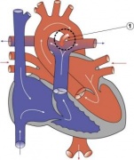
===Double Outlet Right Ventricle=== | ===Double Outlet Right Ventricle=== | ||
{| | |||
| [[File:Double_Outlet_Right_Ventricle.jpg|300px|link=Cardiovascular System - Double Outlet Right Ventricle]] | |||
| | |||
De-oxygenated blood enters the aorta from the right ventricle and is returned to the body. | De-oxygenated blood enters the aorta from the right ventricle and is returned to the body. | ||
:'''Links:''' [[Cardiovascular_System_-_Double Outlet Right Ventricle|Double Outlet Right Ventricle]] | [http://www.ncbi.nlm.nih.gov/sites/entrez?db=pubmed&cmd=search&term=Double_Outlet_Right_Ventricle Search PubMed] | |||
|} | |||
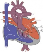
===Tricuspid Atresia=== | ===Tricuspid Atresia=== | ||
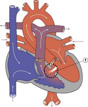
[[File:Tricuspid Atresia.jpg| | {| | ||
Blood is shunted through an atrial septal defect to the left atrium and through the ventricular septal defect to the pulmonary artery. The shaded arrows indicate mixing of the blood. | | [[File:Tricuspid Atresia.jpg|300px|link=Cardiovascular System - Tricuspid Atresia]] | ||
| Blood is shunted through an atrial septal defect to the left atrium and through the ventricular septal defect to the pulmonary artery. The shaded arrows indicate mixing of the blood. | |||
Fontan Procedure: a surgical procedure developed by Fontan and Baudet (1971) to restore a circulation in patients with tricuspid atresia. | Fontan Procedure: a surgical procedure developed by Fontan and Baudet (1971) to restore a circulation in patients with tricuspid atresia. | ||
Search PubMed: | |||
:'''Links:''' [http://www.ncbi.nlm.nih.gov/sites/entrez?db=pubmed&cmd=search&term=Tricuspid%20Atresia Search PubMed] | [http://www.ncbi.nlm.nih.gov/sites/entrez?db=pubmed&cmd=search&term=Fontan%20procedure Fontan procedure] | |||
|} | |||
===Dextrocardia=== | ===Dextrocardia=== | ||
{| | |||
| [[File:Dextrocardia heart position.jpg|300px]] | |||
| [[File:Dextrocardia.jpg|300px]] | |||
|- | |||
| '''Dextrocardia anatomical heart position'''{{#pmid:19142355|PMID19142355}} | |||
| '''Dextrocardia''' (postnatal 1 year old){{#pmid:19142355|PMID19142355}} | |||
|} | |||
Initial malrotation of the heart tube bending left instead of right. Results in heart and greater vessels reversed. Can also occur with ''situs invertus'', where viscera are transposed LR. | |||
Anatomical left-right normal asymmetry is called ''situs solitus''. The alternative heterotaxy can be either randomization (''situs ambiguus'') or a complete reversal (''situs inversus'') of normal organ position. | |||
Links: | :'''Links:''' [[Movie_-_Abnormal_Chick_Heart|Chicken Abnormal Heart Movie]] | [http://www.ncbi.nlm.nih.gov/sites/entrez?db=pubmed&cmd=search&term=Dextrocardia Search PubMed] | ||
===Abnormalities of Conducting System=== | ===Abnormalities of Conducting System=== | ||
[[File:Cardiac Conduction System.jpg|thumb|alt=Cardiac Conduction System|Cardiac Conduction System]] | |||
Also variously called the cardiac conduction system (CCS), cardiac pacemaking and conduction system (CPCS), or atrioventricular conduction system (AVCS). Recently animal models (CCS-lacZ transgenic mouse) have helped identify key processes in the development of this specialized conduction system. | Also variously called the cardiac conduction system (CCS), cardiac pacemaking and conduction system (CPCS), or atrioventricular conduction system (AVCS). Recently animal models (CCS-lacZ transgenic mouse) have helped identify key processes in the development of this specialized conduction system. | ||
"Known arrhythmogenic areas including Bachmann's bundle, the pulmonary veins, and sinus venosus derived internodal structures, demonstrate lacZ expression." (Jongbloed et al, 2004) | "Known arrhythmogenic areas including Bachmann's bundle, the pulmonary veins, and sinus venosus derived internodal structures, demonstrate lacZ expression." (Jongbloed et al, 2004) | ||
===Long QT Syndrome=== | ===Long QT Syndrome=== | ||
Congenital long QT syndrome (LQTS) is a group of rare genetic disorders with prolonged ventricular repolarization and a risk of ventricular tachyarrhythmias. Cause is mutations in genes encoding either cardiac ion channels or channel interacting proteins. | Congenital long QT syndrome ({{LQTS}}) is a group of rare genetic disorders with prolonged ventricular repolarization and a risk of ventricular tachyarrhythmias. Cause is mutations in genes encoding either cardiac ion channels or channel interacting proteins. | ||
* Tachyarrhythmia is defined as a heart rhythm with a ventricular rate of 100 beats/min or greater. | |||
:'''Links:''' [http://www.ncbi.nlm.nih.gov/sites/entrez?db=pubmed&cmd=search&term=Congenital%20long-QT%20syndrome Search PubMed LQTS] | |||
==Heart Vessel Abnormalities== | ==Heart Vessel Abnormalities== | ||
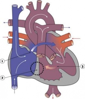
=== | ===Transposition of the Great Vessels=== | ||
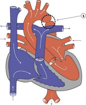
[[File:Transposition of the Great Vessels.jpg| | {| | ||
Characterized by aorta arising from right ventricle and pulmonary artery from the left ventricle and often associated with other cardiac abnormalities (e.g. ventricular septal defect). | | [[File:Transposition of the Great Vessels.jpg|300px|link=Cardiovascular System - Transposition of the Great Vessels]] | ||
| Characterized by aorta arising from right ventricle and pulmonary artery from the left ventricle and often associated with other cardiac abnormalities (e.g. ventricular septal defect). | |||
{| | |||
| | |||
* Australian national rate (1982-1992) 3.6/10,000 births. | |||
* Of 988 infants 4.1% were stillborn and 23.2% liveborn died during neonatal period. | |||
* slightly more common in twin births than singleton. | |||
* Congenital Malformations Australia 1981-1992 P. Lancaster and E. Pedisich ISSN 1321-8352 | |||
* Neonates with transposed great arteries die without an arterial switch operation, first carried out in 1975. | |||
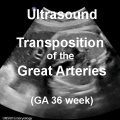
| {{Ultrasound Transposition Great Arteries}} | |||
|} | |||
:'''LInks:''' {{transposition of the great vessels}} | [http://www.ncbi.nlm.nih.gov/sites/entrez?db=pubmed&cmd=search&term=Transposition_of_the_Great_Vessels Search PubMed] | PMID 15987628 | PMID 18851735 | |||
|} | |||
===Coarctation of the Aorta=== | |||
{| | |||
| [[File:Coarctation of the Aorta.jpg|300px|link=Cardiovascular System - Coarctation of the Aorta]] | |||
| | |||
* 5-8% of Congenital Heart Disease | |||
* Aortic constriction. | |||
* Treatment aims at maintaining the ductus arteriosus via prostaglandins and by surgical intervention. | |||
* Recent review{{#pmid:21947983|PMID21947983}} suggests it may: | |||
** affect the aortic arch in a highly variable manner | |||
** be associated with a host of other left sided heart lesions | |||
** represent a wider vasculopathy within the pre-coarctation arterial tree | |||
:'''LInks:''' {{coarctation of the aorta}} | [http://www.ncbi.nlm.nih.gov/sites/entrez?db=pubmed&cmd=search&term=Coarctation_of_the_Aorta Search PubMed] | PMID 21947983 | [http://www.cardiologyjournal.org/en/darmowy_pdf.phtml?id=105&indeks_art=1488 2011 Review PDF] | |||
[ | |} | ||
===Interrupted Aortic Arch=== | ===Interrupted Aortic Arch=== | ||
:'''LInks:''' [http://www.ncbi.nlm.nih.gov/sites/entrez?db=pubmed&cmd=search&term=Interrupted_Aortic_Arch Search PubMed] | |||
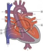
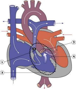
===Pulmonary Atresia=== | ===Pulmonary Atresia=== | ||
[[File:Pulmonary Atresia.jpg| | {| | ||
Abnormal blood flow (as indicated by the shaded blue arrow) is from the right atrium and right ventricle through an atrial septal defect to the left side of the heart. Blood can reach the pulmonary arteries only through a patent ductus arteriosus. | | [[File:Pulmonary Atresia.jpg|300px]] | ||
| | |||
* Abnormal blood flow (as indicated by the shaded blue arrow) is from the right atrium and right ventricle through an atrial septal defect to the left side of the heart. | |||
* Blood can reach the pulmonary arteries only through a patent ductus arteriosus. | |||
* 10% of Congenital Heart Disease | |||
* Unequal division of trunks causes cusps to fuse to form a dome with a narrow/non-existent lumen. | |||
* Heart-lung transplantation may be the only therapy. | |||
:'''LInks:''' [http://www.ncbi.nlm.nih.gov/sites/entrez?db=pubmed&cmd=search&term=Pulmonary_Atresia Search PubMed] | |||
|} | |||
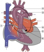
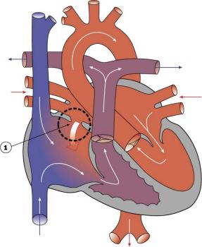
===Total Anomalous Pulmonary Venous Connection=== | ===Total Anomalous Pulmonary Venous Connection=== | ||
[[File:Total Anomalous Pulmonary Venous Connection.jpg| | {| | ||
| [[File:Total Anomalous Pulmonary Venous Connection.jpg|300px]] | |||
| Total Anomalous Pulmonary Venous Connection (TAPVC) or Total Anomalous Pulmonary Venous Return (TAPVR) occurs when pulmonary veins connect to the right atrium (RA) and not the left atrium (LA). This abnormal connection returns oxygenated pulmonary blood from the lungs back to the right atrium or a vein flowing into the right atrium. | |||
Search PubMed: | * < 4% of Congenital Heart Disease | ||
* more common in females | |||
* Total or partial lack of connection of the pulmonary veins with LA. | |||
* They open into RA, one of the systemic veins or both. | |||
* The overloaded pulmonary circuit leads to cyanosis, tachypnoea and dyspnoea. | |||
* Treatment is via surgical redirection | |||
:'''LInks:''' [http://www.ncbi.nlm.nih.gov/sites/entrez?db=pubmed&cmd=search&term=Total%20Anomalous%20Pulmonary%20Venous%20Connection Search PubMed] | |||
|} | |||
===Complete Atrioventricular Canal=== | |||
{| | |||
| [[File:Complete atrioventricular canal.jpg|300px]] | |||
| | |||
:'''LInks:''' [http://www.ncbi.nlm.nih.gov/sites/entrez?db=pubmed&cmd=search&term=Complete%20atrioventricular%20canal Search PubMed] | |||
|} | |||
===Partial Anomalous Pulmonary Venous Drainage=== | ===Partial Anomalous Pulmonary Venous Drainage=== | ||
[[File:Partial Anomalous Pulmonary Venous | {| | ||
| [[File:Partial Anomalous Pulmonary Venous Drainage.jpg|300px]] | |||
| | |||
* < 4% of Congenital Heart Disease | |||
* more common in females | |||
* Total or partial lack of connection of the pulmonary veins with left atria (LA). | |||
* They open into right atria (RA), one of the systemic veins or both. | |||
* The overloaded pulmonary circuit leads to cyanosis, tachypnoea and dyspnoea. | |||
* Treatment is via surgical redirection | |||
:'''LInks:''' [http://www.ncbi.nlm.nih.gov/sites/entrez?db=pubmed&cmd=search&term=Partial%20Anomalous%20Pulmonary%20Venous%20Drainage Search PubMed] | PMID 22119121 | PMID 12652510 | |||
|} | |||
===Aortic Stenosis=== | ===Aortic Stenosis=== | ||
[[File:Aortic Stenosis.jpg| | {| | ||
| [[File:Aortic Stenosis.jpg|300px]] | |||
| | |||
* 7% of Congenital Heart Disease | |||
* Persistence of tissue that normally degenerates. | |||
* Results in left ventricle hypertrophy, heart murmurs. | |||
:'''LInks:''' [http://www.ncbi.nlm.nih.gov/sites/entrez?db=pubmed&cmd=search&term=Aortic%20Stenosis Search PubMed] | |||
|} | |||
===Pulmonary Stenosis=== | ===Pulmonary Stenosis=== | ||
[[File:Pulmonary Stenosis.jpg| | {| | ||
| [[File:Pulmonary Stenosis.jpg|300px]] | |||
| | |||
* 10% of Congenital Heart Disease | |||
* Unequal division of trunks causes cusps to fuse to form a dome with a narrow/non-existent lumen. | |||
* Heart-lung transplantation may be the only therapy. | |||
:'''LInks:''' [http://www.ncbi.nlm.nih.gov/sites/entrez?db=pubmed&cmd=search&term=Pulmonary%20Stenosis Search PubMed] | |||
|} | |||
===Hypertrophic Cardiomyopathy=== | |||
Hypertrophic cardiomyopathy (HCM) is a postnatal cardiac abnormality with left ventricular hypertrophy following pre-hypertrophic crypts, abnormal mitral leaflets and trabeculae. This heart anomaly is due to mutations in the sarcomeric protein genes, most commonly myosin-binding protein C (MYBPC3). | |||
==Blood Disorders== | |||
===Sickle Cell Anemia=== | |||
{| | |||
| People who have this form of sickle cell disease inherit two sickle cell genes (“S”), one from each parent. This is commonly called “sickle cell anemia”, and is usually the most severe form of the disease. The name comes from the "sickle" shape of the RBC compared to the normal "donut" shape. | |||
The scanning electron micrograph (SEM) on the right shows the ultrastructural morphology of a sickle cell RBC found in a blood specimen of an 18 year old female patient with sickle cell anemia, (HbSS). | |||
| [[File:Sickle cell RBC SEM01.jpg|300px]] | |||
Sickle cell RBC (Image CDC) | |||
|} | |||
===Thalassemia=== | |||
Thalassemia is a group of inherited (genetic) blood disorders most frequently in people of Italian, Greek, Middle Eastern, Southern Asian and African Ancestry. The most severe form of alpha thalassemia, affecting mainly people of Southeast Asian, Chinese and Filipino ancestry, results in fetal or newborn death. | |||
The two main types of thalassemia are called "alpha" and "beta," depending on which part of an oxygen-carrying protein in the red blood cells is lacking. Both types of thalassemia are inherited in the same manner. A child who inherits one mutated gene is a carrier, which is sometimes called "thalassemia trait." Most carriers lead completely normal, healthy lives. | |||
:'''Links:''' [[Cardiovascular System - Blood Development|Blood Development]] | [http://www.nlm.nih.gov/medlineplus/sicklecellanemia.html Medline Plus - sickle cell anemia] | [http://www.nlm.nih.gov/medlineplus/thalassemia.html Medline Plus - thalassemia] | |||
==Statistics== | |||
===Australia=== | |||
[[File:Abnormal81-92-heart.png|400px]] | |||
{{Template:AbnormalDataPie1981-1992}} | |||
===Belgium=== | |||
Congenital heart disease in 111 225 births in Belgium 2008 study{{#pmid:19046347|PMID19046347}}: | |||
* 921 children with congenital heart disease (birth prevalence of 8.3 per 1000). | |||
* 33% ventricular septal defects most frequently occurring condition | |||
* 18% ostium secundum atrial septal defects | |||
* 10% pulmonary valve abnormalities | |||
* 39% had either cardiosurgical operation or catheter intervention. | |||
* 4% of the children died (higher in univentricular physiology, pulmonary atresia with VSD, left ventricle outflow obstruction and tetralogy of Fallot). | |||
===South America=== | |||
Paediatric and congenital heart disease in South America: an overview.{{#pmid:19174420|PMID19174420}} | |||
* Ventricular septal defects - Surgical mortality with repair in infancy, mainly in the muscular septum, probably slightly higher in South America than in North America and Europe. | |||
* Secundum atrial septal defects - trans-catheter closure is the preferred method of treating most patients. | |||
* Pulmonary valve stenosis and atresia with intact ventricular septum - balloon pulmonary valvuloplasty for PVS. | |||
* Aortic stenosis - balloon valvuloplasty. | |||
* Coarctation of the aorta - children balloon angioplasty, adolescents and adults a trend towards the use of bare and covered stents. | |||
* Tetralogy of Fallot - patients with severe hypoxaemia have a modified Blalock–Taussig shunt. | |||
===USA=== | |||
"Baltimore-Washington Infant Study data on live-born cases and controls (1981-1989) was reanalyzed for potential environmental and genetic risk-factor associations in complete atrioventricular septal defects AVSD (n = 213), with separate comparisons to the atrial (n = 75) and the ventricular (n = 32) forms of partial AVSD. ...Maternal diabetes constituted a potentially preventable risk factor for the most severe, complete form of AVSD."{{#pmid:11241431|PMID11241431}} | |||
((USA 1997-2009 Abnormalities Sex Ratio table}} | |||
==References== | ==References== | ||
<references/> | <references/> | ||
{{ | |||
{{ | ===Articles=== | ||
{{#pmid:27873373}} | |||
{{#pmid:17967198}} | |||
===Search Pubmed=== | |||
'''Search Pubmed:''' [http://www.ncbi.nlm.nih.gov/sites/entrez?db=pubmed&cmd=search&term=Cardiovascular%20System%20Abnormalities Cardiovascular System Abnormalities] | |||
==Terms== | |||
{{Cardiovascular terms}} | |||
==External Links== | |||
{{External Links}} | |||
* [http://ipccc.net International Society for Nomenclature of Paediatric and Congenital Heart Disease (ISNPCHD)] | |||
{{Glossary}} | |||
{{Footer}} | |||
[[Category:Cardiovascular]] [[Category:Heart]] [[Category:Abnormal Development]] | [[Category:Cardiovascular]] [[Category:Heart]] [[Category:Abnormal Development]] | ||
[[Category:Atrial Septal Defect]][[Category:Ventricular Septal Defect]] | |||
[[Category:Patent Ductus Arteriosus]] | |||
Latest revision as of 11:05, 18 February 2020
| Embryology - 14 Jun 2024 |
|---|
| Google Translate - select your language from the list shown below (this will open a new external page) |
|
العربية | català | 中文 | 中國傳統的 | français | Deutsche | עִברִית | हिंदी | bahasa Indonesia | italiano | 日本語 | 한국어 | မြန်မာ | Pilipino | Polskie | português | ਪੰਜਾਬੀ ਦੇ | Română | русский | Español | Swahili | Svensk | ไทย | Türkçe | اردو | ייִדיש | Tiếng Việt These external translations are automated and may not be accurate. (More? About Translations) |
Introduction
Heart defects and preterm birth are the most common causes of neonatal and infant death. The long-term development of the heart combined with extensive remodelling and post-natal changes in circulation lead to an abundance of abnormalities associated with this system. A good recent review of cardiac abnormalities is available online.[1]
A UK study literature showed that preterm infants have more than twice as many cardiovascular malformations (5.1 / 1000 term infants and 12.5 / 1000 preterm infants) as do infants born at term and that 16% of all infants with cardiovascular malformations are preterm. (0.4% of live births occur at greater than 28 weeks of gestation, 0.9% at 28 to 31 weeks, and 6% at 32 to 36 weeks. Overall, 7.3% of live-born infants are preterm)[2]
"Baltimore-Washington Infant Study data on live-born cases and controls (1981-1989) was reanalyzed for potential environmental and genetic risk-factor associations in complete atrioventricular septal defects AVSD (n = 213), with separate comparisons to the atrial (n = 75) and the ventricular (n = 32) forms of partial AVSD. ...Maternal diabetes constituted a potentially preventable risk factor for the most severe, complete form of AVSD."[3]
In addition, there are in several congenital abnormalities that exist in adults (bicuspid aortic valve, mitral valve prolapse, and partial anomalous pulmonary venous connection) which may not be clinically recognized.
Developmental abnormalities of the cardiovascular system are classified under ICD-11 includes a coding based upon the International Pediatric Cardiac Code.[4][5]


| System Abnormalities | ||||
|---|---|---|---|---|
|
Some Recent Findings

|
| More recent papers |
|---|
 This table allows an automated computer search of the external PubMed database using the listed "Search term" text link.
More? References | Discussion Page | Journal Searches | 2019 References | 2020 References Search term: Cardiovascular Abnormality |
| Older papers |
|---|
| These papers originally appeared in the Some Recent Findings table, but as that list grew in length have now been shuffled down to this collapsible table.
See also the Discussion Page for other references listed by year and References on this current page.
|
International Classification of Diseases
The International Classification of Diseases (ICD) World Health Organization's classification used worldwide as the standard diagnostic tool for epidemiology, health management and clinical purposes. This includes the analysis of the general health situation of population groups. It is used to monitor the incidence and prevalence of diseases and other health problems.
| ICD-11 | |||||
|---|---|---|---|---|---|
|
| ICD10 Congenital malformations of the circulatory system (Q20-Q28) |
|---|
| Cardiovascular System - Abnormalities |
| Q20 Congenital malformations of cardiac chambers and connections
Excl.: dextrocardia with situs inversus (Q89.3) mirror-image atrial arrangement with situs inversus (Q89.3)
|
| Q21 Congenital malformations of cardiac septa
Excl.: acquired cardiac septal defect (I51.0)
|
Q22 Congenital malformations of pulmonary and tricuspid valves
|
Q23 Congenital malformations of aortic and mitral valves
|
| Q24 Other congenital malformations of heart
Excl.: endocardial fibroelastosis (I42.4)
|
Q25 Congenital malformations of great arteries
|
Q26 Congenital malformations of great veins
|
| Q27 Other congenital malformations of peripheral vascular system
Excl.: anomalies of: cerebral and precerebral vessels (Q28.0-Q28.3) coronary vessels (Q24.5) pulmonary artery (Q25.5-Q25.7) congenital retinal aneurysm (Q14.1) haemangioma and lymphangioma (D18.-)
|
| Q28 Other congenital malformations of circulatory system
Excl.: congenital aneurysm: NOS (Q27.8) coronary (Q24.5) peripheral (Q27.8) pulmonary (Q25.7) retinal (Q14.1) ruptured: cerebral arteriovenous malformation (I60.8) malformation of precerebral vessels (I72.-)
|
| ICD-10 |
- Links: Neonatal Development | International Society for Nomenclature of Paediatric and Congenital Heart Disease (ISNPCHD)
Heart Abnormalities
Cardiac Critical Periods
| Lesion | Start of Susceptibility to Malformation |
End of Susceptibility to Malformation | |||
|---|---|---|---|---|---|
| Interatrial communications | |||||
| Oval fossa defect | 6 weeks (E13.5) | Term | |||
| Sinus venosus defect | 8 weeks | 12 weeks | |||
| Coronary sinus defect | 8 weeks | Term | |||
| Vestibular defect | 7 weeks | 8 weeks | |||
| Ventricular Septal Defect | |||||
| Muscular | 8 weeks | Difficult to predict | |||
| Perimembranous | 6 weeks | 8 weeks | |||
| Doubly committed | 7 weeks | 8 weeks | |||
| Atrioventricular Septal Defect | |||||
| Ostium primum | 5 weeks | 6 weeks | |||
| “Complete” | 5 weeks | 6 weeks | |||
| Aortic coarctation | |||||
| With VSD | 5 weeks | 8 weeks | |||
| With intact ventricular septum | 8 weeks | Term | |||
| Double Outlet Right Ventricle | 6 weeks | 8 weeks | |||
| Transposition of Great Arteries | 6 weeks | 8 weeks | |||
| Ebstein’s malformation | 6 weeks | 8 weeks | |||
| Hypoplastic left heart syndrome | |||||
| With mitral atresia | 5 weeks | 8 weeks | |||
| With mitral stenosis | 8 weeks | Term | |||
| Pulmonary atresia | |||||
| With VSD | 6 weeks | 8 weeks | |||
| With intact ventricular septum | 8 weeks | Term | |||
| Other | |||||
| Functionally single ventricle | 5 weeks | 6 weeks | |||
| Tetralogy of Fallot | 7 weeks | 8 weeks | |||
| Totally anomalous pulmonary venous return | 8 weeks | 12 weeks | |||
| Tricuspid atresia | 5 weeks | 6 weeks | |||
| Common arterial trunk | 5 weeks | 7 weeks | |||
| Bicuspid aortic valve | 6 weeks | Term | |||
| Notes | |||||
| For approximate clinical Gestational Age GA add 2 weeks; number in brackets is mouse equivalent. Data Reference[1] | Links: heart | abnormal development | timeline | Category:Timeline | ||||
| Table 2. Summary of Timing (Post Fertilization) of Cardiac Susceptibility to a Drug-Induced Malformation | ||
|---|---|---|
| Lesion | Start of Susceptibility to Malformation |
End of Susceptibility to Malformation |
| Interatrial communications | ||
| Oval fossa defect | 6 weeks (E13.5) | Term |
| Sinus venosus defect | 8 weeks | 12 weeks |
| Coronary sinus defect | 8 weeks | Term |
| Vestibular defect | 7 weeks | 8 weeks |
| Ventricular Septal Defect | ||
| Muscular | 8 weeks | Difficult to predict |
| Perimembranous | 6 weeks | 8 weeks |
| Doubly committed | 7 weeks | 8 weeks |
| Atrioventricular Septal Defect | ||
| Ostium primum | 5 weeks | 6 weeks |
| “Complete” | 5 weeks | 6 weeks |
| Aortic coarctation | ||
| With VSD | 5 weeks | 8 weeks |
| With intact ventricular septum | 8 weeks | Term |
| Double Outlet Right Ventricle | 6 weeks | 8 weeks |
| Transposition of Great Arteries | 6 weeks | 8 weeks |
| Ebstein’s malformation | 6 weeks | 8 weeks |
| Hypoplastic left heart syndrome | ||
| With mitral atresia | 5 weeks | 8 weeks |
| With mitral stenosis | 8 weeks | Term |
| Pulmonary atresia | ||
| With VSD | 6 weeks | 8 weeks |
| With intact ventricular septum | 8 weeks | Term |
| Other | ||
| Functionally single ventricle | 5 weeks | 6 weeks |
| Tetralogy of Fallot | 7 weeks | 8 weeks |
| Totally anomalous pulmonary venous return | 8 weeks | 12 weeks |
| Tricuspid atresia | 5 weeks | 6 weeks |
| Common arterial trunk | 5 weeks | 7 weeks |
| Bicuspid aortic valve | 6 weeks | Term |
|
Notes: For approximate clinical Gestational Age GA add 2 weeks; number in brackets is mouse equivalent. Reference: Anderson RH. Teratogenecity in the setting of cardiac development and maldevelopment. (2016) | ||
Sex Ratios
| Male preponderance | Female preponderance |
|---|---|
|
|
| Cardiac defects | |
|
|
| Table data[16] Links: abnormal development | cardiovascular abnormalities | USA | Male | Female | cleft lip and palate | |
- Links: sex bias
Ventricular Septal Defect
|
The Ventricular Septal Defect (VSD) usually occurs in the membranous (perimembranous) rather than muscular interventricular septum, and is more frequent in males that females.
Perimembranous defects are located close to the aortic and tricuspid valves and adjacent to atrioventricular conduction bundle.
|
Atrial Septal Defects
|
Atrial Septal Defects (ASD) are a group of common (1% of cardiac) congenital anomolies defects occuring in a number of different forms and more often in females.
Treatment: The surgical repair requires a cardiopulmonary bypass and is recommended in most cases of ostium secundum ASD, even though there is a significant risk involved. Ostium primum defects tend to present earlier and are often associated with endocardial cushion defects and defective mitral or tricuspid valves. In such cases, valve replacement may be necessary and the extended operation has a considerable chance of mortality.
|
Patent Ductus Arteriosus

|
Patent ductus arteriosus (PDA), or Patent arterial duct (PAD), or common truncus, occurs commonly in preterm infants, and at approximately 1 in 2000 full term infants and more common in females (to male ratio is 2:1). Can also be associated with specific genetic defects, Trisomy 21 and Trisomy 18, and the Rubinstein-Taybi syndrome and CHARGE syndrome. The opening is asymptomatic when the duct is small and can close spontaneously (by day three in 60% of normal term neonates), the remainder are ligated simply and with little risk, with transcatheter closure of the duct generally indicated in older children. The operation is always recommended even in the absence of cardiac failure and can often be deferred until early childhood.
|


Tetralogy of Fallot
|
Named after Etienne-Louis Arthur Fallot (1888) who described it as "la maladie blue" and is a common developmental cardiac defect. The syndrome consists of a number of a number of cardiac defects possibly stemming from abnormal neural crest migration.
|
Hypoplastic Left Heart

|
Characterized by hypoplasia (underdevelopment or absence) of the left ventricle obstructive valvular and vascular lesion of the left side of the heart.
|
Double Outlet Right Ventricle

|
De-oxygenated blood enters the aorta from the right ventricle and is returned to the body.
|
Tricuspid Atresia
|
Blood is shunted through an atrial septal defect to the left atrium and through the ventricular septal defect to the pulmonary artery. The shaded arrows indicate mixing of the blood.
Fontan Procedure: a surgical procedure developed by Fontan and Baudet (1971) to restore a circulation in patients with tricuspid atresia.
|
Dextrocardia

|

|
| Dextrocardia anatomical heart position[18] | Dextrocardia (postnatal 1 year old)[18] |
Initial malrotation of the heart tube bending left instead of right. Results in heart and greater vessels reversed. Can also occur with situs invertus, where viscera are transposed LR.
Anatomical left-right normal asymmetry is called situs solitus. The alternative heterotaxy can be either randomization (situs ambiguus) or a complete reversal (situs inversus) of normal organ position.
Abnormalities of Conducting System

Also variously called the cardiac conduction system (CCS), cardiac pacemaking and conduction system (CPCS), or atrioventricular conduction system (AVCS). Recently animal models (CCS-lacZ transgenic mouse) have helped identify key processes in the development of this specialized conduction system.
"Known arrhythmogenic areas including Bachmann's bundle, the pulmonary veins, and sinus venosus derived internodal structures, demonstrate lacZ expression." (Jongbloed et al, 2004)
Long QT Syndrome
Congenital long QT syndrome (LQTS) is a group of rare genetic disorders with prolonged ventricular repolarization and a risk of ventricular tachyarrhythmias. Cause is mutations in genes encoding either cardiac ion channels or channel interacting proteins.
- Tachyarrhythmia is defined as a heart rhythm with a ventricular rate of 100 beats/min or greater.
- Links: Search PubMed LQTS
Heart Vessel Abnormalities
Transposition of the Great Vessels

|
Characterized by aorta arising from right ventricle and pulmonary artery from the left ventricle and often associated with other cardiac abnormalities (e.g. ventricular septal defect).
|
Coarctation of the Aorta
|
|
Interrupted Aortic Arch
- LInks: Search PubMed
Pulmonary Atresia

|
|
Total Anomalous Pulmonary Venous Connection

|
Total Anomalous Pulmonary Venous Connection (TAPVC) or Total Anomalous Pulmonary Venous Return (TAPVR) occurs when pulmonary veins connect to the right atrium (RA) and not the left atrium (LA). This abnormal connection returns oxygenated pulmonary blood from the lungs back to the right atrium or a vein flowing into the right atrium.
|
Complete Atrioventricular Canal

|
|
Partial Anomalous Pulmonary Venous Drainage

|
|
Aortic Stenosis

|
|
Pulmonary Stenosis

|
|
Hypertrophic Cardiomyopathy
Hypertrophic cardiomyopathy (HCM) is a postnatal cardiac abnormality with left ventricular hypertrophy following pre-hypertrophic crypts, abnormal mitral leaflets and trabeculae. This heart anomaly is due to mutations in the sarcomeric protein genes, most commonly myosin-binding protein C (MYBPC3).
Blood Disorders
Sickle Cell Anemia
| People who have this form of sickle cell disease inherit two sickle cell genes (“S”), one from each parent. This is commonly called “sickle cell anemia”, and is usually the most severe form of the disease. The name comes from the "sickle" shape of the RBC compared to the normal "donut" shape.
|

Sickle cell RBC (Image CDC) |
Thalassemia
Thalassemia is a group of inherited (genetic) blood disorders most frequently in people of Italian, Greek, Middle Eastern, Southern Asian and African Ancestry. The most severe form of alpha thalassemia, affecting mainly people of Southeast Asian, Chinese and Filipino ancestry, results in fetal or newborn death.
The two main types of thalassemia are called "alpha" and "beta," depending on which part of an oxygen-carrying protein in the red blood cells is lacking. Both types of thalassemia are inherited in the same manner. A child who inherits one mutated gene is a carrier, which is sometimes called "thalassemia trait." Most carriers lead completely normal, healthy lives.
Statistics
Australia

Data shown as a percentage of all major abnormalities based upon published statistics using the same groupings as Congenital Malformations Australia 1981-1992 P. Lancaster and E. Pedisich ISSN 1321-8352.
Belgium
Congenital heart disease in 111 225 births in Belgium 2008 study[20]:
- 921 children with congenital heart disease (birth prevalence of 8.3 per 1000).
- 33% ventricular septal defects most frequently occurring condition
- 18% ostium secundum atrial septal defects
- 10% pulmonary valve abnormalities
- 39% had either cardiosurgical operation or catheter intervention.
- 4% of the children died (higher in univentricular physiology, pulmonary atresia with VSD, left ventricle outflow obstruction and tetralogy of Fallot).
South America
Paediatric and congenital heart disease in South America: an overview.[21]
- Ventricular septal defects - Surgical mortality with repair in infancy, mainly in the muscular septum, probably slightly higher in South America than in North America and Europe.
- Secundum atrial septal defects - trans-catheter closure is the preferred method of treating most patients.
- Pulmonary valve stenosis and atresia with intact ventricular septum - balloon pulmonary valvuloplasty for PVS.
- Aortic stenosis - balloon valvuloplasty.
- Coarctation of the aorta - children balloon angioplasty, adolescents and adults a trend towards the use of bare and covered stents.
- Tetralogy of Fallot - patients with severe hypoxaemia have a modified Blalock–Taussig shunt.
USA
"Baltimore-Washington Infant Study data on live-born cases and controls (1981-1989) was reanalyzed for potential environmental and genetic risk-factor associations in complete atrioventricular septal defects AVSD (n = 213), with separate comparisons to the atrial (n = 75) and the ventricular (n = 32) forms of partial AVSD. ...Maternal diabetes constituted a potentially preventable risk factor for the most severe, complete form of AVSD."[3]
((USA 1997-2009 Abnormalities Sex Ratio table}}
References
- ↑ 1.0 1.1 Anderson RH. Teratogenecity in the setting of cardiac development and maldevelopment. (2016)
- ↑ Tanner K, Sabrine N & Wren C. (2005). Cardiovascular malformations among preterm infants. Pediatrics , 116, e833-8. PMID: 16322141 DOI.
- ↑ 3.0 3.1 Loffredo CA, Hirata J, Wilson PD, Ferencz C & Lurie IW. (2001). Atrioventricular septal defects: possible etiologic differences between complete and partial defects. Teratology , 63, 87-93. PMID: 11241431 <87::AID-TERA1014>3.0.CO;2-5 DOI.
- ↑ Franklin RC, Jacobs JP, Krogmann ON, Béland MJ, Aiello VD, Colan SD, Elliott MJ, William Gaynor J, Kurosawa H, Maruszewski B, Stellin G, Tchervenkov CI, Walters Iii HL, Weinberg P & Anderson RH. (2008). Nomenclature for congenital and paediatric cardiac disease: historical perspectives and The International Pediatric and Congenital Cardiac Code. Cardiol Young , 18 Suppl 2, 70-80. PMID: 19063777 DOI.
- ↑ Giroud JM, Jacobs JP, Spicer D, Backer C, Martin GR, Franklin RC, Béland MJ, Krogmann ON, Aiello VD, Colan SD, Everett AD, William Gaynor J, Kurosawa H, Maruszewski B, Stellin G, Tchervenkov CI, Walters HL, Weinberg P, Anderson RH & Elliott MJ. (2010). Report from the international society for nomenclature of paediatric and congenital heart disease: creation of a visual encyclopedia illustrating the terms and definitions of the international pediatric and congenital cardiac code. World J Pediatr Congenit Heart Surg , 1, 300-13. PMID: 23804886 DOI.
- ↑ Australian Institute of Health and Welfare 2019. Congenital heart disease in Australia. Cat. no. CDK 14. Canberra: AIHW.
- ↑ Alanen J, Korpimaki T, Kouru H, Sairanen M, Leskinen M, Gissler M, Ryynanen M & Nevalainen J. (2019). First trimester combined screening biochemistry in detection of congenital heart defects. J. Matern. Fetal. Neonatal. Med. , 32, 3272-3277. PMID: 29683008 DOI.
- ↑ Behiry EG, Al-Azzouny MA, Sabry D, Behairy OG & Salem NE. (2019). Association of NKX2-5, GATA4, and TBX5 polymorphisms with congenital heart disease in Egyptian children. Mol Genet Genomic Med , , e612. PMID: 30834692 DOI.
- ↑ Behar JA, Bonnemains L, Shulgin V, Oster J, Ostras O & Lakhno I. (2019). Non-invasive fetal electrocardiography for the detection of fetal arrhythmias. Prenat. Diagn. , , . PMID: 30602066 DOI.
- ↑ García Fernández S, Arenas Ramirez J, Otero Chouza MT, Rodriguez-Vijande Alonso B & Llaneza Coto ÁP. (2018). Early fetal ultrasound screening for major congenital heart defects without Doppler. Eur. J. Obstet. Gynecol. Reprod. Biol. , 233, 93-97. PMID: 30580230 DOI.
- ↑ Dong SZ & Zhu M. (2018). Utility of fetal cardiac magnetic resonance imaging to assess fetuses with right aortic arch and right ductus arteriosus. J. Matern. Fetal. Neonatal. Med. , 31, 1627-1631. PMID: 28438064 DOI.
- ↑ Zhu JY, Fu Y, Nettleton M, Richman A & Han Z. (2017). High throughput in vivo functional validation of candidate congenital heart disease genes inDrosophila. Elife , 6, . PMID: 28084990 DOI.
- ↑ Best KE & Rankin J. (2016). Long-Term Survival of Individuals Born With Congenital Heart Disease: A Systematic Review and Meta-Analysis. J Am Heart Assoc , 5, . PMID: 27312802 DOI.
- ↑ Miyake T, Shinohara T, Inoue T, Marutani S & Takemura T. (2011). Spontaneous closure of muscular trabecular ventricular septal defect: comparison of defect positions. Acta Paediatr. , 100, e158-62. PMID: 21517965 DOI.
- ↑ Rydeen AB & Waxman JS. (2016). Cyp26 Enzymes Facilitate Second Heart Field Progenitor Addition and Maintenance of Ventricular Integrity. PLoS Biol. , 14, e2000504. PMID: 27893754 DOI.
- ↑ Michalski AM, Richardson SD, Browne ML, Carmichael SL, Canfield MA, VanZutphen AR, Anderka MT, Marshall EG & Druschel CM. (2015). Sex ratios among infants with birth defects, National Birth Defects Prevention Study, 1997-2009. Am. J. Med. Genet. A , 167A, 1071-81. PMID: 25711982 DOI.
- ↑ Pendse N, Gupta S, Geelani MA, Minhas HS, Agarwal S, Tomar A & Banerjee A. (2009). Repair of atrial septal defects on the perfused beating heart. Tex Heart Inst J , 36, 425-7. PMID: 19876418
- ↑ 18.0 18.1 Faig-Leite FS & Faig-Leite H. (2008). Anatomy of a dextrocardia case with situs solitus. Arq. Bras. Cardiol. , 91, e64-6. PMID: 19142355
- ↑ Kenny D & Hijazi ZM. (2011). Coarctation of the aorta: from fetal life to adulthood. Cardiol J , 18, 487-95. PMID: 21947983
- ↑ Moons P, Sluysmans T, De Wolf D, Massin M, Suys B, Benatar A & Gewillig M. (2009). Congenital heart disease in 111 225 births in Belgium: birth prevalence, treatment and survival in the 21st century. Acta Paediatr. , 98, 472-7. PMID: 19046347 DOI.
- ↑ Pedra CA, Haddad J, Pedra SF, Peirone A, Pilla CB & Marin-Neto JA. (2009). Paediatric and congenital heart disease in South America: an overview. Heart , 95, 1385-92. PMID: 19174420 DOI.
Articles
Godfrey ME, Friedman KG, Drogosz M, Rudolph AM & Tworetzky W. (2017). Cardiac output and blood flow redistribution in fetuses with D-loop transposition of the great arteries and intact ventricular septum: insights into pathophysiology. Ultrasound Obstet Gynecol , 50, 612-617. PMID: 27873373 DOI.
Kumar SD, Dheen ST & Tay SS. (2007). Maternal diabetes induces congenital heart defects in mice by altering the expression of genes involved in cardiovascular development. Cardiovasc Diabetol , 6, 34. PMID: 17967198 DOI.
Search Pubmed
Search Pubmed: Cardiovascular System Abnormalities
Terms
| Cardiovascular Terms |
|---|
Cardiovascular System Development See also Heart terms, Immune terms and Blood terms.
|
| Other Terms Lists |
|---|
| Terms Lists: ART | Birth | Bone | Cardiovascular | Cell Division | Endocrine | Gastrointestinal | Genital | Genetic | Head | Hearing | Heart | Immune | Integumentary | Neonatal | Neural | Oocyte | Palate | Placenta | Radiation | Renal | Respiratory | Spermatozoa | Statistics | Tooth | Ultrasound | Vision | Historic | Drugs | Glossary |
External Links
External Links Notice - The dynamic nature of the internet may mean that some of these listed links may no longer function. If the link no longer works search the web with the link text or name. Links to any external commercial sites are provided for information purposes only and should never be considered an endorsement. UNSW Embryology is provided as an educational resource with no clinical information or commercial affiliation.
Glossary Links
- Glossary: A | B | C | D | E | F | G | H | I | J | K | L | M | N | O | P | Q | R | S | T | U | V | W | X | Y | Z | Numbers | Symbols | Term Link
Cite this page: Hill, M.A. (2024, June 14) Embryology Cardiovascular System - Abnormalities. Retrieved from https://embryology.med.unsw.edu.au/embryology/index.php/Cardiovascular_System_-_Abnormalities
- © Dr Mark Hill 2024, UNSW Embryology ISBN: 978 0 7334 2609 4 - UNSW CRICOS Provider Code No. 00098G