|
|
| (15 intermediate revisions by 2 users not shown) |
| Line 8: |
Line 8: |
|
| |
|
| Development of the epidermis (integumentary) and sensory placodes (sensory) and later development of neural, neural crest, will be covered in future topic specific lectures. The current lecture will also introduce the significance of environmental factors, such as maternal diet, to embryonic development. | | Development of the epidermis (integumentary) and sensory placodes (sensory) and later development of neural, neural crest, will be covered in future topic specific lectures. The current lecture will also introduce the significance of environmental factors, such as maternal diet, to embryonic development. |
| | |
| | |
| | <br> |
| | |
| | |
| | {| class="wikitable mw-collapsible mw-collapsed" |
| | ! colspan=2|2017 Medicine Lecture Video - Neural |
| | |- |
| | | I think this is the lecture with poor audio quality. While I do not have a Science Lecture available I have added the Medicine equivalent content. Note that the lecture slides from Dr Beverdam's lecture are the examinable theory content. |
| | | {{2017BGDLecture-Neural}} |
| | |} |
|
| |
|
| ==Objectives== | | ==Objectives== |
| Line 19: |
Line 30: |
|
| |
|
| ==Lecture Resources== | | ==Lecture Resources== |
| | |
| {| class="wikitable mw-collapsible mw-collapsed" | | {| class="wikitable mw-collapsible mw-collapsed" |
| ! Movies | | ! Movies |
| Line 187: |
Line 199: |
| |} | | |} |
|
| |
|
| ==Secondary Neuralation== | | ==Secondary Neurulation== |
| {| border='0px' | | {| border='0px' |
| |- | | |- |
| Line 355: |
Line 367: |
|
| |
|
|
| |
|
| '''Secondary neurulation''' begins at stage 12 - is the differentiation of the caudal part of the neural tube from the caudal eminence (or end-bud) without the intermediate phase of a neural plate. Above text modified from<ref><pubmed>8005032</pubmed></ref> | | '''Secondary neurulation''' begins at stage 12 - is the differentiation of the caudal part of the neural tube from the caudal eminence (or end-bud) without the intermediate phase of a neural plate. Above text modified from{{#pmid:8005032|PMID8005032}} |
|
| |
|
| {{Carnegie stages}} | | {{Carnegie stages}} |
| Line 372: |
Line 384: |
|
| |
|
| ===Maternal Diet=== | | ===Maternal Diet=== |
| | [[File:Monitoring the health impacts of mandatory folic acid and iodine fortification 2016.jpg|thumb|150px|alt=AIHW Report - Folic acid and iodine fortification (2016)|Folic acid and iodine fortification (2016)]] |
| ====Folate==== | | ====Folate==== |
| Found that supplementation of maternal diet with folate reduces incidence of NTDs. | | Found that supplementation of maternal diet with folate reduces incidence of NTDs. |
| Line 556: |
Line 569: |
| {{Neural terms}} | | {{Neural terms}} |
|
| |
|
| | | {{2017ANAT2341 footer}} |
| | |
| {{2016ANAT2341}} | |
|
| |
|
| [[Category:Ectoderm]] [[Category:Neural]] [[Category:Neural Crest]] [[Category:Week 3]] [[Category:Week 4]] | | [[Category:Ectoderm]] [[Category:Neural]] [[Category:Neural Crest]] [[Category:Week 3]] [[Category:Week 4]] |
Introduction
Covering the same period as the previous mesoderm lecture, lets now look at changes to the ectoderm.
The ectoderm will from the entire nervous system (both central and peripheral), the epidermis of the skin and in the head region specialised placodes.
Development of the epidermis (integumentary) and sensory placodes (sensory) and later development of neural, neural crest, will be covered in future topic specific lectures. The current lecture will also introduce the significance of environmental factors, such as maternal diet, to embryonic development.
| 2017 Medicine Lecture Video - Neural
|
| I think this is the lecture with poor audio quality. While I do not have a Science Lecture available I have added the Medicine equivalent content. Note that the lecture slides from Dr Beverdam's lecture are the examinable theory content.
|
|
Objectives
- Understanding of events during the third and fourth week of development
- Understanding the process of early neural development
- Brief understanding of neural crest formation
- Brief understanding of epidermis formation
- Understanding of the adult components derived from ectoderm
- Brief understanding of early neural abnormalities
Lecture Resources
| References
|
| Hill, M.A. (2020). UNSW Embryology (20th ed.) Retrieved June 10, 2024, from https://embryology.med.unsw.edu.au
|
|
| Moore, K.L., Persaud, T.V.N. & Torchia, M.G. (2015). The developing human: clinically oriented embryology (10th ed.). Philadelphia: Saunders.
|
The following chapter links only work with a UNSW connection.
|
| Schoenwolf, G.C., Bleyl, S.B., Brauer, P.R., Francis-West, P.H. & Philippa H. (2015). Larsen's human embryology (5th ed.). New York; Edinburgh: Churchill Livingstone.
|
The following chapter links only work with a UNSW connection.
|
Development Overview
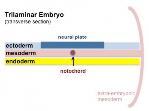
| 1 trilaminar - neural plate
|
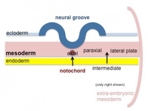
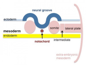
2 neural plate to groove
|
| 3 groove folding
|
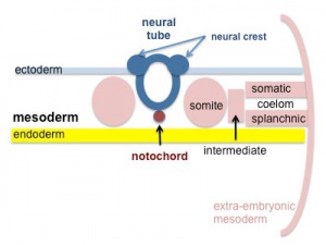
4 neural tube and neural crest
|
see also the cartoon animation
Notochord
- forms initially as the Axial Process, a hollow tube which extends from the primitive pit , cranially to the oral membrane
- the axial process then allow transient communication between the amnion and the yolk sac through the neuroenteric canal.
- the axial process then merges with the Endodermal layer to form the Notochordal Plate.
- the notochordal plate then rises back into the Mesodermal layer as a solid column of cells which is the Notochord.
Ectoderm
Two parts
- midline columnar epithelium - neural plate (central nervous system)
- lateral cuboidal epithelium - surface ectoderm (epidermis) and sensory placodes Placodes
|

|
Neural Plate
| <html5media height="520" width="320">File:Neuralplate_001.mp4</html5media>
Click Here to play on mobile device
|
Development of the neural plate region at the embryonic disc stage.
Dorsal view of the embryonic disc from the amniotic cavity side showing the ectoderm with the central region developing into the neural plate.
The neural plate extends from buccopharyngeal membrane to primitive node and forms above the notochord and paraxial mesoderm.The neuroectodermal cells form a broad brain plate and narrower spinal cord region.
Three specific regions medial to lateral can also be identified: midline region floor plate, neural plate, edge of neural plate neural crest
|
- extends from buccopharyngeal membrane to primitive node
- forms above notochord and paraxial mesoderm
- neuroectodermal cells
- broad brain plate
- narrower spinal cord
- 3 components form: floor plate, neural plate, neural crest
|
Neural Determination- neuronal populations are specified before plate folds
- signals from notochord and mesoderm - secrete noggin, chordin,follistatin
- all factors bind BMP-4 an inhibitor of neuralation
- bone morphogenic protein acts through membrane receptor
- lateral inhibition generates at spinal cord level 3 strips of cells
- expression of delta inhibits nearby cells, which express notch receptor, from becoming neurons
- Delta-Notch inetraction- generates Neural strips
|
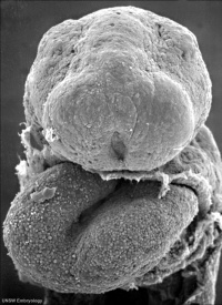
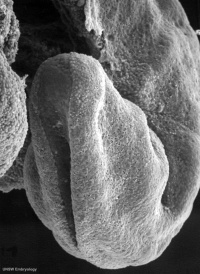
Neural Groove
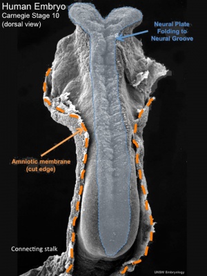
Embryo dorsal view (week 4, stage10)
Embryo transverse view (week 4, stage10)
<html5media height="480" width="480">File:Neuraltube_001.mp4</html5media>
Click Here to play on mobile device
This animation of early neural development from week 3 onward shows the neural groove fusing to form the neural tube.
View - Dorsolateral of the whole early embryo and yolk sac. Cranial (head) to top and caudal (tail) to bottom. Yolk sac is shown to the left.
Beginning with the neural groove initially fusing at the level of the 4th somite to form the neural tube and closing in both directions to leave 2 openings or neuropores: a cranial neuropore (anterior neuropore) and a caudal neuropore (posterior neuropore).
The animation also shows as the embryo grows and folds it increases in size relative to the initial yolk sac. Note also the increasing number of somites over time.
- forms in the midline of the neural plate (day 18-19)
- either side of which are the neural folds which continues to deepen until about week 4
- neural folds begins to fuse, beginning at 4th somite level
Neural Tube
| <html5media height="440" width="380">File:Mouse neural tube 01.mp4</html5media>
Click Here to play on mobile device
|
|
|
| Mouse neural tube closure
|
Human Stage 10 neural groove to tube
|
Human Stage 11 forming neuropores
|
- the neural tube forms the brain and spinal cord
- fusion of neural groove extends rostrally and caudally
- begins at the level of 4th somite
- closes neural groove "zips up" in some species.
- humans appear to close at multiple points along the tube.
Last parts of neural groove to close are the Neuropores
- two openings at either end of the embryo
- cranial (rostral, anterior) neuropore closes (day 25) about 2 days before caudal
| Neuropores
|
|
|
| Anterior Neuropore (cranial)
|
Posterior Neuropore (caudal)
|
|
About Neuropores
- Anatomically - lamina terminals is the location of the cranial neuropore.
- Failure of neural tube closure - a neural tube defect.
- Maternal dietary folate is required for this closure.
|
Secondary Neurulation
| <html5media height="320" width="320">File:Secondary_neurulation_01.mp4</html5media>
Click Here to play on mobile device
|
This animation shows the early developmental process often described as secondary neurulation.
Red - site of secondary neurulation | Blue - neural tube
- caudal end of neural tube formed by secondary neuralation
- solid cord canalized by extension of neural canal
- filum terminale and ventriculus terminalis and part of the conus medullaris.
- Links: MP4 version | Neural System Development | PMID 8351645
|
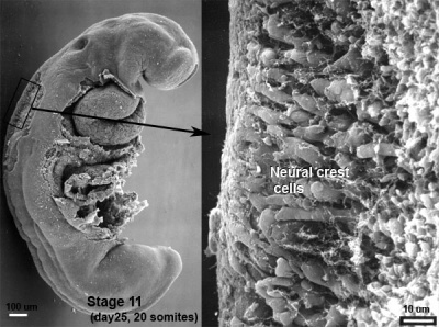
Neural Crest
- a population of cells at the edge of the neural plate that lie dorsally when the neural tube fuses
- dorsal to the neural tube, as a pair of streaks
- pluripotential (forms many different types of cells)
- cells migrate throughout the embryo
- migration studied by quail-chick chimeras - transplanted quail cells have obvious nucleoli compared with chicken
|
|
Neural Crest Derivitives
- dorsal root ganglia (DRG)
- autonomic ganglia
- adrenal medulla
- drg sheath cells, glia
- pia-arachnoid sheath
- skin melanocytes
- connective tissue of cardiac outflow
- thyroid parafollicular cells
- craniofacial skeleton
- teeth odontoblasts
- Links: Lecture - Neural Crest Development | Neural Crest Development
Early Brain Structure
Primary Vesicles
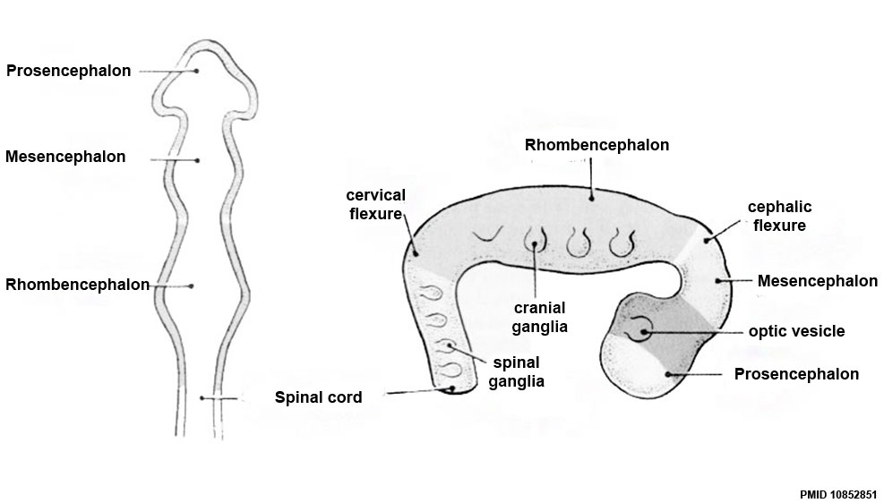
- rostral neural tube forms 3 primary brain vesicles (week 4)
- 3 primary vesicles: prosencephalon (forebrain), mesencephalon (midbrain), rhombencephalon (hindbrain)
Secondary Vesicles
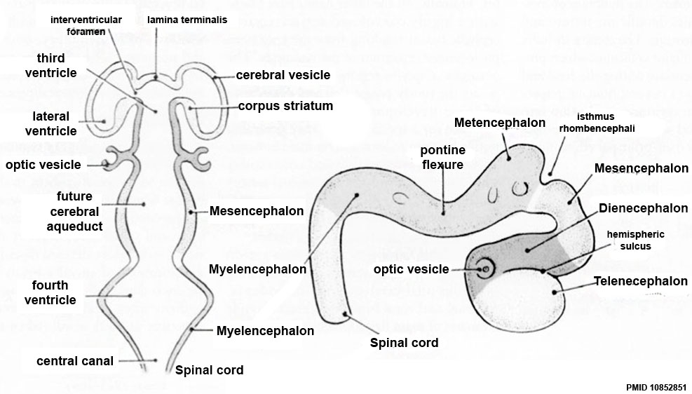
From the 3 primary vesicles developing in week 5 to form 5 secondary vesicles
- prosencephalon- telencephalon (endbrain, forms cerebral hemispheres), diencephalon (betweenbrain, forms optic outgrowth)
- mesencephalon
- rhombencephalon- metencephalon (behindbrain), myelencephalon (medullabrain)
Ventricles
MH - this will be covered in detail in later neural development
- cavity within tube will form the contiguious space of the ventricules of the brain and central canal of spinal cord
- space is filled initially with amniotic fluid, later with CerebroSpinal Fluid (CSF)
- CSF is secreted by a modified vascular structure, the chorioid plexus, lying within the ventricles (More? Ventricular System)
Brain Flexures
Rapid growth folds the neural tube forming 3 brain flexures
- cephalic flexure - pushes mesencephalon upwards
- cervical flexure - between brain stem and spinal cord
- pontine flexure - generates 4th ventricle
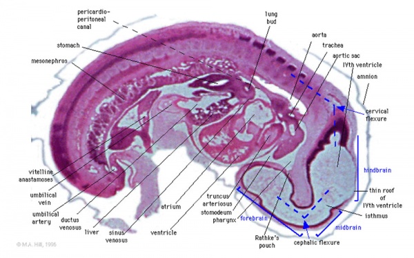
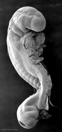
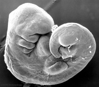
Carnegie stage 13 Embryo showing neural tube and brain flexures.
Neural Layers
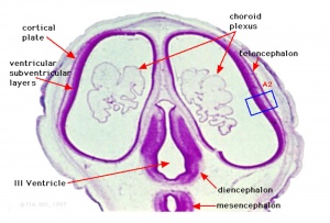
Stage 22 developing head cross section
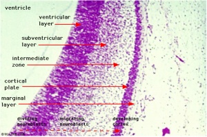
Stage 22 developing cortex
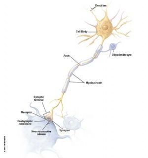
Neuron and supporting glial cells
- neural stem cells lie in the layer closest to the ventricular space, the ventricular layer
- this layer generates both neuroblasts and glioblasts
Neuroblasts - neurons arise first as neuroblasts and migrate along radial gial, their migration stops at cortical plate.
Glioblasts - glia arise later as glioblasts
Both neurons and glia undergo a complex process of growth, differentiation and interaction over a long developmental time period.
Spinal Cord Axes
Identified by experimental manipulation of interactions.
- Initial experiments looked at how isolated tissues may influence the development of the spinal cord.
- Repositionining of specific tissues both in vivo and in vitro
- specific markers of or alteration of differentiation.
- today molecular signals - Sonic Hedgehog (ventral), Dorsalin (dorsal), Hox (rostrocaudal)
| Additional Information - Spinal Cord Axes
|
| Students do not need to know the details within this table.
Ventral Axis
 Notochord secreting sonic hedgehog - Sonic Hedgehog (SHH) - notochord secretes sonic hedgehog
- Gene expression studies (ISH) showed shh gene expression occured in a subset of inducing tissues
- has a patterning role elsewhere (limb, sclerotome, lung)
- 2 signaling activities acting (locally and at a distance) Ventral- Sonic Hedgehog
- Binds to cell surface receptor patched
- without shh, patched (Ptc) binds smoothened (Smo)
- with shh shh-Ptc releases Smo activating G protein pathway Gene Diseases
- shh Human mutation- holoprosencephaly 3
- characteristic faces of the severe form of HPE which included a single fused eye (cyclopia) and a nose-like structure (proboscis) above the eye
- Downstream targets of Sonic hedgehog signalling:
- transcription factors like Gli3 (responsible for Greigs polycephalosyndactyly in humans)
- d Hoxd13 (responsible for polysyndactyly)
Dorsal Axis
- Dorsalin - ectoderm secretes a growth factor shown to controls patterning in embryonic mesoderm (frog)
- Transforming Growth factor beta, (TGF b), related factors BMP-2, BMP-4, BMP-7, radar (flies related protein determines dorsoventral)
- homology search of vertebrate library identified protein of same family.
- dorsalin-1 (dsl-1) (Basler, Cell 73, p687, 1993) Dorsalin-1
- From overlying ectoderm
- Naming comes from the obvious reason that it promotes the differentiation of neural crest cells.
- Also signal for dorsal signal of neural tube.
- Inhibits the differentiation of motoneurons.
- Implication is that dsl-1 and shh act antagonistically, or competitively to establish d-v axis of neural tube.
Rostro-Caudal Axis
- Brain rostro-caudal axis is generated by differential expression of Hox genes (transcriptional activators)
- corresponding to genetic order on chromosome. (Wilkinson, Nature, 341, p405, 1989) Hox Genes
- Stands for Homeobox domain Genes
- A family of transcription factors
- Discovered in flies and conserved between all species. [../OtherEmb/fly.htm#antennapedia antennapedia]
- Expressed in sequence along the embryo rostro-caudal axis.
- Regulate many other aspects of development.
- 180aa region binds DNA and regulate gene expression
- large family of genes organized and expressed in sequence on the chromosome
- Nkx-2.2 first detected at 1 somite stage
- Lim hox gene expressed at spinal cord level
|
Ectodermal Placodes
- Specialized ectodermal "patches" in the head region
- Contribute sensory structures - otic placode (otocyst), nasal placode, lens placode
- Contribute teeth
- Covered in Head and Sensory Development Lectures
Human Neuralation - Early Stages
|
|
|
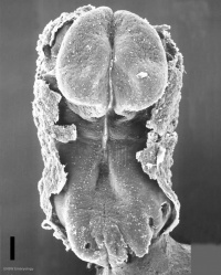
| Carnegie stage 8 (about 18 postovulatory days) neural groove and folds are first seen.
|
Carnegie stage 9 the three main divisions of the brain, which are not cerebral vesicles, can be distinguished while the neural groove is still completely open.
|
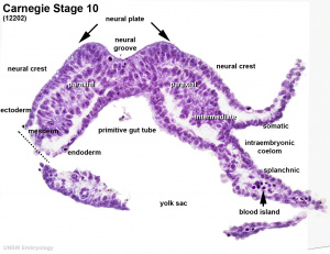
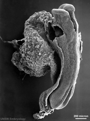
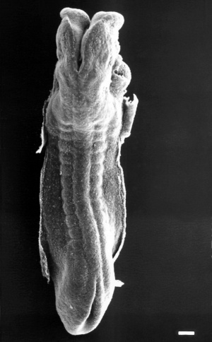
Carnegie stage 10 (two days later) neural folds begin to fuse near the junction between brain and spinal cord, when neural crest cells are arising mainly from the neural ectoderm.
|

|
|
|
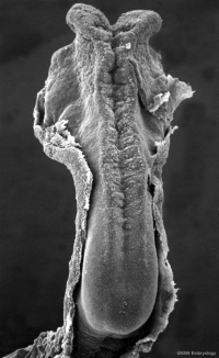
| Carnegie stage 11 (about 24 days) the rostral (or cephalic) neuropore closes within a few hours; closure is bidirectional, it takes place from the dorsal and terminal lips and may occur in several areas simultaneously. The two lips, however, behave differently.
|
Carnegie stage 12 (about 26 days) The caudal neuropore takes a day to close. Level of final closure is approximately at future somitic pair 31, corresponds to the level of sacral vertebra 2.
|
Carnegie stage 13 (4 weeks) the neural tube is normally completely closed.
|
Secondary neurulation begins at stage 12 - is the differentiation of the caudal part of the neural tube from the caudal eminence (or end-bud) without the intermediate phase of a neural plate. Above text modified from[1]
- Carnegie Stages: 1 | 2 | 3 | 4 | 5 | 6 | 7 | 8 | 9 | 10 | 11 | 12 | 13 | 14 | 15 | 16 | 17 | 18 | 19 | 20 | 21 | 22 | 23 | About Stages | Timeline
Abnormalities
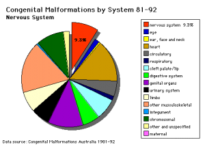
Australian Birth Statistics
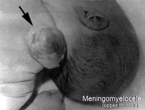
Neural tube defect - Meningomyelocele
- Links: Neural System - Abnormalities | Folic Acid and Neural Tube Defects | Iodine Deficiency
Neural Tube Defects (NTD)
Failure of neural tube closure either incorrectly or incomplete
- Dysraphism is the term often used to describe the defective fusion of the neural folds. The position and degree of failure of fusion will result in either embryonic death or a range of different neural defects. The way (mode) in which the human neural tube fuses has been a source of contention. In humans, fusion appears to initiate at multiple sites but the mode is different from that found in many animal species used in developmental studies.
- severity dependent upon level within the tube and degree of failure
- caudal failure - spina bifida cranial failure - anancephaly
Maternal Diet
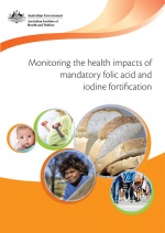
Folic acid and iodine fortification (2016)
Folate
Found that supplementation of maternal diet with folate reduces incidence of NTDs.
- A randomised controlled trial conducted by the Medical Research Council of the United Kingdom demonstrated a 72% reduction in risk of recurrence by periconceptional (ie before and after conception) folic acid supplementation (4mg daily).
- Women who have one infant with a neural tube defect have a significantly increased risk of recurrence (40-50 per thousand compared with 2 per thousand for all births)
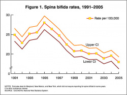
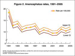
In the U.S.A. the Food and Drug Administration in 1996 authorized that all enriched cereal grain products be fortified with folic acid, with optional fortification beginning in March 1996 and mandatory fortification in January 1998. The data in the above graphs show the subsequent changes in anencephaly and spina bifida rate over that period.
Iodine
A second maternal diet requirement for later neural growth and development.
- Iodine is required for fetal thyroid hormone synthesis.
Holoprosencephaly
Holoprosencephaly (HPE) is developmental abnormality where the forebrain does not divide into the two separate hemispheres and ventricles.
Critical Periods of Human Development
Exposure to teratogens during these "critical periods" results in specific abnormalities.
- most systems are susceptible during embryonic development (first trimester)
- the earlier the exposure the more severe the effects
- each system has a different critical period
- longest critical periods
- longest developing systems (neural, genital)
- complicated developmental origins (sensory systems)
The table below identifies approximate windows of time, "critical periods", that following exposure to teratogens can lead to developmental abnormalities (anomalies, congenital). In general, the effects for each system are more severe (major anomalies) in the embryonic period during organogenesis in the first trimester. Later teratogen exposure are less severe (minor anomalies) in the fetal period during continued growth and differentiation in the second and third trimester.
| Conceptus
|
Embryonic development (weeks)
|
Fetal period (weeks)
|
| 1
|
2
|
3
|
4
|
5
|
6
|
7
|
8
|
9
|
16
|
20-36
|
38
|

|

|

|

|

|

|

|

|
|
|
|
|
|
|
|
Neural
|
|

|
|
|
Heart
|
|
|
|
|
|
|
|
|
|
|
|
Upper limbs
|
|
|
|
|
|
|
|
|
|
|
|
|
Lower limbs
|
|
|
|
|
|
|
|
|
|
|
|
|
Ear
|
|
|
|
|
|
|
|
|
|
|
Eye
|
|

|
|
|
|
|
|
|
|
|
Palate
|
|
|
|
|
|
|
|
|
|
|
|
|
|
|
Teeth
|
|
|
|
|
|
|
|
|
|
|
|
|
|
External genitalia
|
|
| Loss
|
Major abnormalities
|
Functional and Minor abnormalities
|
- ↑ O'Rahilly R & Müller F. (1994). Neurulation in the normal human embryo. Ciba Found. Symp. , 181, 70-82; discussion 82-9. PMID: 8005032
Neural Development Terms
Only brief descriptions are given below, more complete definitions can be found in the glossary.
| Neural Terms
|
Neural Development
- 3DMRI - Three-dimensional magnetic resonance imaging. A new technique that allows 3D analysis of embryonic structures. (More? Magnetic Resonance Imaging)
- 3rd ventricle - a fluid-filled space formed from neural tube lumen, located within the diencephalon (from the primary vesicle prosencephalon, forebrain).
- 4th ventricle - a fluid-filled space formed from neural tube lumen, located within the rhombencephalon (from the primary vesicle, hindbrain).
- adenohypophysis - (anterior pituitary) = 3 parts pars distalis, pars intermedia, pars tuberalis.
- afferent - refers to the direction of conduction from the periphery toward the central nervous system. Efferent is in the opposite direction.
- alar plate - embryonic dorsolateral region of the neural tube forming at spinal cord level dorsal horns (afferent) and brain level different structures.
- anlage - (German = primordium) structure or cells that will form a future adult structure.
- arachnoid mater - (G.) spider web-like used in reference to the middle layer of the brain meninges.
- astrocytes - cells named by their "star-like" branching appearance, are the most abundant glial cells in the brain, important for the blood-brain barrier.
- basal ganglia - (basal nuclei) neural structure derived from the secondary vesicle telencephalon (endbrain) structure from the earlier primary vesicle prosencephalon (forebrain).
- basal plate - embryonic ventrolateral region of the neural tube forming at spinal cord level ventral horns (efferent) and brain level different structures.
- brachial plexus - mixed spinal nerves innervating the upper limb form a complex meshwork (crossing).
- brain - general term for the central nervous system formed from 3 primary vesicles.
- buccopharyngeal membrane - (oral membrane) at cranial (mouth) end of gastrointestinal tract (GIT) where surface ectoderm and GIT endoderm meet. (see also cloacal membrane).
- cauda equina - (horse's tail) caudal extension of the mature spinal cord.
- central canal - lumen, cavity of neural tube within the spinal cord. Space is continuous with ventricular system of the brain.
- central cerebral sulcus - (central fissure, fissure of Rolando, Rolandic fissure) fold in the cerebral cortex associated with the sensorimotor cortex.
- cerebral aqueduct - ventricular cavity within the mesencephalon.
- cervical flexure - most caudal brain flexure (of 3) between spinal cord and rhompencephalon.
- choroid plexus - specialized vascular plexus responsible for secreting ventricular fluid that with further additions becomes cerebrospinal fluid (CSF).
- cloacal membrane - at caudal (anal) end of gastrointestinal tract (GIT) where surface ectoderm and GIT endoderm meet forms the openings for GIT, urinary, reproductive tracts. (see also buccopharyngeal membrane).
- connectome - term describing the detailed map of neural connections in the central nervous system.
- cortex - - CNS structure derived from the secondary vesicle telencephalon (endbrain) from the earlier primary vesicle prosencephalon (forebrain).
- cortical plate - outer neural tube region which post-mitotic neuroblasts migrate too along radial glia to form adult cortical layers.
- cranial flexure - (=midbrain flexure) most cranial brain flexure (of 3) between mesencephalon and prosencephalon.
- diencephalon - the caudal portion of forebrain after it divides into 2 parts in the 5 secondary vesicle brain (week 5). (cavity- 3rd ventricle) Forms the thalmus and other nuclei in the adult brain. (sc-My-Met-Mes-Di-Tel)
- dorsal root ganglia - (spinal ganglia) sensory ganglia derived from the neural crest lying laterally paired and dorsally to the spinal cord (in the embryo found ventral to the spinal cord). Connects centrally with the dorsal horn of the spinal cord.
- dura mater- "tough" (Latin, mater = mother) used in reference to the tough outer layer of the brain meninges.
- efferent - refers to the direction of conduction from the central nervous system toward the periphery. Afferent is in the opposite direction.
- ependyma - epithelia of remnant cells after neurons and glia have been generated and left the ventricular zone.
- floorplate - early forming thin region of neural tube closest to the notochord.
- ganglia - (pl. of ganglion) specialized neural cluster within either the CNS or PNS.
- glia - supporting, non-neuronal cells of the nervous system. Generated from the same neuroepithelial stem cells that form neurons in ventricular zone of neural tube. Form astrocytes, oligodendrocytes.
- grey matter - neural regions containing cell bodies (somas) of neurons. In the brain it is the outer layer, in the spinal cord it is inner layer. (see white matter white matter).
- growth factor - usually a protein or peptide that will bind a cell membrane receptor and then activates an intracellular signaling pathway. The function of the pathway will be to alter the cell directly or indirectly by changing gene expression. (eg SHH).
- HOX - (homeobox) family of transcription factors that bind DNA and activate gene expression. Expression of different Hox genes along neural tube defines rostral-caudal axis and segmental levels.
- hydrocephalus - abnormality as the result of an imbalance between the rate at which the CSF is being formed and the rate at which the CSF is passing through the arachnoidal villi back into the blood (hydrocephalus rate is a function of the degree of imbalance in these two). Very small imbalance exhibit subtle, if any, symptoms. Large imbalances will have rapidly evolving symptoms of unmistakable import.
- isthmus- (G. narrow passage).
- lamina terminalis - anterior region of brain where cranial neuropore closes.
- lumbar plexus - mixed spinal nerves innervating the lower limb form a complex meshwork (crossing).
- mantle layer - layer of cells generated by first neuroblasts migrating from the ventricular zone of the neural tube. Layers are rearranged during development of the brain and spinal cord. (Ven-Man-Mar-CP)
- marginal zone - layer of processes from neuroblasts in mantle layer. (Ven-Man-Mar-CP)
- mater - (Latin, mater = mother) used in relation to the 3 layers of the meninges.
- meninges - mesenchyme surrounding neural tube forms 3 layer (Dura-, pia-, arachnoid- mater) connective tissue sheath of nervous system. (D-P-A-cns)
- mesencephalon - (midbrain), the middle portion of the 3 primary vesicle brain (week 4). (sc-R-M-P)
- metencephalon - the cranial portion of hindbrain after it divides into 2 parts in the 5 secondary vesicle brain (week 5). Forms the pons and cerebellum in the adult brain. (sc-My-Met-Mes-Di-Tel)
- microglia - CNS innate immune cells that have a macrophage function, derive from yolk sac progenitor cells migrating into the CNS. microglia
- myelencephalon - the caudal portion of hindbrain after it divides into 2 parts in the 5 secondary vesicle brain (week 5). Forms the medulla in the adult brain. (sc-My-Met-Mes-Di-Tel)
- neural tube - neural plate region of ectoderm pinched off to form hollow ectodermal tube above notochord in mesoderm.
- neural tube defect - (NTD) any developmental abnormality that affects neural tube development. Commonly failure of neural tube closure.
- neuroblast - undifferentiated neuron found in ventricular layer of neural tube.
- neurohypophysis - (posterior pituitary; pas nervosa)
- neuromere - (prosomere) the model units for segmental brain development regions based upon a series of neural tube transverse subunits.
- neuron - The cellur "unit" of the nervous system, transmitting signals between neurons and other cells. The post-mitotic cells generated from neuroepithelial stem cells (neuroblasts) in ventricular zone of neural tube.
- neuropore - opening at either end of neural tube cranial (rostral, anterior) neuropore closes (day 25) about 2 days before caudal (posterior) that closes at somite level 32 to 34. Neural Tube Defects (NTDs) can be due to failure of these two neuropores to close.
- notochord - rod of cells lying in mesoderm layer ventral to the neural tube, induces neural tube and secretes sonic hedgehog which "ventralizes" the neural tube.
- olfactory bulb - (cranial nerve I, CN I) bipolar neurons from nasal epithelium project axons through cribiform palate into olfactory bulb of the brain associated with smell.
- optic nerve - (cranial nerve II, CN II) retinal ganglion neurons project from the retina as a tract into the brain (at the level of the diencephalon) associated with vision.
- optic vesicle - diencephalon region of neural tube outgrowth that forms the primordia of the retina associated with vision.
- opercularization - during fetal development of the sensorimotor cortex, the insula (located deep within the lateral sulcus) begins to invaginate from the surface of the immature cerebrum, until at term, the opercula completely cover the insula.
- otocyst - (otic vesicle) sensory placode that sinks into mesoderm to form spherical vesicle (stage 13/14 embryo) that will form components of the inner ear associated with hearing.
- pharyngeal arch - (branchial arch, Gk. gill) form the main structures of the head and neck. Humans have 5 arches appearing in week 4 that form 4 external swellings, each arch has a pouch, membrane and cleft.
- pharynx - uppermost end of GIT, beginning at the buccopharyngeal membrane and at the level of the pharyngeal arches.
- pia mater - (G.) (L. pius = soft, faithful + mater = mother) delicate vascular membrane which adheres to surface of brain and spinal cord, faithfully following their contours, the inner layer of the brain meninges.
- placode - specialized regions of ectoderm which form components of the sensory apparatus.
- pontine flexure - middle brain flexure (of 3) between cervical and cranial flexure in opposite direction, also generates thin roof of rhombencephalon and divides it into myelencephalon and metencephalon. ( sc-^V^ )
- posterior insula - during sensorimotor cortex development this region is composed of the anterior and posterior long insular gyri and the postcentral insular sulcus, which separates them.
- prosencephalon - (forebrain), the most cranial portion of the 3 primary vesicle brain (week 4). (sc-R-M-P)
- prosomere - (neuromere) a model for segmental brain development based upon a series of neural tube transverse subunits. PMID 12948657
- Rathke's pouch - a portion of the roof of the pharynx pushes upward towards the floor of the brain forming the anterior pituitary (adenohypophysis, pars distalis, pars tuberalis pars intermedia). Where it meets a portion of the brain pushing downward forming the posterior pituitary (neurohypophysis, pars nervosa). Rathke's pouch eventually looses its connection with the pharynx.
- rhombencephalon - (hindbrain), the most caudal portion of the 3 primary vesicle brain (week 4). (sc-R-M-P)
- rhombic lip - metencephalon posterior part extending from the roof of the fourth ventricle to dorsal neuroepithelial cells that contributes to the cerebellum.
- roofplate - early forming thin region of neural tube closest to the overlying ectoderm.
- spinal cord - caudal end of neural tube that does not contribute to brain. Note: the process of secondary neuralation contributes the caudal end of the spinal cord.
- spinal ganglia - (dorsal root ganglia, drg) sensory ganglia derived from the neural crest lying laterally paired and dorsally to the spinal cord (in the embryo found ventral to the spinal cord). Connects centrally with the dorsal horn of the spinal cord.
- spinal nerve - mixed nerve (motor and sensory) arising as latera pairs at each vertebral segmental level.
- sonic hedgehog - (shh) secreted growth factor that binds patched (ptc) receptor on cell membrane. SHH function is different for different tissues in the embryo. In the nervous system, it is secreted by the notochord, ventralizes the neural tube, inducing the floor plate and motor neurons.
- sulcus - (L. furrow) groove.
- sulcus limitans - longitudinal lateral groove in neural tube approx. midway between roofplate and floorplate. Groove divides alar (dorsal) and basal (ventral) plate regions.
- telencephalon - the cranial portion of forebrain after it divides into 2 parts in the 5 secondary vesicle brain (week 5). (cavity- lateral ventricles and some of 3rd ventricle) Forms the cerebral hemispheres in the adult brain. (sc-My-Met-Mes-Di-Tel)
- thalamus - (G. thalamos= bedchamber) cns nucleus, lateral to 3rd ventricle, paired (pl thalami).
- thyroid hormone - hormone required for brain development. T3 (3,5,3′-triiodothyronine) binding to nuclear receptors then act as a transcription factor in both neurons and glial cells. iodine deficiency
- transcription factor - a factor (protein or protein with steroid) that binds to DNA to alter gene expression, usually to activate. (eg steroid hormone+receptor, Retinoic acid+Receptor, Hox, Pax, Lim, Nkx-2.2)
- trigeminal ganglion - (cranial nerve V, CN V) first arch ganglion, very large and has 3 portions.
- vagal ganglion - (cranial nerve X, CN X) fourth and sixth arch ganglion, innervates the viscera and heart.
- ventricles - the fluid-filled interconnected cavity system with the brain. Fluid (cerebrospinal fluid, CSF) is generated by the specialized vascular network, the choroid plexus. The ventricles are directly connected to the spinal canal (within the spinal cord).
- ventricular zone - Neuroepithelial cell layer of neural tube closest to lumen. Neuroepithelial cells generate neurons, glia and ependymal cells. (Ven-Man-Mar-CP)
- vestibulocochlear nerve - (cranial nerve VIII, CN VIII, also called statoacoustic)
- white matter - - neural regions containing processes (axons) of neurons. In the brain it is the inner layer, in the spinal cord it is outer layer. (see grey matter).
|


