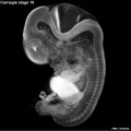
Carnegie stage 19
| Embryology - 29 Jul 2026 |
|---|
| Google Translate - select your language from the list shown below (this will open a new external page) |
|
العربية | català | 中文 | 中國傳統的 | français | Deutsche | עִברִית | हिंदी | bahasa Indonesia | italiano | 日本語 | 한국어 | မြန်မာ | Pilipino | Polskie | português | ਪੰਜਾਬੀ ਦੇ | Română | русский | Español | Swahili | Svensk | ไทย | Türkçe | اردو | ייִדיש | Tiếng Việt These external translations are automated and may not be accurate. (More? About Translations) |
Introduction

Facts
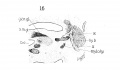
Week 7, 48 - 51 days, 16 - 18 mm
Gestational Age GA - week 9
Summary
- Ectoderm: sensory placodes, lens pit, otocyst, nasal pits moved ventrally, fourth ventricle of brain
- Mesoderm: heart prominence, ossification continues
- Head: forebrain, eye, external acoustic meatus
- Body: straightening of trunk, heart, liver, umbilical cord
See also Stage 19 Events
Features
- eyelid, eye, external acoustic meatus, auricle of external ear, digital ray, wrist, liver prominence
- Identify: straightening of trunk, pigmented eye, eyelid, external acoustic meatus, digital rays, liver prominance, thigh, ankle, foot plate, umbilical cord
| Week: | 1 | 2 | 3 | 4 | 5 | 6 | 7 | 8 |
| Carnegie stage: | 1 2 3 4 | 5 6 | 7 8 9 | 10 11 12 13 | 14 15 | 16 17 | 18 19 | 20 21 22 23 |
- Carnegie Stages: 1 | 2 | 3 | 4 | 5 | 6 | 7 | 8 | 9 | 10 | 11 | 12 | 13 | 14 | 15 | 16 | 17 | 18 | 19 | 20 | 21 | 22 | 23 | About Stages | Timeline
Bright Field

|
Virtual Slide
|
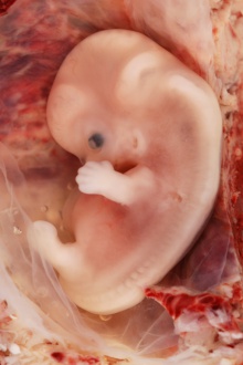
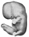
- Facts: Week 7, 48 - 51 days, 16 - 18 mm
- View: Lateral view. Amniotic membrane removed.
- Features: eyelid, eye, external acoustic meatus, auricle of external ear, digital ray, wrist, liver prominence
- Identify: straightening of trunk, pigmented eye, eyelid, external acoustic meatus, digital rays, liver prominance, thigh, ankle, foot plate, umbilical cord
Image: Dr Steven O'Connor (Houston, Texas) - Other embryo images.
- Carnegie Stages: 1 | 2 | 3 | 4 | 5 | 6 | 7 | 8 | 9 | 10 | 11 | 12 | 13 | 14 | 15 | 16 | 17 | 18 | 19 | 20 | 21 | 22 | 23 | About Stages | Timeline
Cite this page: Hill, M.A. (2026, July 29) Embryology Carnegie stage 19. Retrieved from https://embryology.med.unsw.edu.au/embryology/index.php/Carnegie_stage_19
- © Dr Mark Hill 2026, UNSW Embryology ISBN: 978 0 7334 2609 4 - UNSW CRICOS Provider Code No. 00098G
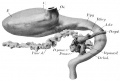
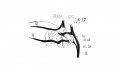
Scanning EM


Ventral view of head showing upper lip, maxilla and nasal region.
Image Source: Prof Virginia Diewert
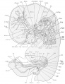
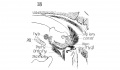
Kyoto Collection

View: This is a dorsolateral view of embryo. Amniotic membrane removed.
Image source: Embryology page Created: 19.03.1999
Image source: The Kyoto Collection images are reproduced with the permission of Prof. Kohei Shiota and Prof. Shigehito Yamada, Anatomy and Developmental Biology, Kyoto University Graduate School of Medicine, Kyoto, Japan for educational purposes only and cannot be reproduced electronically or in writing without permission.
Carnegie Collection


- Carnegie stage 19: 4501 Right | 4501 Anterior | 4501 Left | 6824 Right | 6824 Anterior | 6824 Left | 8092 Right | 8092 Anterior | 8092 Left

|

|
| Lens[1] | Eye and Optic Nerve[1] |
| Carnegie Collection - Stage 19 | |||||||||||
|---|---|---|---|---|---|---|---|---|---|---|---|
| Serial No. | Size (mm) | Grade | Fixative | Embedding Medium | Plane | Thinness (µm) | Stain | Score | Sex | Year | Notes |
| 17 | E, 18 Ch, 40x30x20 | Poor | Alc. | P | 50, 100 | Al. carm. | 16.5 | Male | 1894 | ||
| 43 | E, 16 | Good | Alc. | P | 50 | Al. coch. | 10 | Male | 1894 | ||
| 293 | E, 19 | Poor | Ale. | P | Sagittal | 50 | Coch. | 16.5 | S | 1905 | |
| 390 | E, 19 | Good | Formol? | P | Sagittal | 20, | (Stain - Haematoxylin Eosin) | 11.5 | Male | 1906 | Tubal Injected
50 |
| 409 | E.18 Ch, 50x40x40 | Good | Formalin | P | Transverse | 20 | Copper, iron H. & erythrosin | 14.5 | Male | 1907 | |
| 432 | E..18.5 Ch , 45x35x20 | Good | Formalin | P | Sagittal | 20 | H. & Congo red | 13.5 | Male | 1910 | Tubal |
| 576 | E. 17 Ch, 60x40 | Good | Formalin | P | Sagittal | 15, 20 | (Stain - Haematoxylin Eosin) | 14.5 | d | 1912 | Tubal |
| 626 | E., 21.5 Ch., 40x30x21 | Good | Formalin | P | Transverse | 100 | Al. coch. | 14_5 | 6 | 1913 | |
| 6??8 | E, 20 Ch, ca. 30 | Poor | Formalin | P | Sagittal | 50 | Al. coch. | 12 | 9 | 1913 | Head damaged |
| 709 | E, 19 Ch. 40x35x25 | Poor | Alc. | P | Coronal | 40 | Al. coch, Lyons blue | 15 | 49 | 1913 | |
| 837 | E. 21 Ch. 65x45x | Good | Formalin | P | Sagittal | 40 | Al. coch. | 14.5 | P | 1914 | |
| 1324 | E., 18 50x30x18 | Good | Formalin | C | Coronal | 40 | (Stain - Haematoxylin Eosin), aur, or. G | 125 | 79 | 1915 | |
| 1332 | E., 19 Ch., 40x43x22 | Poor | Formalin | C | Coronal | 40 | (Stain - Haematoxylin Eosin) aur, or. G. | 15 | Male | 1915 | |
| 1390 | E., 18 Ch, 40x38x15 | Good | Formalin | P | Sagittal | 20 | Al. coch. | 10_5 | Male | 1915 | Tubal |
| 1534 | E., 13 Ch.,35x31x25 | Poor | Formalin | P | 53% | 50 | Al. coch. | 13.5 | F | 1916 | Protruding midbrain |
| 2114 | E., 19.3 Ch., 49x42x33 | Good | Formol | P | Transverse | 40 | A1. coch. | 12 | M | 1918 | |
| 4405 | E., 15.5 | Good | Formalin | P | Transverse | 10 | Coch, Mallory | 13.5 | <3 | 1923 | Midbrain injured |
| 4501 | E, 18 | Exc. | Bouin | P | Transverse | 15 | Coch, or. G. | 14.6 | 1924 | Cystic left kidney | |
| 5609 | E., 18 | Exc. | Formalin | P | Coronal | 25 | A1. coch. | 13.5 | Male | ||
| 6150 | E., 17 Ch., 40x39x30 | Good | Bouin | C-P | Transverse | 15 | (Stain - Haematoxylin Eosin) | 16.5 | Male | 1930 | Tubal |
| 6824 | E., 18.5 Ch., 45x40x25 | Good | Formalin | C-P | Sagittal | 12 | (Stain - Haematoxylin Eosin) | 14.5 | Female | 1933 | |
| 7900 | E., 16.5 | Good | Bouin | C-P | Sagittal | 20 | (Stain - Haematoxylin Eosin), phlox. | 11.5 | . . | 1941 | Tubal |
| 8092 | E., 16.3 Ch., 52 x 47 | Exc. | Bouin | C-P | Transverse | 20 | (Stain - Haematoxylin Eosin), phlox. | 13 | Male | 1942 | |
| 8913 | E.,? Ch, 34 | Poor | Formalin | p | Transverse | 10 | Alan . | 7 | 1951 | rubella. Medical abortion. Isolated head damaged | |
| 8965 | E, 19.1 Ch, 42x32x19 | Good | Formol—Zenker | C-P | Transverse | 10 | Borax, carm, or. G. | 1952 | Univ. Chicago No. H 173 | ||
| 9097 | E, 21 | Exc. | Formol—glucose | C-P | Coronal | 10 | Azan ? . | ? | 1930 | Univ. Chicago No H 1380 | |
| 9113 | E, 185 Ch, 24 | Exc. | Formalin | C-P | Transverse | 10 | Alan > 6 | 1953 | Rubella. Medical abortion | ||
| 9325 | E, 17.0 Ch, 32x28x20 | Good | Formalin | —acetic p | Transverse | 15& 8-10 | Azan & Ag | ? | - | 1955 | Tubal |
Abbreviations
| |||||||||||
| iBook - Carnegie Embryos | |
|---|---|

|
|
Blechschmidt Collection

Embryo (170452)
- Links: Blechschmidt Collection
Photographs

|

|
| Virtual Slide | |
| Image - Dr Ed Uthman | Image - Dr Steven O'Connor |
Embryo Virtual Slides
| Stage 19 - Left Lateral
|
| Stage 19 | Embryo Slides |
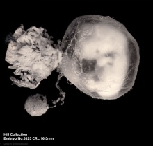
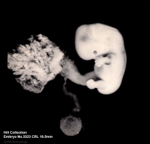
Hill Collection
| Righthand view | Lefthand view |
|---|---|

|

|
Image source: The images from the Hill Collection (part of the Embryological Collection) are reproduced with the permission of the Museum für Naturkunde, Leibniz Institute for Research on Evolution and Biodiversity. Images are for educational purposes only and must not be reproduced electronically or in writing without permission from the Museum für Naturkunde Berlin.
Embryo Virtual Slides
|
|
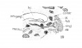
Hinrichsen Collection

|
Hinrichsen collection Human Embryo ME28 (stage19).
Embryo left lateral view.
SEM upper limb showing digital ray development. |

Image source: The Hinrichsen Collection images are reproduced with the permission of Prof. Beate Brand-Saberi, Head, Department of Anatomy and Molecular Embryology, Ruhr-Universität Bochum. Images are for educational purposes only and cannot be reproduced electronically or in writing without permission.
Madrid Collection
| Madrid Collection Embryos | ||||||
|---|---|---|---|---|---|---|
| Carnegie Stage |
Embryo | Days | CRL (mm) | Section thickness |
Staining | Section plane |
| 19 | PA | 47 | 17 | 10 | (Stain - Haematoxylin Eosin) | transverse |
| 19 | Civ1 | 48 | 18 | 10 | (Stain - Haematoxylin Eosin)-Azan | transverse |
Events
- vision - (stage 19 -22) the eyelid folds develop into the eyelids and cover more of the eye as the palpebral fissure takes shape. The upper and the lower eyelids meet at the outer canthus in Stage 19.[2]
- hearing - otic capsule now cartilaginous. Cochlea tip becomes curled. Malleus and incus present.
- smell - individualization of the olfactory bulb and nuclei. The distinction between olfactory structures and terminal and vomeronasal ones begins to be clear. [3] Stages Description | primitive olfactory bulb
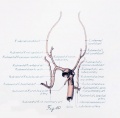
- Cardiovascular
- Cerebral artery the middle cerebral artery becomes more prominent, the plexi fuse into a single artery and further branches pierce the cerebral hemisphere.[4]
- arterial system[5] Chapter 18 fig. 447).
- aortic arches [6] stages 11–19 (figs. 29–40).
- heart fusion of aortic and mitral endocardial cushion material</ref>Teal SI., Moore GW. and Hutchins GM. Development of aortic and mitral valve continuity in the human embryonic heart. (1986) Amer. J. Anat., 176:447-460.</ref>
- respiratory - first generation of subsegmental bronchi now complete, see bronchial tree reconstruction[7] (plates 3 and 4).
- Gastrointestinal - anal membrane defined.
- submandibular gland - Short, club-like duct entering mesenchymal primordium of gland.[1]
- renal - Cloacal membrane ruptures from urinary pressure at stage 18 or stage 19,
- Musculoskeletal - Sternum right and left sternal bars are present.[8] (figs. 7-17 and 7-22)
- neural
- rhombencephalon migration for olivary and arcuate nuclei begins.
- choroid plexus of the fourth ventricle present.
- stria medullaris thalami reaches the habenular nuclei.
- habenular commissure begins to develop.
- endocrine[9]
- Hypophysis - the caudal part of the craniopharyngeal pouch is reduced to a closed epithelial stem (Andersen et al. 1971).
- Epiphysis - the "anterior lobe" of the pineal body shows a characteristic step and wedge appearance (Stadium 4 of Turkewitsch 1933) (O'Rahilly 1968).
- parathyroid - 3 become detached from the pharyngeal endo- derm (Jirfisek 1980).
- Adrenal Cortex - C2 cells lie on the surface of the gland and form a "capsule".[10]
- Adrenal Medulla - Sympathicoblasts penetrate the cortex at stages 19 and 20, and form scattered islets of medullary tissue throughout the cortex (Jirfisek 1980).
- meninges (spinal cord) - increased chondrification ventral vertebral wall and greater concentration in the dorsal vertebral wall (the membrana reuniens), further ventral migration of the ganglia, and formation of large cavities in the meninx primitiva and a reticular arrangement of its cells. Layers of stratified cells overlying the dorsal surfaces of the centra and intervertebral disks, distinguishable in stage 17, 18, are now separated from these structures by a narrow network of loose cells. This stratified concentration of cells is the rudiment of the ventral dura mater, and may be traced both laterally and medially to the ganglia. The part lateral to the ganglia passes over into, and becomes indistinguishable from, the dense stratum of the meninx primitiva that forms the more dorsal canal wall. Passing caudad, the ventral dura mater rudiment lies in ever closer proximity to the centra and intervertebral disks, becoming continuous with these structures in the lower cervical segments. It can, however, be identified throughout the thoracic regions.[11]
- genital[12]
References
- ↑ 1.0 1.1 1.2 Streeter GL. Developmental Horizons In Human Embryos Description Or Age Groups XIX, XX, XXI, XXII, And XXIII, Being The Fifth Issue Of A Survey Of The Carnegie Collection. (1957) Carnegie Instn. Wash. Publ. 611, Contrib. Embryol., 36: 167-196.
- ↑ Pearson AA. The development of the eyelids. Part I. External features. (1980) J. Anat.: 130(1): 33-42. PMID 7364662 PDF
- ↑ Bossy J. Development of olfactory and related structures in staged human embryos. (1980) Anat. Embryol., 161(2);225-36 PMID 7469043
- ↑ Menshawi K, Mohr JP & Gutierrez J. (2015). A Functional Perspective on the Embryology and Anatomy of the Cerebral Blood Supply. J Stroke , 17, 144-58. PMID: 26060802 DOI.
- ↑ Keibel F. and Mall FP. Manual of Human Embryology II. (1912) J. B. Lippincott Company, Philadelphia.
- ↑ Congdon ED. Transformation of the aortic-arch system during the development of the human embryo. (1922) Contrib. Embryol., Carnegie Inst. Wash. Publ 277, 14:47-110.
- ↑ Wells LJ. Development of the human diaphragm and pleural sacs. (1954) Contrib. Embryol., Carnegie Inst. Wash. Publ. 603, 35: 107-134.
- ↑ Gasser RL. Atlas of Human Embryos. (1975) Harper & Row, Hagerstown, Maryland.
- ↑ O'Rahilly R. The timing and sequence of events in the development of the human endocrine system during the embryonic period proper. (1983) Anat. Embryol., 166: 439-451. PMID 6869855
- ↑ Crowder RE. The development of the adrenal gland in man, with special reference to origin and ultimate location of cell types and evidence in favor of the "cell migration" theory. (1957) Contrib. Embryol., Carnegie Inst. Wash. 36, 193-210.
- ↑ Sensenig EC. The early development of the meninges of the spinal cord in human embryos. (1951) Contrib. Embryol., Carnegie Inst. Wash. Publ. 611.
- ↑ O'Rahilly R. (1983). The timing and sequence of events in the development of the human reproductive system during the embryonic period proper. Anat. Embryol. , 166, 247-61. PMID: 6846859
- ↑ Lee S. High incidence of true hermaphroditism in the early human embryos. (1971) Biol Neonate 18: 2418-2425. PMID 5163617
- ↑ Jirásek JE. Development of the Genital System and Male Pseudohermaphroditism. (1971) Johns Hopkins Press, Baltimore.
- ↑ Wilson KM. Origin and development of the rete ovarii and the rete testis in the human embryo. (1926) Carnegie Instn. Wash. Publ. 362, Contrib. Embryol., Carnegie Inst. Wash., 17:69-88.
Additional Images
Stage 19 Optical Projection Tomography

External ear Stages 14-23 and adult


Historic
| Historic Disclaimer - information about historic embryology pages |
|---|
 |
1914 Thyng HEC839 human embryo 17.8 mm external appearance
1914 Thyng HEC839 human embryo 17.8 mm gastrointestinal
1914 Thyng Plate 2
1922 Embryo 18 mm reconstruction model, Carnegie Embryo 1390
1922 Carnegie Embryo 1390
1911 Cloacal model
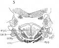
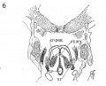
1911 Frontal section to show epiglottis thyreoid cartilage, and hyoid bone
1911 Frontal section to show superior laryngeal nerve
1911 Frontal section to show M. crico-thyreoideus and arytaenoid masses.
1911 Frontal section to show M. cricoary taenoideus posterior, interarytaenoideus and nerve recurrens.
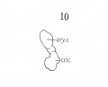
1911 Graphic reconstruction of cricoid and arytaenoid cartilages
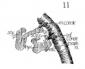
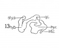
1911 Graphic reconstructions of pharyngeal constrictors
1911 Graphic reconstruction of laryngeal musculature
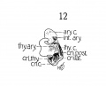
1911 Graphic reconstruction of thyreoid cartilage hyoid bone, and styloid process
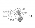
1911 Graphic reconstructions of larynx cartilages
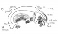
1911 Sagittal section to show laryngeal musculature and nerve recurrens
1911 Sagittal section laryngeal region
1911 Graphic reconstruction of 9th, 10th, and 12th cranial nerves in larynx region
1911 Sagittal section laryngeal region.
1911 Sagittal section laryngeal region, submaxillary ganglion.
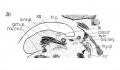
Fig. 20 Sagittal section laryngeal region.


- Carnegie Stages: 1 | 2 | 3 | 4 | 5 | 6 | 7 | 8 | 9 | 10 | 11 | 12 | 13 | 14 | 15 | 16 | 17 | 18 | 19 | 20 | 21 | 22 | 23 | About Stages | Timeline
Cite this page: Hill, M.A. (2026, July 29) Embryology Carnegie stage 19. Retrieved from https://embryology.med.unsw.edu.au/embryology/index.php/Carnegie_stage_19
- © Dr Mark Hill 2026, UNSW Embryology ISBN: 978 0 7334 2609 4 - UNSW CRICOS Provider Code No. 00098G