Mouse Stages: Difference between revisions
mNo edit summary |
|||
| (26 intermediate revisions by 2 users not shown) | |||
| Line 1: | Line 1: | ||
{{Header}} | |||
== Introduction == | == Introduction == | ||
[[File:Mouse.jpg|left]] | [[File:Mouse.jpg|left]] | ||
'''Theiler Stages''' divide mouse development into 26 prenatal and 2 postnatal stages and is based upon the publication '''The House Mouse: Atlas of Mouse Development''' by Theiler Springer-Verlag, NY (1972, 1989). There is also a newer 1993 Downs and Davies staging of the mouse embryo included in each Theiler stage (Staging of gastrulating mouse embryos by morphological landmarks in the dissecting microscope. | '''Theiler Stages''' divide mouse development into 26 prenatal and 2 postnatal stages and is based upon the publication '''The House Mouse: Atlas of Mouse Development''' by Theiler Springer-Verlag, NY (1972, 1989). There is also a newer 1993 Downs and Davies staging of the mouse embryo included in each Theiler stage (Staging of gastrulating mouse embryos by morphological landmarks in the dissecting microscope.{{#pmid:8269852|PMID8269852}} | ||
{{ | Note there have been early online atlases{{#pmid:9007211|PMID9007211}} and several new staging papers and online atlases released.{{#pmid:26487781|PMID26487781}}{{#pmid:28185240|PMID28185240}} | ||
{{Mouse links}} | |||
{{Mouse E days}} | |||
== Theiler Stage 1 == | == Theiler Stage 1 == | ||
[[File:Mouse_pronuclei_02.jpg|thumb|alt=Mouse zygote|Mouse zygote]] | |||
0-0.9 dpc (range 0-2.5 dpc) | * '''Zygote''' | ||
* One-cell stage embryo (fertilised egg) | |||
* 1 cell. Zona pellucida present. | |||
* 0-0.9 dpc (range 0-2.5 dpc) | |||
== Theiler Stage 2 == | == Theiler Stage 2 == | ||
[[File:Spermatozoa mitochondria 2cell.jpg|thumb|alt=Mouse 2 blastomeres|Mouse 2 blastomeres]] | |||
2-4 cells. Zona pellucida present. First cleavage occurs at about 24 hours. | * '''Blastomeres''' | ||
* Dividing egg stage | |||
Embryonic age = 1 dpc (range 1-2.5 dpc) | * 2-4 cells. | ||
* Zona pellucida present. First cleavage occurs at about 24 hours. | |||
* Embryonic age = 1 dpc (range 1-2.5 dpc) | |||
== Theiler Stage 3 == | == Theiler Stage 3 == | ||
Morula (early to fully compacted) | {| width= 100% | | ||
| row span=3| | |||
4-16 cells. Zona pellucida present. Usually found in the oviduct towards the utero-tubal junction. | * '''Morula''' (early to fully compacted) | ||
* 4-16 cells. | |||
Embryonic age = 2 dpc (range 1-3.5 dpc) | * Zona pellucida present. Usually found in the oviduct towards the utero-tubal junction. | ||
* Embryonic age = 2 dpc (range 1-3.5 dpc) | |||
| [[File:Spermatozoa_mitochondria_4cell.jpg|300px]] | |||
|- | |||
| | |||
| [[File:Spermatozoa_mitochondria_8cell.jpg|300px]] | |||
|- | |||
| | |||
| [[File:Spermatozoa_mitochondria_morula.jpg|300px]] | |||
|} | |||
== Theiler Stage 4 == | == Theiler Stage 4 == | ||
Blastocyst (ICM apparent) | [[File:Mouse-early_blastocyst_01.jpg|thumb|alt=Mouse Blastocyst|Mouse Blastocyst]] | ||
* '''Blastocyst''' (ICM apparent) | |||
* 16-40 compacted cells. Zona pellucida present. | |||
* Embryo progresses from morula to the blastocyst. Early evidence of the blastocoelic cavity. | |||
* In the blastocyst stage (zona-intact) there is a distinct inner cell mass and an outer layer of trophectoderm cells. Usually located in the uterine lumen. Embryonic age = 3 dpc (range 2-4 dpc) | |||
== Theiler Stage 5 == | == Theiler Stage 5 == | ||
Blastocyst (Zona pellucida absent) | [[File:Mouse-early blastocyst 02.jpg|thumb|alt=Mouse Hatched Blastocyst|Mouse Hatched Blastocyst]] | ||
* Blastocyst (Zona pellucida absent) | |||
* Zona free blastocyst. Invariably located within the uterine lumen. | |||
* Embryonic age = 4 dpc (range 3-5.5 dpc) | |||
[[File:Mouse-hatching blastocyst.jpg|300px]] | |||
== Theiler Stage 6 == | == Theiler Stage 6 == | ||
Attachment of blastocyst | * Attachment of blastocyst | ||
* Blastocyst implants, first evidence of embryonic endoderm cells covering the blastocoelic surface of the inner cell mass. | |||
Blastocyst implants, first evidence of embryonic endoderm cells covering the blastocoelic surface of the inner cell mass. | * Embryonic age = 4.5 dpc (range 4-5.5 dpc) | ||
* Equivalent Witschi Stage in rat = 8 | |||
Embryonic age = 4.5 dpc (range 4-5.5 dpc) | * Equivalent Carnegie Stage in human = 4 | ||
Equivalent Witschi Stage in rat = 8 | |||
Equivalent Carnegie Stage in human = 4 | |||
== Theiler Stage 7 == | == Theiler Stage 7 == | ||
Implantation and formation of egg cylinder | * Implantation and formation of egg cylinder | ||
* Ectoplacental cone appears. | |||
Ectoplacental cone appears. | * Rapid increase in the number of inner cell mass cells leading to the formation of the epiblast with subsequent growth to form the egg cylinder. | ||
* The proximal or visceral cells (opposite side from the trophoblastic cap) are cuboidal in shape. | |||
Rapid increase in the number of inner cell mass cells leading to the formation of the epiblast with subsequent growth to form the egg cylinder. | * Primary endoderm lines the mural trophectoderm. | ||
* Embryonic age = 5 dpc (range 4.5-6 dpc) | |||
The proximal or visceral cells (opposite side from the trophoblastic cap) are cuboidal in shape. | * Equivalent Witschi Stage in rat = 10 | ||
* Equivalent Carnegie Stage in human = 5 | |||
Primary endoderm lines the mural trophectoderm. | |||
Embryonic age = 5 dpc (range 4.5-6 dpc) | |||
Equivalent Witschi Stage in rat = 10 | |||
Equivalent Carnegie Stage in human = 5 | |||
== Theiler Stage 8 == | == Theiler Stage 8 == | ||
Differentiation of egg cylinder | * Differentiation of egg cylinder | ||
* Implantation site 2x3mm. | |||
Implantation site 2x3mm. | * The maternal tissue is invaded by trophoblast (primary) giant cells and the ectoplacental cone is invaded by maternal blood. | ||
* Differentiation of the egg cylinder into embryonic and extra-embryonic regions and the formation of the pro-amniotic cavity. | |||
The maternal tissue is invaded by trophoblast (primary) giant cells and the ectoplacental cone is invaded by maternal blood. | * Reichert's membrane, which is non-cellular and secreted by the distal endoderm, first appears. | ||
* Embryonic age = 6 dpc (range 5-6.5 dpc) | |||
Differentiation of the egg cylinder into embryonic and extra-embryonic regions and the formation of the pro-amniotic cavity. | * Equivalent Witschi Stage in rat = 10-11 | ||
* Equivalent Carnegie Stage in human = 5 | |||
Reichert's membrane, which is non-cellular and secreted by the distal endoderm, first appears. | |||
Embryonic age = 6 dpc (range 5-6.5 dpc) | |||
Equivalent Witschi Stage in rat = 10-11 | |||
Equivalent Carnegie Stage in human = 5 | |||
== Theiler Stage 9 == | == Theiler Stage 9 == | ||
===Stage 9a=== | |||
* Advanced Endometrial Reaction | |||
Advanced Endometrial Reaction | * Advanced egg-cylinder stage with the first evidence of an embryonic axis. | ||
* Clear morphological distinction between the embryonic and extra-embryonic ectoderm. | |||
Advanced egg-cylinder stage with the first evidence of an embryonic axis. | * The ectoplacental cone is further invaded by maternal blood and the original lumen of the uterine crypt has disappeared. | ||
* Equivalent Downs and Davies Stage : PS (pre-streak) | |||
Clear morphological distinction between the embryonic and extra-embryonic ectoderm. | ===Stage 9b=== | ||
* Advanced Endometrial Reaction | |||
The ectoplacental cone is further invaded by maternal blood and the original lumen of the uterine crypt has disappeared. | * Late in this stage gastrulation begins, producing the first mesodermal cells. | ||
* Equivalent Downs and Davies Stage : ES (early streak) | |||
Equivalent Downs and Davies Stage : PS (pre-streak) | |||
Advanced Endometrial Reaction | |||
Late in this stage gastrulation begins, producing the first mesodermal cells. | |||
Equivalent Downs and Davies Stage : ES (early streak) | |||
== Theiler Stage 10 == | == Theiler Stage 10 == | ||
===Stage 10a=== | |||
* Amnion | |||
Amnion | * Tissue at the posterior end of the primitive streak bulges into the pro-amniotic cavity and forms the amniotic fold | ||
* Equivalent Downs and Davies stage: MS (mid-streak) | |||
Tissue at the posterior end of the primitive streak bulges into the pro-amniotic cavity and forms the amniotic fold | ===Stage 10b=== | ||
* Amnion | |||
Equivalent Downs and Davies stage: MS (mid-streak) | * In the mesoderm of the posterior amniotic fold small cavities coalesce to form a single cavity, the exocoelom | ||
* Embryonic age = 7.0 dpc (range 6.5-7.5 dpc) | |||
* Equivalent Downs and Davies stages: MS - LS (mid-streak to late streak) | |||
* Equivalent Witschi Stage in rat = 12 | |||
Amnion | * Equivalent Carnegie Stage in humans = 8 | ||
===Stage 10c=== | |||
In the mesoderm of the posterior amniotic fold small cavities coalesce to form a single cavity, the exocoelom | * Amnion | ||
* The allantoic bud first appears, gastrulation continues and the node becomes visible. | |||
Embryonic age = 7.0 dpc (range 6.5-7.5 dpc) | * Embryonic age = 7.0 dpc (range 6.5-7.5 dpc) | ||
* Equivalent Downs and Davies stages: MS - LS (mid-streak to late streak) | |||
Equivalent Downs and Davies stages: MS - LS (mid-streak to late streak) | * Equivalent Witschi Stage in rat = 12 | ||
* Equivalent Carnegie Stage in humans = 8 | |||
Equivalent Witschi Stage in rat = 12 | |||
Equivalent Carnegie Stage in humans = 8 | |||
Amnion | |||
The allantoic bud first appears, gastrulation continues and the node becomes visible. | |||
Embryonic age = 7.0 dpc (range 6.5-7.5 dpc) | |||
Equivalent Downs and Davies stages: MS - LS (mid-streak to late streak) | |||
Equivalent Witschi Stage in rat = 12 | |||
Equivalent Carnegie Stage in humans = 8 | |||
== Theiler Stage 11 == | == Theiler Stage 11 == | ||
Neural Plate, Presomite stage | ===Stage 11a=== | ||
* Neural Plate, Presomite stage | |||
* The amniotic cavity is now sealed off into three distinct cavities - the amniotic cavity, the exocoelom and the ectoplacental cleft. The neural plate is defined anteriorly and the head process is developing. In the midline, subjacent to the neural groove, the notochodal plate is visible. | |||
* Embryonic age = 7.5 dpc (range 7.25-8 dpc) | |||
* Equivalent Downs and Davies stages: OB-EB (no allantoic bud to early allantoic bud); LB-EHF-LHF (late allantoic bud to early head fold to late head fold) | |||
* Equivalent Witschi Stage in rat = 12-13 | |||
* Equivalent Carnegie Stage in humans = 9 | |||
Head folds continue to enlarge and the foregut pocket begins to form. | ===Stage 11b=== | ||
* Neural Plate, Presomite stage | |||
Embryonic age = 7.5 dpc (range 7.25-8 dpc) ; LB-EHF-LHF (late allantoic bud to early head fold to late head fold) . | * The allantoic bud elongates. | ||
* Embryonic age = 7.5 dpc (range 7.25-8 dpc) . | |||
Equivalent Downs & Davies stages: OB-EB (no allantoic bud to early allantoic bud) | * Equivalent Downs & Davies stages: OB-EB (no allantoic bud to early allantoic bud); LB-EHF-LHF (late allantoic bud to early head fold to late head fold) | ||
* Equivalent Witschi Stage in rat = 12-13 | |||
Equivalent Witschi Stage in rat = 12-13 | * Equivalent Carnegie Stage in humans = 9 | ||
===Stage 11c=== | |||
Equivalent Carnegie Stage in humans = 9 | * Neural Plate, Presomite stage | ||
* The rostral part of the neural plate begins to enlarge to form the head folds. The neural groove is visible. | |||
* Embryonic age = 7.5 dpc (range 7.25-8 dpc) | |||
* Equivalent Downs and Davies stages: OB-EB (no allantoic bud to early allantoic bud); LB-EHF-LHF (late allantoic bud to early head fold to late head fold) | |||
* Equivalent Witschi Stage in rat = 12-13. | |||
* Equivalent Carnegie Stage in humans = 9 | |||
===Stage11d=== | |||
* Neural Plate, Presomite stage | |||
* Head folds continue to enlarge and the foregut pocket begins to form. | |||
* Embryonic age = 7.5 dpc (range 7.25-8 dpc) ; LB-EHF-LHF (late allantoic bud to early head fold to late head fold) . | |||
* Equivalent Downs & Davies stages: OB-EB (no allantoic bud to early allantoic bud) | |||
* Equivalent Witschi Stage in rat = 12-13 | |||
* Equivalent Carnegie Stage in humans = 9 | |||
== Theiler Stage 12 == | == Theiler Stage 12 == | ||
===Theiler Stage 12a=== | |||
* First Somites Unturned embryo with first appearance of somite pairs 1-4 somites. | |||
* The allantois extends further into the exocoelom and the maxillary components of the 1st branchial arch become prominent. | |||
* The preotic sulcus is visible in the 2-3 somite embryo. The cardiogenic plate begins to form and the foregut pocket is clearly visible. | |||
* Embryonic age = 8 dpc (range 7.5-8.75 dpc) | |||
* 1-7 somite pairs | |||
* Equivalent Witschi Stage in rat = 14-15 | |||
* Equivalent Carnegie Stage in humans = 9 | |||
===Theiler Stage 12b=== | |||
* First Somites Unturned embryo with first appearance of somite pairs 5-7 somites. | |||
* The headfolds are particularly prominent and neural closure occurs in the region of the 4th and 5th somites, extending in both directions from this site. | |||
* The optic placodes are first evident and become indented to form the optic pits. The heart rudiment develops rapidly. The allantois contacts the chorion at the end of this stage. Absent: The 2nd branchial arch and >7 somites. | |||
* Embryonic age = 8 dpc (range 7.5-8.75 dpc) | |||
* 1-7 somite pairs | |||
* Equivalent Witschi Stage in rat = 14-15 | |||
* Equivalent Carnegie Stage in humans = 9 | |||
== Theiler Stage 13 == | == Theiler Stage 13 == | ||
Turning of the embryo | Turning of the embryo | ||
* This is a short period with turning initiated in embryos with 6-8 pairs of somites and usually completed in embryos with 14-16 pairs of somites. | |||
This is a short period with turning initiated in embryos with 6-8 pairs of somites and usually completed in embryos with 14-16 pairs of somites. | * The first branchial arch has maxillary and mandibular components but the maxillary process is not visible until later (TS16). | ||
* A second branchial arch is now evident. | |||
The first branchial arch has maxillary and mandibular components but the maxillary process is not visible until later (TS16). | * There is evidence of regionalisation of the heart and the neural tube is closed from a point opposite the outflow tract to the proximal part of the tail. | ||
* Notochord and dorsal prepancreatic endoderms remain in contact until about the 13-somite stage. | |||
A second branchial arch is now evident. | * Absent: 3rd branchial arch. | ||
* Embryonic age = 8.5 dpc (range 8-9.25 dpc) | |||
There is evidence of regionalisation of the heart and the neural tube is closed from a point opposite the outflow tract to the proximal part of the tail. | * 8-12 somite pairs | ||
* Equivalent Witschi Stage in rat = 15 | |||
Notochord and dorsal prepancreatic endoderms remain in contact until about the 13-somite stage. | * Equivalent Carnegie Stage in humans = 10 | ||
Absent: 3rd branchial arch. | |||
Embryonic age = 8.5 dpc (range 8-9.25 dpc) | |||
8-12 somite pairs | |||
Equivalent Witschi Stage in rat = 15 | |||
Equivalent Carnegie Stage in humans = 10 | |||
== Theiler Stage 14 == | == Theiler Stage 14 == | ||
Formation and closure of anterior neuropore. | Formation and closure of anterior neuropore. | ||
* The rostral extremity of the neural tube closes in embryos with usually about 15-18 somite pairs and defines this stage. | |||
The rostral extremity of the neural tube closes in embryos with usually about 15-18 somite pairs and defines this stage. | * The otic pit becomes progressively more indented but not closed, the mandibular process of the 1st branchial arch is clearly visible. | ||
* The 3rd branchial arch becomes visible late in the stage. | |||
The otic pit becomes progressively more indented but not closed, the mandibular process of the 1st branchial arch is clearly visible. | * An increasingly prominent ridge on the lateral body wall, approximately at the level of the 8th-12th somite, indicates the site of the future forelimb bud. | ||
* Absent: forelimb bud. | |||
The 3rd branchial arch becomes visible late in the stage. | * Embryonic age = 9 dpc (range 8.5-9.75 dpc) | ||
* 13-20 somite pairs | |||
An increasingly prominent ridge on the lateral body wall, approximately at the level of the 8th-12th somite, indicates the site of the future forelimb bud. | * Equivalent Witschi Stage in rat = 16 | ||
* Equivalent Carnegie Stage in humans = 11 | |||
Absent: forelimb bud. | |||
Embryonic age = 9 dpc (range 8.5-9.75 dpc) | |||
13-20 somite pairs | |||
Equivalent Witschi Stage in rat = 16 | |||
Equivalent Carnegie Stage in humans = 11 | |||
== Theiler Stage 15 == | == Theiler Stage 15 == | ||
[[File:Mouse-E9.5.jpg|thumb|Mouse E9.5]] | [[File:Mouse-E9.5.jpg|thumb|Mouse E9.5]] | ||
Formation of posterior neuropore, forelimb bud | Formation of posterior neuropore, forelimb bud | ||
* The posterior neuropore forms and the condensation of the forelimb bud becomes apparent near the 8th-12th somite pairs. | |||
The posterior neuropore forms and the condensation of the forelimb bud becomes apparent near the 8th-12th somite pairs. | * A distinct condensation of the hind limb bud appears just at the end of the stage. | ||
* The forebrain vesicle subdivides into telencephalic and diencephalic vesicles. | |||
A distinct condensation of the hind limb bud appears just at the end of the stage. | * Lung development begins. | ||
* Pancreas first indication of morphogenesis, dorsal pancreatic bud (22-25 somites). | |||
The forebrain vesicle subdivides into telencephalic and diencephalic vesicles. | * Absent: hindlimb bud, Rathke's pouch. | ||
* Embryonic age = 9.5 dpc (range 9-10.25 dpc) | |||
Lung development begins. | * 21-29 somite pairs | ||
* Equivalent Witschi Stage in rat = 17-19 | |||
Pancreas first indication of morphogenesis, dorsal pancreatic bud (22-25 somites). | * Equivalent Carnegie Stage in humans = 12 | ||
Absent: hindlimb bud, Rathke's pouch. | |||
Embryonic age = 9.5 dpc (range 9-10.25 dpc) | |||
21-29 somite pairs | |||
Equivalent Witschi Stage in rat = 17-19 | |||
Equivalent Carnegie Stage in humans = 12 | |||
== Theiler Stage 16 == | == Theiler Stage 16 == | ||
Closure of posterior neuropore. Hind limb bud and tail bud | Closure of posterior neuropore. Hind limb bud and tail bud | ||
* The hind limb bud becomes visible at the level of the 23rd-28th somites. | |||
The hind limb bud becomes visible at the level of the 23rd-28th somites. | * The tail bud appears as a short stump and the 3rd and 4th branchial arches are distinctly concave. | ||
* Rathke's pouch and the nasal processes start to form. At the end of this stage the posterior neuropore begins to close. | |||
The tail bud appears as a short stump and the 3rd and 4th branchial arches are distinctly concave. | * Pancreas ventral pancreatic bud appears (30 somites, 10.25-10.5). | ||
* Absent: thin and long tail. | |||
Rathke's pouch and the nasal processes start to form. At the end of this stage the posterior neuropore begins to close. | * Embryonic age = 10 dpc (range 9.5-10.75 dpc) | ||
* 30-34 somite pairs | |||
Pancreas ventral pancreatic bud appears (30 somites, 10.25-10.5). | * Equivalent Witschi Stage in rat = 20-21 | ||
* Equivalent Carnegie Stage in humans = 13-15 | |||
Absent: thin and long tail. | |||
Embryonic age = 10 dpc (range 9.5-10.75 dpc) | |||
30-34 somite pairs | |||
Equivalent Witschi Stage in rat = 20-21 | |||
Equivalent Carnegie Stage in humans = 13-15 | |||
== Theiler Stage 17 == | == Theiler Stage 17 == | ||
[[File:Mouse CT E10.5.jpg|thumb|Mouse E10.5]] | [[File:Mouse CT E10.5.jpg|thumb|Mouse E10.5]] | ||
Deep Lens Indentation | * Deep Lens Indentation | ||
* The most obvious distinguishing features are the deepening of the lens pit, with a narrowing of its outer pore-like opening, and the first appearance of the physiological umbilical hernia. | |||
The most obvious distinguishing features are the deepening of the lens pit, with a narrowing of its outer pore-like opening, and the first appearance of the physiological umbilical hernia. | * The 1st branchial arch is conspicuously divided into maxillary and mandibular components. | ||
* There is advanced development of the brain tube and the tail elongates and thins. | |||
The 1st branchial arch is conspicuously divided into maxillary and mandibular components. | * Absent: nasal pits. | ||
* Embryonic age = 10.5 dpc (range 10-11.25 dpc) | |||
There is advanced development of the brain tube and the tail elongates and thins. | * 35-39 somite pairs | ||
* Equivalent Witschi Stage in rat = 24-25 | |||
Absent: nasal pits. | * Equivalent Carnegie Stage in humans = 13-15 | ||
Embryonic age = 10.5 dpc (range 10-11.25 dpc) | |||
35-39 somite pairs | |||
Equivalent Witschi Stage in rat = 24-25 | |||
Equivalent Carnegie Stage in humans = 13-15 | |||
== Theiler Stage 18 == | == Theiler Stage 18 == | ||
Closure of Lens Vesicle | [[File:Mouse_embryo_E11_and_tomography_01.jpg|thumb|300px|alt=Theiler Stage 18|Theiler Stage 18]] | ||
Closure of Lens Vesicle | |||
The primary externally recognisable feature is the progressive closure of the lens vesicle. | * The primary externally recognisable feature is the progressive closure of the lens vesicle. | ||
* The somites in the cervical region are no longer visible and the rapid growth of the brain is striking. | |||
The somites in the cervical region are no longer visible and the rapid growth of the brain is striking. | * The nasal pits start to form. Absent: auditory hillocks, anterior footplate. | ||
* Embryonic age = 11 dpc (range 10.5-11.25 dpc) | |||
The nasal pits start to form. Absent: auditory hillocks, anterior footplate. | * 40-44 somite pairs | ||
* Equivalent Witschi Stage in rat = 25-26 | |||
Embryonic age = 11 dpc (range 10.5-11.25 dpc) | * Equivalent Carnegie Stage in humans = 13-15 | ||
40-44 somite pairs | |||
Equivalent Witschi Stage in rat = 25-26 | |||
Equivalent Carnegie Stage in humans = 13-15 | |||
== Theiler Stage 19 == | == Theiler Stage 19 == | ||
[[File:Mouse CT E11.5.jpg|thumb|Mouse E11.5]] | [[File:Mouse CT E11.5.jpg|thumb|Mouse E11.5]] | ||
[[File:Mouse CT axes E11.5.jpg|thumb|Mouse E11.5]] | [[File:Mouse CT axes E11.5.jpg|thumb|Mouse E11.5]] | ||
Lens vesicle completely separated from surface | Lens vesicle completely separated from surface | ||
* The lens vesicle becomes completely closed and detached from the ectoderm. | |||
The lens vesicle becomes completely closed and detached from the ectoderm. | * The peripheral margins of the eye become well defined. | ||
* The forelimbs are seen to be divided into two regions, the proximal part consisting of the future limb-girdle and 'arm' and the more peripheral part which forms a circular or paddle-shaped 'handplate' (anterior footplate). The medial and lateral margins of the otic pit are coming together reducing the entrance to a narrow slit and the auditory hillocks become visible. Absent: retinal pigmentation, signs of 'fingers'. | |||
The peripheral margins of the eye become well defined. | * Embryonic age = 11.5 dpc (range 11-12.25 dpc) | ||
* 45-47 somite pairs | |||
The forelimbs are seen to be divided into two regions, the proximal part consisting of the future limb-girdle and 'arm' and the more peripheral part which forms a circular or paddle-shaped 'handplate' (anterior footplate). The medial and lateral margins of the otic pit are coming together reducing the entrance to a narrow slit and the auditory hillocks become visible. Absent: retinal pigmentation, signs of 'fingers'. | * Equivalent Witschi Stage in rat = 26-27 | ||
* Equivalent Carnegie Stage in humans = 16 | |||
Embryonic age = 11.5 dpc (range 11-12.25 dpc) | |||
45-47 somite pairs | |||
Equivalent Witschi Stage in rat = 26-27 | |||
Equivalent Carnegie Stage in humans = 16 | |||
== Theiler Stage 20 == | == Theiler Stage 20 == | ||
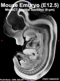
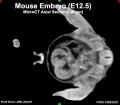
[[File:Mouse CT E12.5 scalebar.jpg|thumb|Mouse CT E12.5]] | [[File:Mouse CT E12.5 scalebar.jpg|thumb|Mouse CT E12.5]] | ||
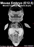
[[File:Mouse CT E12.5.jpg|thumb|Mouse E12.5]] | [[File:Mouse CT E12.5.jpg|thumb|Mouse E12.5]] | ||
Earliest signs of fingers | Earliest signs of fingers | ||
* The 'handplate' (anterior footplate) is no longer circular but develops angles which correspond to the future digits. | |||
The 'handplate' (anterior footplate) is no longer circular but develops angles which correspond to the future digits. | * The posterior footplate is also distinguishable from the lower part of the leg. | ||
* It is possible to see the pigmentation of the pigmented layer of the retina through the transparent cornea. | |||
The posterior footplate is also distinguishable from the lower part of the leg. | * The tongue and brain vesicles are clearly visible. | ||
* Absent:5 rows of whiskers, indented handplate. | |||
It is possible to see the pigmentation of the pigmented layer of the retina through the transparent cornea. | * Embryonic age = 12 dpc (range 11.5-13 dpc) | ||
* 48-51 somite pairs | |||
The tongue and brain vesicles are clearly visible. | * Equivalent Witschi Stage in rat = 28 | ||
* Equivalent Carnegie Stage in humans = 17 | |||
Absent:5 rows of whiskers, indented handplate. | |||
Embryonic age = 12 dpc (range 11.5-13 dpc) | |||
48-51 somite pairs | |||
Equivalent Witschi Stage in rat = 28 | |||
Equivalent Carnegie Stage in humans = 17 | |||
== Theiler Stage 21 == | == Theiler Stage 21 == | ||
Anterior footplate indented, marked pinna | Anterior footplate indented, marked pinna | ||
* The distal borders of the anterior and posterior footplates are now indented and the digit widths and locations can be discerned. | |||
The distal borders of the anterior and posterior footplates are now indented and the digit widths and locations can be discerned. | * The 'elbow' and 'wrist' are now identifiable. | ||
* The pinna rapidly develops and forms a crest at right angles to the head. | |||
The 'elbow' and 'wrist' are now identifiable. | * Five rows of vibrissae are visible as well as a prominant hair follicle over the eye and another over the ear. | ||
* The lens vesicle has lost its lumen. | |||
The pinna rapidly develops and forms a crest at right angles to the head. | * The physiological umbilical hernia is prominent. Absent: hair follicles, distally separate fingers. | ||
* Embryonic age = 13 dpc (range 12.5-14) | |||
Five rows of vibrissae are visible as well as a prominant hair follicle over the eye and another over the ear. | * 52-55 somite pairs | ||
* Equivalent Witschi Stage in rat = 29-30 | |||
The lens vesicle has lost its lumen. | * Equivalent Carnegie Stage in humans = 18-19 | ||
The physiological umbilical hernia is prominent. Absent: hair follicles, distally separate fingers. | |||
Embryonic age = 13 dpc (range 12.5-14) | |||
:'''Links:''' [[Quicktime_Movie_-_Mouse_E13_microCT|MicroCT embryo]] | [[:Category:Mouse E13|Mouse E13]] | |||
== Theiler Stage 22 == | == Theiler Stage 22 == | ||
[[File:Mouse CT E14.5.jpg|thumb|Mouse CT E14.5]] | [[File:Mouse CT E14.5.jpg|thumb|Mouse CT E14.5]] | ||
Fingers separate distally | Fingers separate distally | ||
* Individual 'fingers' are visible in the anterior footplate and there are deep indentations between the 'toes' which are not yet separated. | |||
* The long bones of the limbs are present and there are hair follicles in the pectoral, pelvic and trunk regions. | |||
* The pinna is turned forwards and the umbilical hernia is conspicuous. | |||
* Absent: hair follicles in the cephalic region. | |||
* Embryonic age = 14 dpc (range 13.5-15 dpc) | |||
* 56-60 somite pairs | |||
* Equivalent Witschi Stage in rat = 31 | |||
* Equivalent Carnegie Stage in humans = 20-23 | |||
:'''Links:''' [[Quicktime_Movie_-_Mouse_E14_sectioned_microCT|MicroCT sectioned embryo]] | [[Quicktime_Movie_-_Mouse_E14_microCT|MicroCT embryo]] | |||
== Theiler Stage 23 == | == Theiler Stage 23 == | ||
Toes separate | [[File:Mouse_embryo_E15_microCT_icon.jpg|thumb|Mouse E15 CT]] | ||
Toes separate | |||
The 'toes' separate and are clearly divergent, not becoming parallel until later. | * The 'toes' separate and are clearly divergent, not becoming parallel until later. | ||
* Hair follicles are present in the cephalic region but not at the periphery of the vibrissae. | |||
Hair follicles are present in the cephalic region but not at the periphery of the vibrissae. | * The pinna covers more than half of the external auditory meatus and the eyelids are still open. | ||
* Absent: nail primordia, 'fingers' 2-5 parallel. | |||
The pinna covers more than half of the external auditory meatus and the eyelids are still open. | * Embryonic age = 15 dpc | ||
* >60 somite pairs | |||
Absent: nail primordia, 'fingers' 2-5 parallel. | * Equivalent Witschi Stage in rat = 32 | ||
* Human fetal period. | |||
Embryonic age = 15 dpc | |||
>60 somite pairs | |||
Equivalent Witschi Stage in rat = 32 | |||
Human | |||
== Theiler Stage 24 == | == Theiler Stage 24 == | ||
[[File:Mouse CT E16.5.jpg|thumb|Mouse CT E16.5]] | [[File:Mouse CT E16.5.jpg|thumb|Mouse CT E16.5]] | ||
Reposition of umbilical hernia | Reposition of umbilical hernia | ||
* 'Fingers' 2-5 are nearly parallel. Nail primordia are visible on the 'toes'. | |||
'Fingers' 2-5 are nearly parallel. Nail primordia are visible on the 'toes'. | * The eyelids have fused in most cases by the end of the stage and the pinna almost completely covers the external auditory meatus. | ||
* The umbilical hernia is disappearing and there is a corresponding increase in the size of the peritoneal sac. | |||
The eyelids have fused in most cases by the end of the stage and the pinna almost completely covers the external auditory meatus. | * Absent: 'fingers' and 'toes' joined together. | ||
* Embryonic age = 16 dpc | |||
The umbilical hernia is disappearing and there is a corresponding increase in the size of the peritoneal sac. | * > 60 Somite pairs | ||
* Equivalent Witschi Stage in rat = 33 | |||
Absent: 'fingers' and 'toes' joined together. | |||
Embryonic age = 16 dpc | |||
> 60 Somite pairs | |||
Equivalent Witschi Stage in rat = 33 | |||
== Theiler Stage 25 == | == Theiler Stage 25 == | ||
'''Skin wrinkled''' The skin has thickened and formed wrinkles and the subcutaneous veins are less visible. | '''Skin wrinkled''' The skin has thickened and formed wrinkles and the subcutaneous veins are less visible. | ||
* * The 'fingers' and 'toes' have become parallel and the umbilical hernia has disappeared. | |||
The 'fingers' and 'toes' have become parallel and the umbilical hernia has disappeared. | * The eyelids have fused. Whiskers are just visible. | ||
* Absent: ear extending over auditory meatus, long whiskers. | |||
The eyelids have fused. Whiskers are just visible. | * Embryonic age = 17 dpc | ||
* Equivalent Witschi Stage in rat = 34 | |||
Absent: ear extending over auditory meatus, long whiskers. | * == Theiler Stage 26 == | ||
Embryonic age = 17 dpc | |||
Equivalent Witschi Stage in rat = 34 | |||
== Theiler Stage 26 == | |||
[[File:Mouse CT E18.5.jpg|thumb|Mouse CT E18.5]] | [[File:Mouse CT E18.5.jpg|thumb|Mouse CT E18.5]] | ||
'''Long whiskers''' The whiskers that were present at stage 25 are definitely longer and the skin has thickened. | '''Long whiskers''' The whiskers that were present at stage 25 are definitely longer and the skin has thickened. | ||
* The pinna is larger and such that virtually none of the lumen of the auditory meatus is visible. The eyes are barely visible through the closed eyelids. | |||
The pinna is larger and such that virtually none of the lumen of the auditory meatus is visible. The eyes are barely visible through the closed eyelids. | * Embryonic age = 18 dpc | ||
* Equivalent Witschi Stage in rat = 35 | |||
Embryonic age = 18 dpc | |||
Equivalent Witschi Stage in rat = 35 | |||
== Theiler Stage 27 == | == Theiler Stage 27 == | ||
* New born Mouse | |||
New born Mouse | |||
== Theiler Stage 28 == | == Theiler Stage 28 == | ||
Post-natal development | * Post-natal development | ||
== Data == | == Data == | ||
| Line 529: | Line 354: | ||
The table below gives an approximate comparison of human, mouse and rat embryos based upon Carnegie staging. | The table below gives an approximate comparison of human, mouse and rat embryos based upon Carnegie staging. | ||
{ | {{CarnegieComparisonHRM}} | ||
==Somitogenesis== | |||
{{Mouse Somitogenesis table}} | |||
:'''Links:''' [[Somitogenesis]] | |||
==Timed Pregnancy== | ==Timed Pregnancy== | ||
* Presence of the vaginal plug indicates that the mating occurred. | * Presence of the vaginal plug indicates that the mating occurred. | ||
| Line 620: | Line 372: | ||
<references/> | <references/> | ||
{{ | |||
{{Animals}} | |||
{{Glossary}} | |||
{{Footer}} | |||
[[Category:Mouse]] | [[Category:Mouse]] | ||
Latest revision as of 08:20, 24 April 2018
| Embryology - 16 Jun 2024 |
|---|
| Google Translate - select your language from the list shown below (this will open a new external page) |
|
العربية | català | 中文 | 中國傳統的 | français | Deutsche | עִברִית | हिंदी | bahasa Indonesia | italiano | 日本語 | 한국어 | မြန်မာ | Pilipino | Polskie | português | ਪੰਜਾਬੀ ਦੇ | Română | русский | Español | Swahili | Svensk | ไทย | Türkçe | اردو | ייִדיש | Tiếng Việt These external translations are automated and may not be accurate. (More? About Translations) |
Introduction

Theiler Stages divide mouse development into 26 prenatal and 2 postnatal stages and is based upon the publication The House Mouse: Atlas of Mouse Development by Theiler Springer-Verlag, NY (1972, 1989). There is also a newer 1993 Downs and Davies staging of the mouse embryo included in each Theiler stage (Staging of gastrulating mouse embryos by morphological landmarks in the dissecting microscope.[1]
Note there have been early online atlases[2] and several new staging papers and online atlases released.[3][4]
| Mouse Links: Introduction | Mouse Stages | Mouse Timeline | Mouse Timeline Detailed | Mouse Estrous Cycle | Mouse Heart | Mouse Knockout | Movie - Cephalic Plexus | Movie - Blastocyst Cdx2 | ANAT2341 Project 2009 | Category:Mouse | |||||||||||||||||||||||||||||||||||||||||||||||||||||||||||||||||||||||||||||||||||||||||||||||||||||||||||||||||||||||||||
|
| ||||||||||||||||||||||||||||||||||||||||||||||||||||||||||||||||||||||||||||||||||||||||||||||||||||||||||||||||||||||||||
- Mouse Stages: E1 | E2.5 | E3.0 | E3.5 | E4.5 | E5.0 | E5.5 | E6.0 | E7.0 | E7.5 | E8.0 | E8.5 | E9.0 | E9.5 | E10 | E10.5 | E11 | E11.5 | E12 | E12.5 | E13 | E13.5 | E14 | E14.5 | E15 | E15.5 | E16 | E16.5 | E17 | E17.5 | E18 | E18.5 | E19 | E20 | Timeline | About timed pregnancy
| Carnegie | Stage | |||||||||||||||||||||||
| Human | Days | 1 | 2-3 | 4-5 | 5-6 | 7-12 | 13-15 | 15-17 | 17-19 | 20 | 22 | 24 | 28 | 30 | 33 | 36 | 40 | 42 | 44 | 48 | 52 | 54 | 55 | 58 |
| Mouse | Days | 1 | 2 | 3 | E4.5 | E5.0 | E6.0 | E7.0 | E8.0 | E9.0 | E9.5 | E10 | E10.5 | E11 | E11.5 | E12 | E12.5 | E13 | E13.5 | E14 | E14.5 | E15 | E15.5 | E16 |
| Rat | Days | 1 | 3.5 | 4-5 | 5 | 6 | 7.5 | 8.5 | 9 | 10.5 | 11 | 11.5 | 12 | 12.5 | 13 | 13.5 | 14 | 14.5 | 15 | 15.5 | 16 | 16.5 | 17 | 17.5 |
| Note these Carnegie stages are only approximate day timings for average of embryos. Links: Carnegie Stage Comparison | ||||||||||||||||||||||||
| ||||||||||||||||||||||||
| Timeline Links: human timeline | mouse timeline | mouse detailed timeline | chicken timeline | rat timeline | Medaka | Category:Timeline |
Theiler Stage 1

- Zygote
- One-cell stage embryo (fertilised egg)
- 1 cell. Zona pellucida present.
- 0-0.9 dpc (range 0-2.5 dpc)
Theiler Stage 2

- Blastomeres
- Dividing egg stage
- 2-4 cells.
- Zona pellucida present. First cleavage occurs at about 24 hours.
- Embryonic age = 1 dpc (range 1-2.5 dpc)
Theiler Stage 3
|

|

| |

|
Theiler Stage 4

- Blastocyst (ICM apparent)
- 16-40 compacted cells. Zona pellucida present.
- Embryo progresses from morula to the blastocyst. Early evidence of the blastocoelic cavity.
- In the blastocyst stage (zona-intact) there is a distinct inner cell mass and an outer layer of trophectoderm cells. Usually located in the uterine lumen. Embryonic age = 3 dpc (range 2-4 dpc)
Theiler Stage 5

- Blastocyst (Zona pellucida absent)
- Zona free blastocyst. Invariably located within the uterine lumen.
- Embryonic age = 4 dpc (range 3-5.5 dpc)

Theiler Stage 6
- Attachment of blastocyst
- Blastocyst implants, first evidence of embryonic endoderm cells covering the blastocoelic surface of the inner cell mass.
- Embryonic age = 4.5 dpc (range 4-5.5 dpc)
- Equivalent Witschi Stage in rat = 8
- Equivalent Carnegie Stage in human = 4
Theiler Stage 7
- Implantation and formation of egg cylinder
- Ectoplacental cone appears.
- Rapid increase in the number of inner cell mass cells leading to the formation of the epiblast with subsequent growth to form the egg cylinder.
- The proximal or visceral cells (opposite side from the trophoblastic cap) are cuboidal in shape.
- Primary endoderm lines the mural trophectoderm.
- Embryonic age = 5 dpc (range 4.5-6 dpc)
- Equivalent Witschi Stage in rat = 10
- Equivalent Carnegie Stage in human = 5
Theiler Stage 8
- Differentiation of egg cylinder
- Implantation site 2x3mm.
- The maternal tissue is invaded by trophoblast (primary) giant cells and the ectoplacental cone is invaded by maternal blood.
- Differentiation of the egg cylinder into embryonic and extra-embryonic regions and the formation of the pro-amniotic cavity.
- Reichert's membrane, which is non-cellular and secreted by the distal endoderm, first appears.
- Embryonic age = 6 dpc (range 5-6.5 dpc)
- Equivalent Witschi Stage in rat = 10-11
- Equivalent Carnegie Stage in human = 5
Theiler Stage 9
Stage 9a
- Advanced Endometrial Reaction
- Advanced egg-cylinder stage with the first evidence of an embryonic axis.
- Clear morphological distinction between the embryonic and extra-embryonic ectoderm.
- The ectoplacental cone is further invaded by maternal blood and the original lumen of the uterine crypt has disappeared.
- Equivalent Downs and Davies Stage : PS (pre-streak)
Stage 9b
- Advanced Endometrial Reaction
- Late in this stage gastrulation begins, producing the first mesodermal cells.
- Equivalent Downs and Davies Stage : ES (early streak)
Theiler Stage 10
Stage 10a
- Amnion
- Tissue at the posterior end of the primitive streak bulges into the pro-amniotic cavity and forms the amniotic fold
- Equivalent Downs and Davies stage: MS (mid-streak)
Stage 10b
- Amnion
- In the mesoderm of the posterior amniotic fold small cavities coalesce to form a single cavity, the exocoelom
- Embryonic age = 7.0 dpc (range 6.5-7.5 dpc)
- Equivalent Downs and Davies stages: MS - LS (mid-streak to late streak)
- Equivalent Witschi Stage in rat = 12
- Equivalent Carnegie Stage in humans = 8
Stage 10c
- Amnion
- The allantoic bud first appears, gastrulation continues and the node becomes visible.
- Embryonic age = 7.0 dpc (range 6.5-7.5 dpc)
- Equivalent Downs and Davies stages: MS - LS (mid-streak to late streak)
- Equivalent Witschi Stage in rat = 12
- Equivalent Carnegie Stage in humans = 8
Theiler Stage 11
Stage 11a
- Neural Plate, Presomite stage
- The amniotic cavity is now sealed off into three distinct cavities - the amniotic cavity, the exocoelom and the ectoplacental cleft. The neural plate is defined anteriorly and the head process is developing. In the midline, subjacent to the neural groove, the notochodal plate is visible.
- Embryonic age = 7.5 dpc (range 7.25-8 dpc)
- Equivalent Downs and Davies stages: OB-EB (no allantoic bud to early allantoic bud); LB-EHF-LHF (late allantoic bud to early head fold to late head fold)
- Equivalent Witschi Stage in rat = 12-13
- Equivalent Carnegie Stage in humans = 9
Stage 11b
- Neural Plate, Presomite stage
- The allantoic bud elongates.
- Embryonic age = 7.5 dpc (range 7.25-8 dpc) .
- Equivalent Downs & Davies stages: OB-EB (no allantoic bud to early allantoic bud); LB-EHF-LHF (late allantoic bud to early head fold to late head fold)
- Equivalent Witschi Stage in rat = 12-13
- Equivalent Carnegie Stage in humans = 9
Stage 11c
- Neural Plate, Presomite stage
- The rostral part of the neural plate begins to enlarge to form the head folds. The neural groove is visible.
- Embryonic age = 7.5 dpc (range 7.25-8 dpc)
- Equivalent Downs and Davies stages: OB-EB (no allantoic bud to early allantoic bud); LB-EHF-LHF (late allantoic bud to early head fold to late head fold)
- Equivalent Witschi Stage in rat = 12-13.
- Equivalent Carnegie Stage in humans = 9
Stage11d
- Neural Plate, Presomite stage
- Head folds continue to enlarge and the foregut pocket begins to form.
- Embryonic age = 7.5 dpc (range 7.25-8 dpc) ; LB-EHF-LHF (late allantoic bud to early head fold to late head fold) .
- Equivalent Downs & Davies stages: OB-EB (no allantoic bud to early allantoic bud)
- Equivalent Witschi Stage in rat = 12-13
- Equivalent Carnegie Stage in humans = 9
Theiler Stage 12
Theiler Stage 12a
- First Somites Unturned embryo with first appearance of somite pairs 1-4 somites.
- The allantois extends further into the exocoelom and the maxillary components of the 1st branchial arch become prominent.
- The preotic sulcus is visible in the 2-3 somite embryo. The cardiogenic plate begins to form and the foregut pocket is clearly visible.
- Embryonic age = 8 dpc (range 7.5-8.75 dpc)
- 1-7 somite pairs
- Equivalent Witschi Stage in rat = 14-15
- Equivalent Carnegie Stage in humans = 9
Theiler Stage 12b
- First Somites Unturned embryo with first appearance of somite pairs 5-7 somites.
- The headfolds are particularly prominent and neural closure occurs in the region of the 4th and 5th somites, extending in both directions from this site.
- The optic placodes are first evident and become indented to form the optic pits. The heart rudiment develops rapidly. The allantois contacts the chorion at the end of this stage. Absent: The 2nd branchial arch and >7 somites.
- Embryonic age = 8 dpc (range 7.5-8.75 dpc)
- 1-7 somite pairs
- Equivalent Witschi Stage in rat = 14-15
- Equivalent Carnegie Stage in humans = 9
Theiler Stage 13
Turning of the embryo
- This is a short period with turning initiated in embryos with 6-8 pairs of somites and usually completed in embryos with 14-16 pairs of somites.
- The first branchial arch has maxillary and mandibular components but the maxillary process is not visible until later (TS16).
- A second branchial arch is now evident.
- There is evidence of regionalisation of the heart and the neural tube is closed from a point opposite the outflow tract to the proximal part of the tail.
- Notochord and dorsal prepancreatic endoderms remain in contact until about the 13-somite stage.
- Absent: 3rd branchial arch.
- Embryonic age = 8.5 dpc (range 8-9.25 dpc)
- 8-12 somite pairs
- Equivalent Witschi Stage in rat = 15
- Equivalent Carnegie Stage in humans = 10
Theiler Stage 14
Formation and closure of anterior neuropore.
- The rostral extremity of the neural tube closes in embryos with usually about 15-18 somite pairs and defines this stage.
- The otic pit becomes progressively more indented but not closed, the mandibular process of the 1st branchial arch is clearly visible.
- The 3rd branchial arch becomes visible late in the stage.
- An increasingly prominent ridge on the lateral body wall, approximately at the level of the 8th-12th somite, indicates the site of the future forelimb bud.
- Absent: forelimb bud.
- Embryonic age = 9 dpc (range 8.5-9.75 dpc)
- 13-20 somite pairs
- Equivalent Witschi Stage in rat = 16
- Equivalent Carnegie Stage in humans = 11
Theiler Stage 15

Formation of posterior neuropore, forelimb bud
- The posterior neuropore forms and the condensation of the forelimb bud becomes apparent near the 8th-12th somite pairs.
- A distinct condensation of the hind limb bud appears just at the end of the stage.
- The forebrain vesicle subdivides into telencephalic and diencephalic vesicles.
- Lung development begins.
- Pancreas first indication of morphogenesis, dorsal pancreatic bud (22-25 somites).
- Absent: hindlimb bud, Rathke's pouch.
- Embryonic age = 9.5 dpc (range 9-10.25 dpc)
- 21-29 somite pairs
- Equivalent Witschi Stage in rat = 17-19
- Equivalent Carnegie Stage in humans = 12
Theiler Stage 16
Closure of posterior neuropore. Hind limb bud and tail bud
- The hind limb bud becomes visible at the level of the 23rd-28th somites.
- The tail bud appears as a short stump and the 3rd and 4th branchial arches are distinctly concave.
- Rathke's pouch and the nasal processes start to form. At the end of this stage the posterior neuropore begins to close.
- Pancreas ventral pancreatic bud appears (30 somites, 10.25-10.5).
- Absent: thin and long tail.
- Embryonic age = 10 dpc (range 9.5-10.75 dpc)
- 30-34 somite pairs
- Equivalent Witschi Stage in rat = 20-21
- Equivalent Carnegie Stage in humans = 13-15
Theiler Stage 17

- Deep Lens Indentation
- The most obvious distinguishing features are the deepening of the lens pit, with a narrowing of its outer pore-like opening, and the first appearance of the physiological umbilical hernia.
- The 1st branchial arch is conspicuously divided into maxillary and mandibular components.
- There is advanced development of the brain tube and the tail elongates and thins.
- Absent: nasal pits.
- Embryonic age = 10.5 dpc (range 10-11.25 dpc)
- 35-39 somite pairs
- Equivalent Witschi Stage in rat = 24-25
- Equivalent Carnegie Stage in humans = 13-15
Theiler Stage 18

Closure of Lens Vesicle
- The primary externally recognisable feature is the progressive closure of the lens vesicle.
- The somites in the cervical region are no longer visible and the rapid growth of the brain is striking.
- The nasal pits start to form. Absent: auditory hillocks, anterior footplate.
- Embryonic age = 11 dpc (range 10.5-11.25 dpc)
- 40-44 somite pairs
- Equivalent Witschi Stage in rat = 25-26
- Equivalent Carnegie Stage in humans = 13-15
Theiler Stage 19


Lens vesicle completely separated from surface
- The lens vesicle becomes completely closed and detached from the ectoderm.
- The peripheral margins of the eye become well defined.
- The forelimbs are seen to be divided into two regions, the proximal part consisting of the future limb-girdle and 'arm' and the more peripheral part which forms a circular or paddle-shaped 'handplate' (anterior footplate). The medial and lateral margins of the otic pit are coming together reducing the entrance to a narrow slit and the auditory hillocks become visible. Absent: retinal pigmentation, signs of 'fingers'.
- Embryonic age = 11.5 dpc (range 11-12.25 dpc)
- 45-47 somite pairs
- Equivalent Witschi Stage in rat = 26-27
- Equivalent Carnegie Stage in humans = 16
Theiler Stage 20


Earliest signs of fingers
- The 'handplate' (anterior footplate) is no longer circular but develops angles which correspond to the future digits.
- The posterior footplate is also distinguishable from the lower part of the leg.
- It is possible to see the pigmentation of the pigmented layer of the retina through the transparent cornea.
- The tongue and brain vesicles are clearly visible.
- Absent:5 rows of whiskers, indented handplate.
- Embryonic age = 12 dpc (range 11.5-13 dpc)
- 48-51 somite pairs
- Equivalent Witschi Stage in rat = 28
- Equivalent Carnegie Stage in humans = 17
Theiler Stage 21
Anterior footplate indented, marked pinna
- The distal borders of the anterior and posterior footplates are now indented and the digit widths and locations can be discerned.
- The 'elbow' and 'wrist' are now identifiable.
- The pinna rapidly develops and forms a crest at right angles to the head.
- Five rows of vibrissae are visible as well as a prominant hair follicle over the eye and another over the ear.
- The lens vesicle has lost its lumen.
- The physiological umbilical hernia is prominent. Absent: hair follicles, distally separate fingers.
- Embryonic age = 13 dpc (range 12.5-14)
- 52-55 somite pairs
- Equivalent Witschi Stage in rat = 29-30
- Equivalent Carnegie Stage in humans = 18-19
- Links: MicroCT embryo | Mouse E13
Theiler Stage 22

Fingers separate distally
- Individual 'fingers' are visible in the anterior footplate and there are deep indentations between the 'toes' which are not yet separated.
- The long bones of the limbs are present and there are hair follicles in the pectoral, pelvic and trunk regions.
- The pinna is turned forwards and the umbilical hernia is conspicuous.
- Absent: hair follicles in the cephalic region.
- Embryonic age = 14 dpc (range 13.5-15 dpc)
- 56-60 somite pairs
- Equivalent Witschi Stage in rat = 31
- Equivalent Carnegie Stage in humans = 20-23
- Links: MicroCT sectioned embryo | MicroCT embryo
Theiler Stage 23
Toes separate
- The 'toes' separate and are clearly divergent, not becoming parallel until later.
- Hair follicles are present in the cephalic region but not at the periphery of the vibrissae.
- The pinna covers more than half of the external auditory meatus and the eyelids are still open.
- Absent: nail primordia, 'fingers' 2-5 parallel.
- Embryonic age = 15 dpc
- >60 somite pairs
- Equivalent Witschi Stage in rat = 32
- Human fetal period.
Theiler Stage 24

Reposition of umbilical hernia
- 'Fingers' 2-5 are nearly parallel. Nail primordia are visible on the 'toes'.
- The eyelids have fused in most cases by the end of the stage and the pinna almost completely covers the external auditory meatus.
- The umbilical hernia is disappearing and there is a corresponding increase in the size of the peritoneal sac.
- Absent: 'fingers' and 'toes' joined together.
- Embryonic age = 16 dpc
- > 60 Somite pairs
- Equivalent Witschi Stage in rat = 33
Theiler Stage 25
Skin wrinkled The skin has thickened and formed wrinkles and the subcutaneous veins are less visible.
- * The 'fingers' and 'toes' have become parallel and the umbilical hernia has disappeared.
- The eyelids have fused. Whiskers are just visible.
- Absent: ear extending over auditory meatus, long whiskers.
- Embryonic age = 17 dpc
- Equivalent Witschi Stage in rat = 34
- == Theiler Stage 26 ==

Long whiskers The whiskers that were present at stage 25 are definitely longer and the skin has thickened.
- The pinna is larger and such that virtually none of the lumen of the auditory meatus is visible. The eyes are barely visible through the closed eyelids.
- Embryonic age = 18 dpc
- Equivalent Witschi Stage in rat = 35
Theiler Stage 27
- New born Mouse
Theiler Stage 28
- Post-natal development
Data
- Data modified from: Edinburgh Mouse Atlas of Gene Expression- Appendix 4 Theiler Staging] | The Edinburgh Mouse Atlas: Staging Criteria] | Standard Anatomical Nomenclature
CT Images
- Virtual histology of transgenic mouse embryos for high-throughput phenotyping. Johnson JT, Hansen MS, Wu I, Healy LJ, Johnson CR, Jones GM, Capecchi MR, Keller C. PLoS Genet. 2006 Apr;2(4):e61. Epub 2006 Apr 28. PMID: 16683035
- High-resolution magnetic resonance histology of the embryonic and neonatal mouse: a 4D atlas and morphologic database. Petiet AE, Kaufman MH, Goddeeris MM, Brandenburg J, Elmore SA, Johnson GA. Proc Natl Acad Sci U S A. 2008 Aug 26;105(34):12331-6. Epub 2008 Aug 19. PMID: 18713865 | PNAS
Species Stages Comparison
The table below gives an approximate comparison of human, mouse and rat embryos based upon Carnegie staging.
| Carnegie | Stage | |||||||||||||||||||||||
| Human | Days | 1 | 2-3 | 4-5 | 5-6 | 7-12 | 13-15 | 15-17 | 17-19 | 20 | 22 | 24 | 28 | 30 | 33 | 36 | 40 | 42 | 44 | 48 | 52 | 54 | 55 | 58 |
| Mouse | Days | 1 | 2 | 3 | E4.5 | E5.0 | E6.0 | E7.0 | E8.0 | E9.0 | E9.5 | E10 | E10.5 | E11 | E11.5 | E12 | E12.5 | E13 | E13.5 | E14 | E14.5 | E15 | E15.5 | E16 |
| Rat | Days | 1 | 3.5 | 4-5 | 5 | 6 | 7.5 | 8.5 | 9 | 10.5 | 11 | 11.5 | 12 | 12.5 | 13 | 13.5 | 14 | 14.5 | 15 | 15.5 | 16 | 16.5 | 17 | 17.5 |
| Note these Carnegie stages are only approximate day timings for average of embryos. Links: Carnegie Stage Comparison | ||||||||||||||||||||||||
| ||||||||||||||||||||||||
Somitogenesis
| Mouse Somitogenesis | |||||
|---|---|---|---|---|---|
Number |
Stage |
||||
| 12 | 8 | 1 - 7 | 14-15 | 9 | |
| 13 | 8.5 | (8 - 9.25) | 8 - 12 | 15 | 10 |
| 14 | 9 | (8.5 - 9.75) | 13 - 20 | 16 | 11 |
| 15 | 9.5 | (9 - 10.25) | 21 - 29 | 17 - 19 | 12 |
| 16 | 10 | (9.5 - 10.75) | 30 - 34 | 20 - 21 | 13 - 15 |
| 17 | 10.5 | (10 - 11.25) | 35 - 39 | 24 - 25 | 13 - 15 |
| 18 | 11 | (10.5 - 11.25) | 40 - 44 | 25 - 26 | 13 - 15 |
| 19 | 11.5 | (11 - 12.25) | 45 - 47 | 26 - 27 | 16 |
| 20 | 12 | (11.5 - 13) | 48 - 51 | 28 | 17 |
| 21 | 13 | (12.5-14) | 52 - 55 | 29 - 30 | 18 - 19 |
| 22 | 14 | (13.5-15) | 56 - 60 | 31 | 20 - 23 |
| 23 | 15 | 60 + | 32 | Fetal period | |
| Mouse Somitogenesis | |||||
|---|---|---|---|---|---|
Stages |
Number |
Stage |
|||
| 12 | 8 | 1 - 7 | 14-15 | 9 | |
| 13 | 8.5 | (8 - 9.25) | 8 - 12 | 15 | 10 |
| 14 | 9 | (8.5 - 9.75) | 13 - 20 | 16 | 11 |
| 15 | 9.5 | (9 - 10.25) | 21 - 29 | 17 - 19 | 12 |
| 16 | 10 | (9.5 - 10.75) | 30 - 34 | 20 - 21 | 13 - 15 |
| 17 | 10.5 | (10 - 11.25) | 35 - 39 | 24 - 25 | 13 - 15 |
| 18 | 11 | (10.5 - 11.25) | 40 - 44 | 25 - 26 | 13 - 15 |
| 19 | 11.5 | (11 - 12.25) | 45 - 47 | 26 - 27 | 16 |
| 20 | 12 | (11.5 - 13) | 48 - 51 | 28 | 17 |
| 21 | 13 | (12.5-14) | 52 - 55 | 29 - 30 | 18 - 19 |
| 22 | 14 | (13.5-15) | 56 - 60 | 31 | 20 - 23 |
| 23 | 15 | 60 + | 32 | Fetal period | |
- Links: Somitogenesis
Timed Pregnancy
- Presence of the vaginal plug indicates that the mating occurred.
- Assumes that fertilization takes place around midnight during a 7pm to 5am dark cycle.
- Check for vaginal plugs early in the morning, because they fall out or are no longer detectable ~12 hours or sooner after mating.
- Noon of the day on which the vaginal plug is found the embryos are aged "0.5 dpc" (days post coitum).
- Noon on the next day, the embryos are 1.5 dpc, and so on.
References
- ↑ Downs KM & Davies T. (1993). Staging of gastrulating mouse embryos by morphological landmarks in the dissecting microscope. Development , 118, 1255-66. PMID: 8269852
- ↑ Williams BS & Doyle MD. (1996). An Internet atlas of mouse development. Comput Med Imaging Graph , 20, 433-47. PMID: 9007211
- ↑ Wong MD, van Eede MC, Spring S, Jevtic S, Boughner JC, Lerch JP & Henkelman RM. (2015). 4D atlas of the mouse embryo for precise morphological staging. Development , 142, 3583-91. PMID: 26487781 DOI.
- ↑ Geyer SH, Reissig L, Rose J, Wilson R, Prin F, Szumska D, Ramirez-Solis R, Tudor C, White J, Mohun TJ & Weninger WJ. (2017). A staging system for correct phenotype interpretation of mouse embryos harvested on embryonic day 14 (E14.5). J. Anat. , 230, 710-719. PMID: 28185240 DOI.
| Animal Development: axolotl | bat | cat | chicken | cow | dog | dolphin | echidna | fly | frog | goat | grasshopper | guinea pig | hamster | horse | kangaroo | koala | lizard | medaka | mouse | opossum | pig | platypus | rabbit | rat | salamander | sea squirt | sea urchin | sheep | worm | zebrafish | life cycles | development timetable | development models | K12 |
Glossary Links
- Glossary: A | B | C | D | E | F | G | H | I | J | K | L | M | N | O | P | Q | R | S | T | U | V | W | X | Y | Z | Numbers | Symbols | Term Link
Cite this page: Hill, M.A. (2024, June 16) Embryology Mouse Stages. Retrieved from https://embryology.med.unsw.edu.au/embryology/index.php/Mouse_Stages
- © Dr Mark Hill 2024, UNSW Embryology ISBN: 978 0 7334 2609 4 - UNSW CRICOS Provider Code No. 00098G