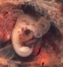
Carnegie stage 15
| Embryology - 29 Jul 2026 |
|---|
| Google Translate - select your language from the list shown below (this will open a new external page) |
|
العربية | català | 中文 | 中國傳統的 | français | Deutsche | עִברִית | हिंदी | bahasa Indonesia | italiano | 日本語 | 한국어 | မြန်မာ | Pilipino | Polskie | português | ਪੰਜਾਬੀ ਦੇ | Română | русский | Español | Swahili | Svensk | ไทย | Türkçe | اردو | ייִדיש | Tiếng Việt These external translations are automated and may not be accurate. (More? About Translations) |
Introduction

Facts
Facts: Week 5, 35 - 38 days, 7 - 9 mm
Gestational Age GA week 7
Summary
- Ectoderm: sensory placodes, lens pit, otocyst, nasal pit, primary/secondary vesicles, fourth ventricle of brain,
- Mesoderm: heart prominence
- Head: 1st, 2nd and 3rd pharyngeal arch, forebrain, site of lens placode, site of otic placode, stomodeum
- Body: heart, liver, umbilical cord, mesonephric ridge
- Limb: upper and lower limb buds, hand plate
See also Carnegie stage 15 Events
Features
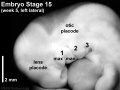
- Identify: midbrain region, nasal pit, lens pit, 1st, 2nd and 3rd pharyngeal arches, 1st pharyngeal groove, maxillary and mandibular components of 1st pharyngeal arch, fourth ventricle of brain, heart prominence, cervical sinus, upper limb bud, mesonephric ridge, lower limb bud, umbilical cord Labelled Stage 15
| Week: | 1 | 2 | 3 | 4 | 5 | 6 | 7 | 8 |
| Carnegie stage: | 1 2 3 4 | 5 6 | 7 8 9 | 10 11 12 13 | 14 15 | 16 17 | 18 19 | 20 21 22 23 |
- Carnegie Stages: 1 | 2 | 3 | 4 | 5 | 6 | 7 | 8 | 9 | 10 | 11 | 12 | 13 | 14 | 15 | 16 | 17 | 18 | 19 | 20 | 21 | 22 | 23 | About Stages | Timeline
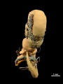
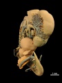
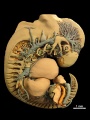
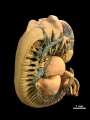
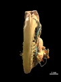
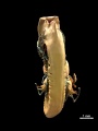
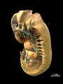
Kyoto Collection

Embryo Lateral view. Amniotic membrane removed.
- Embryo Stage 15 (7207) external view
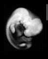
right
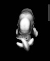
ventral
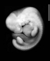
left
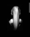
dorsal
left head label


Embryo Sagittal section - upper half
Image source: The Kyoto Collection images are reproduced with the permission of Prof. Kohei Shiota and Prof. Shigehito Yamada, Anatomy and Developmental Biology, Kyoto University Graduate School of Medicine, Kyoto, Japan for educational purposes only and cannot be reproduced electronically or in writing without permission.
Carnegie Collection


| Week: | 1 | 2 | 3 | 4 | 5 | 6 | 7 | 8 |
| Carnegie stage: | 1 2 3 4 | 5 6 | 7 8 9 | 10 11 12 13 | 14 15 | 16 17 | 18 19 | 20 21 22 23 |
| Carnegie Collection Embryos - Stage 15 | ||||||||||
|---|---|---|---|---|---|---|---|---|---|---|
| Serial No. | Size (mm) | Grade | Fixative | Embedding Medium | Plane | Thinness (µm) | Stain | Year | Notes | |
| 2 | E., 7.0 Ch., 25x25x25 | Good | Alc. | P | Transverse | 15 | Al. carm. | 1888 | Least-advanced third | |
| 88 | E.,8 Ch., 30x28x15 | Poor | Alc. | P | Coronal | P | Al. coch. | 1897 | ||
| 113 | E.,8 241 E,6.0 | Poor | P | p | Sagittal | 10 | Borax carmine | ? | ||
| 241 | E,6.0 | Good | Formalin | P | Transverse | 10 | H. & Congo red | 1904 | ||
| 371 | E,6.6 | Good | Formalin | P | Sagittal | 10 | Al. coch. | 1913 | Shrunken and cracked | |
| 389 | E., 9 | Poor | p | p | Sagittal | 20 | (Stain - Haematoxylin Eosin) | 1907 | Tubal | |
| 721 | E., 9.0 Ch., 30x20x10 | Exc. | Zenker formol | P | Transverse | 15 | (Stain - Haematoxylin Eosin) | 1913 | Median in group | |
| 810 | E., 7.0 Ch, 30x25x15 | Good | Alc. | P | Sagittal | 20 | Al. coch | 1913 | ||
| 855 | E.,7.5 | Poor | Formalin | P | Transverse | 100 | Al. coch. | 1914 | Pathological between limbs | |
| 1006 | E,9.0 Ch., 37x26x22 | Poor | Formalin | P | Coronal | 20 | (Stain - Haematoxylin Eosin) or. G. | 1914 | Operative. Most-advanced third | |
| 1091 | E,7.2 Ch., 28x26x20 | Poor | P | P | Coronal | 20 | Al. coch. | 1915 | Macerated | |
| 1354 | E,7.8 Ch, 35x30x25 | Good | Formalin | P | Sagittal | 20 | Al. coch. | 1916 | Least-advanced third | |
| 1767 | E , 11.0 Ch, 41x23x5 | Good | Formalin | P | Sagittal | 40 | (Stain - Haematoxylin Eosin) or. G. | 1917 | Most-advanced third | |
| 2743 | E., 7.2 Ch., 19xl8x14 | Poor | Formalin | P | Transverse | 20 | Al. coch. | 1919 | Macerated. Least-advanced third | |
| 3216 | E, 6.5 Ch, 30x30x5 | Good | Formalin | P | Transverse | 20 | Al. coch. | 1920 | Hysterectomy. Least-advanced third | |
| 3385 | E,83 Ch., 25x20x16 | Exc. | Corros. acetic | P | Trans. | 20 | (Stain - Haematoxylin Eosin) or. G. | 1921 | Some sections lost. Most-advanced third. Ag added | |
| 3441 | E,8.0 Ch., 25x24x20 | Good | Formalin | P | Sag. | 10 | Al. coch. | 1921 | ||
| 3512 | E,8,5 Ch., 33x28x25 | Good | Formalin | P | Trans. | 10 | Al. coch. | 1921 | ||
| 3952 | E,6,7 Ch., 30x25x15 | Good | Formalin | P | Cor. | 15 | Al. coch. | 1922 | Median in group | |
| 4602 | E,9.3 Ch,, 33x30x26 | Good | Formalin | P | Sag. | 15 | Al. coch. | 1924 | Medical abortion | |
| 4782 | E,9.0 Ch., I4xl3x11 | Poor | Formalin | P | Cor. | 20 | Al. coch. | 1924 | ||
| 5772 | E, 8 | Poor | ? | P | Cor. | 15 | Al. coch. eosin | 1928 | ||
| Template:CE5?92 | E, 3 | Good | Corros. acetic | C-P | Cor. | 10 | Al. coch. phlox | 1929 | Transitional to next stage | |
| 6223 | E ? | Poor | Alc. | C-P | Sag. | 8 | Or. G. | 1930 | Fragmented sections. Not saved | |
| 6504 | ||||||||||
| 6506 | ||||||||||
| 6508 | ||||||||||
| 6595 | ||||||||||
| ???? | ||||||||||
Abbreviations
| ||||||||||
| iBook - Carnegie Embryos | |
|---|---|

|
|
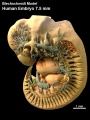
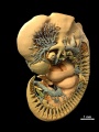
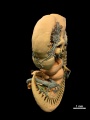
Blechschmidt Collection
| Embryo 7.5mm |
| Page | Play |
left lateral
left ventrolateral
left ventral
ventral
right ventral
right lateral
right dorsolateral
right dorsal
dorsal
left dorsolateral


Image source: The Blechschmidt Collection images are reproduced with the permission of Prof. Christoph Viebahn, director of the Institute of Anatomy and Embryology, , University Medical Center Göttingen. Images are for educational purposes only and cannot be reproduced electronically or in writing without permission.
Madrid Collection
| Madrid Collection Embryos | ||||||
|---|---|---|---|---|---|---|
| Carnegie Stage |
Embryo | Days | CRL (mm) | Section thickness |
Staining | Section plane |
| 15 | GV4 | 36 | 7 | 8 | (Stain - Haematoxylin Eosin) | transverse |
Photograph

|

|
Image: Dr Ed Uthman (Houston, Texas) - other pathology images
Image version links: Virtual Slide | ExtraLarge 1874 x 2000px | Large 959 x 1024px | Medium 468 x 500px
Embryo Virtual Slide
| Stage 15 - Ventral View
|
| Stage 15 | Embryo Slides |
Historic Stage 15 Embryos

- Carnegie No. 2, 7 mm. Described in detail by Mall (1891).[1]
- Hochstetter's embryo I, 7 mm. ( (No. 10 of Hochstetter's series) Described in monographic form by Elze (1907).[2]Includes illustrations of reconstructions. Also shown in Fig. 52 of Keibel and Mall (1910).[3]
- Legg embryo, 7 mm. Described in detail by Thompson (1915).[4]Includes illustrations of reconstructions.
- 8-mm embryo. The peripheral nervous system was described by Volcher (1963)[5] in this embryo of stage 15.
- Keibel No. 1495, 8.5 mm. This advanced example of stage 15 was well described in detail by Barniville (1914)[6].
- Blechschmidt 7.5 mm Blechschmidt Model generated from serial sections of this embryo.
- Newcastle N340 Optical Projection Tomography of this embryo described by J. Kerwin etal., (2004).[7]
Events
- hearing - otic capsule formed by condensed mesenchyme. Ganglion vestibular nerve fibres extend to the otocyst epithelium. External ear auricular hillocks appear, pharyngeal arch 2 forms primordium of the antitragus.
- vision - (about 33 days) the lens pit is closed. The lens vesicle and optic cup lie close to the surface ectoderm and appear to press against the surface.[8]
- smell - the nasal discs form large oval depressions (nasal pits). A continuous cellulovascular strand is observed between the nasal groove and the olfactory field.[9] Stages Description
- limb - upper limb bud visible as a distal hand plate and a proximal forearm, arm, and shoulder region.
- somitogenesis - last stage that somites can be seen from the surface.
- cardiovascular
- heart - atrioventricular bundle appears, atrioventricular cushions present, conotruncal ridges present, foramen secundum appears and semilunar cusps appear.[10]
- Coronary circulation plexus of blind epicardial capillaries appears on the heart begin to acquire coronary sinus connection.[11]
- Cerebral artery vertebral arteries forms from transverse anastomoses between cervical intersegmental arteries, beginning with the proatlantal artery and proceeding downward to the 6th intersegmental artery.[12]
- endocrine[13]
- Epiphysis - pineal body is detectable in the roof of the diencephalon (Stadium I of Turkewitsch 1933) (O'Rahilly 1968).
- thyroid - thyroid primordium may be detached from the pharyngeal epithelium in some instances. "At about the time" when the thyroglossal duct "becomes broken it loses its lumen" (Grosser 1912).
- Adrenal Cortex - primordium is first recognizable. A new type of cell (C1) from the coelomic epithelium is found in the subjacent mesenchyme. New cells (C2) appear in the medial wall of mesonephric glomeruli and begin to migrate into the suprarenal primordium.[14] Jirfisek (1980) denies a mesonephric contribution to the suprarenal.
- Adrenal Medulla - all types of cells (M1, M2, and M3) increase in number. From stage 15 to stage 18, the suprarenal primordium is cigar-shaped and extends from segment T6 to segment L1, lateral to the aorta and mesogastrium.[14]
- Meninges (Spinal Cord) - vertebral rudiments now form concentrations that are distinct from the intermediate zone separating them from the neural tube. This intermediary zone, which is less densely cellular and within which lie the ganglia, is the meninx primitiva, from which the meninges have been generally considered to arise. The neural tube is now completely surrounded by endothelium-lined vascular channels. Directly adjacent to the cord, and particularly evident at its ventrolateral surfaces, is a single-layered cell membrane. This is the first indication of the pia mater, and, where it is present, it lies between the vascular channels and the periphery of the spinal cord.[15]
- ventral body wall - medially vertebrae and ribs formed, and primordia of sternum and hypaxial flank muscle primordium laterally in the body wall.[16]
References
- ↑ Mall FP. A human embryo twenty-six days old. (1891) J Morphol. 5: 459-480.
- ↑ Elze, C. 1907. Beschreibung eines menschlichen Embryo von zirka 7 mm grosster Lange unter besonderer Beriicksichtigung der Frage nach der Enrwickelung der Extremitatenarterien und nach der morphologischen Bedeutung der lateralen Schilddriisenanlage. (Description of a human embryo of about 7 mm greatest length with special reference to the question of the development of the extremities arteries and to the morphological importance of lateral shield primordia). Anat. Hefte, 106, 411-492.
- ↑ Keibel F. and Mall FP. Manual of Human Embryology I. (1910) J. B. Lippincott Company, Philadelphia.
- ↑ Thompson P. Description of a human embryo, 7 mm. greatest length. (1915) Studies in Anatomy. University of Birmingham, pp. 1-50.
- ↑ Volcher, R. 1963. Le systeme nerveux périphérique d'un embryon humain de 8 mm.(The peripheral nervous system of an 8 mm. human embryo) Arch. Biol. (Liege), 74, 95-127. PMID 13997757
- ↑ Barniville HL. The morphology and histology of a human embryo of 8.5 mm. (1914) J Anat. Physiol. 49(1):1-71. PMID 17233012
- ↑ Kerwin J, Scott M, Sharpe J, Puelles L, Robson SC, Martínez-de-la-Torre M, Ferran JL, Feng G, Baldock R, Strachan T, Davidson D & Lindsay S. (2004). 3 dimensional modelling of early human brain development using optical projection tomography. BMC Neurosci , 5, 27. PMID: 15298700 DOI.
- ↑ Pearson AA. (1980). The development of the eyelids. Part I. External features. J. Anat. , 130, 33-42. PMID: 7364662
- ↑ Bossy J. Development of olfactory and related structures in staged human embryos. (1980) Anat. Embryol., 161(2);225-36 PMID 7469043
- ↑ Arráez-Aybar LA, Turrero-Nogués A & Marantos-Gamarra DG. (2008). Embryonic cardiac morphometry in Carnegie stages 15-23, from the Complutense University of Madrid Institute of Embryology Human Embryo Collection. Cells Tissues Organs (Print) , 187, 211-20. PMID: 18057862 DOI.
- ↑ Hutchins GM, Kessler-Hanna A & Moore GW. (1988). Development of the coronary arteries in the embryonic human heart. Circulation , 77, 1250-7. PMID: 3286038
- ↑ Menshawi K, Mohr JP & Gutierrez J. (2015). A Functional Perspective on the Embryology and Anatomy of the Cerebral Blood Supply. J Stroke , 17, 144-58. PMID: 26060802 DOI.
- ↑ O'Rahilly R. The timing and sequence of events in the development of the human endocrine system during the embryonic period proper. (1983) Anat. Embryol., 166: 439-451. PMID 6869855
- ↑ 14.0 14.1 Crowder RE. The development of the adrenal gland in man, with special reference to origin and ultimate location of cell types and evidence in favor of the "cell migration" theory. (1957) Contrib. Embryol., Carnegie Inst. Wash. 36, 193-210.
- ↑ Sensenig EC. The early development of the meninges of the spinal cord in human embryos. (1951) Contrib. Embryol., Carnegie Inst. Wash. Publ. 611.
- ↑ Mekonen HK, Hikspoors JP, Mommen G, Köhler SE & Lamers WH. (2015). Development of the ventral body wall in the human embryo. J. Anat. , 227, 673-85. PMID: 26467243 DOI.
Additional Images
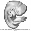
Fig. 52 of Keibel and Mall Manual of Human Embryology I (1910)
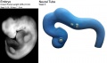
Neural tube model
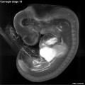
Stage 15 Optical Projection Tomography

External ear Stages 14-23 and adult


Mall, F. P. 1891. A human embryo twenty-six days old. J. Morph, 5, 459-480.
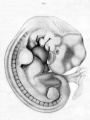
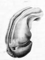
Pl. 1 Fig. 1. External view of the embryo.
Pl. 1 Fig. 2. Corrosion preparation pericardial and pleuro-peritoneal cavities.
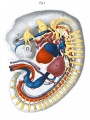
Pl. 2 Fig. 1. Reconstruction viewed from left side.
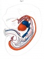
Pl. 2 Fig. 2. Same as Fig. I. Deeper view.


- Carnegie Stages: 1 | 2 | 3 | 4 | 5 | 6 | 7 | 8 | 9 | 10 | 11 | 12 | 13 | 14 | 15 | 16 | 17 | 18 | 19 | 20 | 21 | 22 | 23 | About Stages | Timeline
Cite this page: Hill, M.A. (2026, July 29) Embryology Carnegie stage 15. Retrieved from https://embryology.med.unsw.edu.au/embryology/index.php/Carnegie_stage_15
- © Dr Mark Hill 2026, UNSW Embryology ISBN: 978 0 7334 2609 4 - UNSW CRICOS Provider Code No. 00098G