Mouse Estrous Cycle
| Embryology - 26 Jul 2026 |
|---|
| Google Translate - select your language from the list shown below (this will open a new external page) |
|
العربية | català | 中文 | 中國傳統的 | français | Deutsche | עִברִית | हिंदी | bahasa Indonesia | italiano | 日本語 | 한국어 | မြန်မာ | Pilipino | Polskie | português | ਪੰਜਾਬੀ ਦੇ | Română | русский | Español | Swahili | Svensk | ไทย | Türkçe | اردو | ייִדיש | Tiếng Việt These external translations are automated and may not be accurate. (More? About Translations) |
Introduction

Reproductive processes in female mammals are characterised by cyclic alterations in the female tract and in sexual receptivity. The recurrent period of receptivity, or "heat" is called Estrus. The estrous cycle has been most extensively studied in laboratory rodents (mice and rats). Rats kept separate from males in the laboratory repeat the Estrous cycle throughout the year at intervals of about five days, unless subjected to pregnancy, pseudo-pregnancy (after a sterile mating), or disease. The cycle involves the whole of the reproductive tract, and it is possible to determine the sexual status of the female rat by examination of smears prepared from the vaginal fluid.
Rats and mice are examples of polyestrus mammals (as are cats which are seasonally polyoestrus). Monestrus forms (most wild animals - foxes, bears, wolves etc.) complete a single estrous cycle annually. In the wild, rats and mice probably suspend the cycle for a period during the winter; the reproductive organs are in a state of quiescence, called anestrus.
Note "estrus" the alternate English spelling "oestrus".
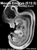
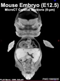
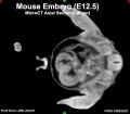
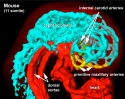
| Mouse Links: Introduction | Mouse Stages | Mouse Timeline | Mouse Timeline Detailed | Mouse Estrous Cycle | Mouse Heart | Mouse Knockout | Movie - Cephalic Plexus | Movie - Blastocyst Cdx2 | ANAT2341 Project 2009 | Category:Mouse | |||||||||||||||||||||||||||||||||||||||||||||||||||||||||||||||||||||||||||||||||||||||||||||||||||||||||||||||||||||||||||
|
| ||||||||||||||||||||||||||||||||||||||||||||||||||||||||||||||||||||||||||||||||||||||||||||||||||||||||||||||||||||||||||
Some Recent Findings

|
| More recent papers |
|---|
 This table allows an automated computer search of the external PubMed database using the listed "Search term" text link.
More? References | Discussion Page | Journal Searches | 2019 References | 2020 References Search term: Mouse Estrous Cycle <pubmed limit=5>Mouse Estrous Cycle</pubmed> |
Mouse Estrous Cycle
| Stage | ||||
|---|---|---|---|---|
| Diestrus | Small follicles only are present with large corpora lutea from the previous ovulation. These secrete for only a very short time unless pregnancy or pseudopregnancy intervene. | Small and anaemic, low motility, lumen small and slit-like. Cells of the uterine mucosa columnar; polymorphonuclear leucocytes in stroma; endometrial glands collapsed, atrophic. | Epithelium thin, mitotic figures infrequent. Leucocytes abundant in stroma, migrate through the epithelium into vaginal lumen. | Stringy mucous in which are entangled many leucocytes and a few nucleated epithelial cells. |
| Proestrus | Some follicles grow rapidly. | Become more vascular, water content increases, organ distends. Contractility more pronounced. Epithelial cells become higher (continuing into estrus). Leucocytes disappear from mucosa. Endometrial glands hypertrophy. | Epithelum thickens, numerous mitoses in inner layers. Old layers of epithelium line the lumen. Leucocytes no longer migrate through the epithelium. Superficial epithelial cells slough off into lumen. | Largely small, round, nucleated epithelial cells, singly or in sheets. None to few leucoytes. |
| Estrus | Ovulation in the rat is spontaneous and occurs about 10 hours after the beginning of estrus. "Heat" (receptivity) lasts about 13 hours. Usually 10-20 eggs ovulated each time. | gains maximum vascularisation. Epithelial cells reach maximum development. No leucocytes. | Outer layer of epithelial cells become cornified and sloughed into the lumen. In early estrus these cells retain their nuclei, but in later stages no nuclei visible and the cells are irregular, flat, cornified plates. The skin around the vaginal orifice becomes swollen. | Contains hundreds of large cornified cells (squames) with degenerate nuclei. Towards the end of estrus the smear becomes "cheesy" - masses of adherent cornified cells. |
| Metestrus | Many corpora lutea, which secrete only for a very short time, and small follicles. | Epithelium continues vacuolar degeneration and replacement. Leucocytes in stroma. Decrease in size and vascularity. | Deeper layers of the estrous epithelium now line the lumen, the older, superficial layers having become cornified and sloughed off. Reduction of mitotic activity in epithelium. Leucocytes in stroma and migrating through the epithelium into the lumen. | Many leucocytes and a few cornified cells. |
| Mouse Estrous Cycle | ||||
Mouse Ovarian Follicle Size

Image Source: High-resolution ultrasound biomicroscopy for monitoring ovarian structures in mice.[7]
Histological Features
For more detailed histological information see [#Champlin Champlin etal., 1973].
Uterus The changes in the uterus may not be particularly well-marked.
- Histological changes in the luminal epithelium
- Histological changes in the glandular epithelium
- Secretory activity of uterine glands
- Changes in stromal cells, e.g. number of leucocytes
- Overall changes in the size distention and shape of the lumen.
Vagina Note the changes in the vaginal epithelium.
- The number of mitotic figures
- The number of layers in the epithelium
- The amount of cornification, and the changes from live to dead cells.
- Surface mucus coating the epithelium
- Presence or absence of infiltrating polymorphonuclear leucocytes.
Vaginal Smears
Relate the appearance of the smears to the sections of the vagina, noting particularly the composition of cells in the smear and the vaginal epithelium.
1. Relative numbers and appearance of cell types - epithelial and polymorphs.
2. Presence of mucus.
Carnegie Stage Species Comparison
- Mouse Stages: E1 | E2.5 | E3.0 | E3.5 | E4.5 | E5.0 | E5.5 | E6.0 | E7.0 | E7.5 | E8.0 | E8.5 | E9.0 | E9.5 | E10 | E10.5 | E11 | E11.5 | E12 | E12.5 | E13 | E13.5 | E14 | E14.5 | E15 | E15.5 | E16 | E16.5 | E17 | E17.5 | E18 | E18.5 | E19 | E20 | Timeline | About timed pregnancy
| Carnegie | Stage | |||||||||||||||||||||||
| Human | Days | 1 | 2-3 | 4-5 | 5-6 | 7-12 | 13-15 | 15-17 | 17-19 | 20 | 22 | 24 | 28 | 30 | 33 | 36 | 40 | 42 | 44 | 48 | 52 | 54 | 55 | 58 |
| Mouse | Days | 1 | 2 | 3 | E4.5 | E5.0 | E6.0 | E7.0 | E8.0 | E9.0 | E9.5 | E10 | E10.5 | E11 | E11.5 | E12 | E12.5 | E13 | E13.5 | E14 | E14.5 | E15 | E15.5 | E16 |
| Rat | Days | 1 | 3.5 | 4-5 | 5 | 6 | 7.5 | 8.5 | 9 | 10.5 | 11 | 11.5 | 12 | 12.5 | 13 | 13.5 | 14 | 14.5 | 15 | 15.5 | 16 | 16.5 | 17 | 17.5 |
| Note these Carnegie stages are only approximate day timings for average of embryos. Links: Carnegie Stage Comparison | ||||||||||||||||||||||||
| ||||||||||||||||||||||||
| Timeline Links: human timeline | mouse timeline | mouse detailed timeline | chicken timeline | rat timeline | Medaka | Category:Timeline |
References
- ↑ 1.0 1.1 Byers SL, Wiles MV, Dunn SL & Taft RA. (2012). Mouse estrous cycle identification tool and images. PLoS ONE , 7, e35538. PMID: 22514749 DOI.
- ↑ Mader SL, Libal NL, Pritchett-Corning K, Yang R & Murphy SJ. (2009). Refining timed pregnancies in two strains of genetically engineered mice. Lab Anim (NY) , 38, 305-10. PMID: 19701181 DOI.
- ↑ Caligioni CS. (2009). Assessing reproductive status/stages in mice. Curr Protoc Neurosci , Appendix 4, Appendix 4I. PMID: 19575469 DOI.
- ↑ Bilinski MJ, Thorne JG, Oh MJ, Leonard S, Murrant C, Tayade C & Croy BA. (2008). Uterine NK cells in murine pregnancy. Reprod. Biomed. Online , 16, 218-26. PMID: 18284876
- ↑ Bachelot A & Binart N. (2005). Corpus luteum development: lessons from genetic models in mice. Curr. Top. Dev. Biol. , 68, 49-84. PMID: 16124996 DOI.
- ↑ Deb K, Reese J & Paria BC. (2006). Methodologies to study implantation in mice. Methods Mol. Med. , 121, 9-34. PMID: 16251731
- ↑ Jaiswal RS, Singh J & Adams GP. (2009). High-resolution ultrasound biomicroscopy for monitoring ovarian structures in mice. Reprod. Biol. Endocrinol. , 7, 69. PMID: 19580664 DOI.
Reviews
Cora MC, Kooistra L & Travlos G. (2015). Vaginal Cytology of the Laboratory Rat and Mouse: Review and Criteria for the Staging of the Estrous Cycle Using Stained Vaginal Smears. Toxicol Pathol , 43, 776-93. PMID: 25739587 DOI.
Chaffin CL & Vandevoort CA. (2013). Follicle growth, ovulation, and luteal formation in primates and rodents: a comparative perspective. Exp. Biol. Med. (Maywood) , 238, 539-48. PMID: 23856905 DOI.
Articles
Yang YJ, Cao YJ, Bo SM, Peng S, Liu WM & Duan EK. (2006). Leptin-directed embryo implantation: leptin regulates adhesion and outgrowth of mouse blastocysts and receptivity of endometrial epithelial cells. Anim. Reprod. Sci. , 92, 155-67. PMID: 16023802 DOI.
Deb K, Reese J & Paria BC. (2006). Methodologies to study implantation in mice. Methods Mol. Med. , 121, 9-34. PMID: 16251731
Lee DS, Yanagimoto Ueta Y, Xuan X, Igarashi I, Fujisaki K, Sugimoto C, Toyoda Y & Suzuki H. (2005). Expression patterns of the implantation-associated genes in the uterus during the estrous cycle in mice. J. Reprod. Dev. , 51, 787-98. PMID: 16210782
Su P, Wu JC, Sommer JR, Gore AJ, Petters RM & Miller WL. (2005). Conditional induction of ovulation in mice. Biol. Reprod. , 73, 681-7. PMID: 15917351 DOI.
Champlin AK, Dorr DL & Gates AH. (1973). Determining the stage of the estrous cycle in the mouse by the appearance of the vagina. Biol. Reprod. , 8, 491-4. PMID: 4736343
Search PubMed
Search PubMed: mouse estrous cycle | estrous cycle | mouse anestrus | oestrous cycle
Terms
- a disintegrin and metalloproteinase - (ADAM) a large family (-8, -9, -10, -12, -15 and -17) of secreted proteins suggested to be involved in remodelling mouse uterine tissue during the oestrous cycle.
- anestrus - lack of a normal estrus cycle.
- Bruce Effect - pheromones from a strange male can prevent embryo implantation in recently bred female.
- calling behaviour - vocalization, vocal communication sounds associated with reproductive behaviour in several species.
- Lee-Boot Effect - female mice housed together (in groups) results in a synchronization of their estrus cycles. In addition, the extended absence of male pheromones leads to a state of anestrus (lack of a normal estrus cycle).
- major urinary proteins - (MUPs) proteins which carry volatile substances, including pheromones, and protect them during their internal passage (liver to kidneys into urine).
- pheromone - a secreted chemical in sweat or urine that causes specific physiological responses.
- Whitten Effect - female mice either singly or housed together (in groups) can be induced into estrus by exposure to male mouse urine or their dirty bedding. The estrous cycle is applicable to many different species, not just the mouse reproductive cycle.
Spelling Note: When searching both American (estrous) and British (oestrous) spellings are used in the literature (More? American and British Spelling Variations). My thanks also to Prof Jill Becker for correcting an obvious spelling error on this page "The adjective is spelled with a 'u' after the o. 'Estrus' refers to the stage of the cycle and is the noun form of the word."
External Links
External Links Notice - The dynamic nature of the internet may mean that some of these listed links may no longer function. If the link no longer works search the web with the link text or name. Links to any external commercial sites are provided for information purposes only and should never be considered an endorsement. UNSW Embryology is provided as an educational resource with no clinical information or commercial affiliation.
- University of California, Irvine - University Laboratory Animal Resources Mouse Breeding Techniques
| Animal Development: axolotl | bat | cat | chicken | cow | dog | dolphin | echidna | fly | frog | goat | grasshopper | guinea pig | hamster | horse | kangaroo | koala | lizard | medaka | mouse | opossum | pig | platypus | rabbit | rat | salamander | sea squirt | sea urchin | sheep | worm | zebrafish | life cycles | development timetable | development models | K12 |
Glossary Links
- Glossary: A | B | C | D | E | F | G | H | I | J | K | L | M | N | O | P | Q | R | S | T | U | V | W | X | Y | Z | Numbers | Symbols | Term Link
Cite this page: Hill, M.A. (2026, July 26) Embryology Mouse Estrous Cycle. Retrieved from https://embryology.med.unsw.edu.au/embryology/index.php/Mouse_Estrous_Cycle
- © Dr Mark Hill 2026, UNSW Embryology ISBN: 978 0 7334 2609 4 - UNSW CRICOS Provider Code No. 00098G