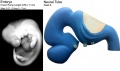
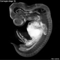
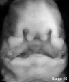
Carnegie stage 16
| Embryology - 6 Jul 2026 |
|---|
| Google Translate - select your language from the list shown below (this will open a new external page) |
|
العربية | català | 中文 | 中國傳統的 | français | Deutsche | עִברִית | हिंदी | bahasa Indonesia | italiano | 日本語 | 한국어 | မြန်မာ | Pilipino | Polskie | português | ਪੰਜਾਬੀ ਦੇ | Română | русский | Español | Swahili | Svensk | ไทย | Türkçe | اردو | ייִדיש | Tiếng Việt These external translations are automated and may not be accurate. (More? About Translations) |
Introduction

Facts
Week 6, 37 - 42 days, 8 - 11 mm
Gestational Age GA - week 8
Summary
- Ectoderm: sensory placodes, lens pit, otocyst,nasal pits moved ventrally, fourth ventricle of brain
- Mesoderm: heart prominence
- Head: 1st, 2nd and 3rd pharyngeal arch, forebrain, eye, auricular hillocks
- Body: heart, liver, umbilical cord, mesonephric ridge
- Limb: upper and lower limb buds, hand plate, developing arm
See also Carnegie stage 16 Events
Features
- Eye showing retinal pigment, nasolacrimal groove, nasal pit, fourth ventricle of brain, umbilical cord, 1st and 2nd pharyngeal arches, cervical sinus, heart, developing arm with hand plate, foot plate
- Identify: nasal pit, nasolacrimal groove, eye, 1st, 2nd and 3rd pharyngeal arches, 1st pharyngeal groove, maxillary and mandibular components of 1st pharyngeal arch, auricular hillocks, fourth ventricle of brain, heart prominence, upper limb bud, mesonephric ridge, lower limb bud, umbilical cord
| Week: | 1 | 2 | 3 | 4 | 5 | 6 | 7 | 8 |
| Carnegie stage: | 1 2 3 4 | 5 6 | 7 8 9 | 10 11 12 13 | 14 15 | 16 17 | 18 19 | 20 21 22 23 |
- Carnegie Stages: 1 | 2 | 3 | 4 | 5 | 6 | 7 | 8 | 9 | 10 | 11 | 12 | 13 | 14 | 15 | 16 | 17 | 18 | 19 | 20 | 21 | 22 | 23 | About Stages | Timeline
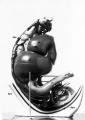
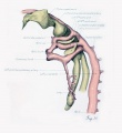
Kyoto Collection

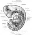
View: This is a dorsolateral view of embryo. Amniotic membrane removed.
Image source: Embryology page Created: 19.03.1999
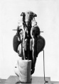
| Head | |
|---|---|

Left lateral view |

Left lateral view (labeled) |

Ventral view of head region. Ventral view of head region (1 mm scale). |
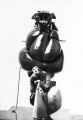
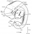
| Central Nervous System | ||||||||||||||||||||||||||||||||||||||||||||||||||||||||||||||||||||||||
|---|---|---|---|---|---|---|---|---|---|---|---|---|---|---|---|---|---|---|---|---|---|---|---|---|---|---|---|---|---|---|---|---|---|---|---|---|---|---|---|---|---|---|---|---|---|---|---|---|---|---|---|---|---|---|---|---|---|---|---|---|---|---|---|---|---|---|---|---|---|---|---|---|

|

|
| ||||||||||||||||||||||||||||||||||||||||||||||||||||||||||||||||||||||
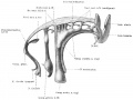
| Right lateral view of the central nervous system of embryo at Carnegie stage 16. Scale bar is 1 mm | ||||||||||||||||||||||||||||||||||||||||||||||||||||||||||||||||||||||||
Movies
|
| ||||||||||
|
|
| |||||||||
|
|
|
Image source: The Kyoto Collection images are reproduced with the permission of Prof. Kohei Shiota and Prof. Shigehito Yamada, Anatomy and Developmental Biology, Kyoto University Graduate School of Medicine, Kyoto, Japan for educational purposes only and cannot be reproduced electronically or in writing without permission.
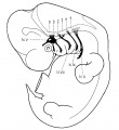
Scanning EM

Ventral view of head showing upper lip, maxilla and nasal region.
Image Source: Prof Virginia Diewert
Carnegie Collection


| Carnegie Collection Embryos - Stage 16 | ||||||||||
|---|---|---|---|---|---|---|---|---|---|---|
| Serial No. | Size (mm) | Grade | Fixative | Embedding Medium | Plane | Thinness (µm) | Stain | Year | Notes | |
| 163 | E., 9.0 Ch., 35x35x20 | Good | Formol | P | Transverse | 20 | Al. coch. | 1899 | Used by Bardeen and Lewis | |
| 221 | E., 7.5 Ch., 40x33x33 | Poor | Formalin | P | Sagittal | 20 | Al. coch. | 1903 | Macerated | |
| 383 | E., 7.0 Ch., 15x15x15 | Poor | Formalin | P | Transverse | 10 | Al. carm., H. & Congo red | 1904 | ||
| 397 | E., 8.0 Ch., 15x15x15 | Poor | Formalin | P | Transverse | 10 | (Stain - Haematoxylin Eosin) | 1907 | ||
| 422 | E., 9.0 Ch., 30x30x30 | Poor | Alc. | P | Transverse | 40 | Al. coch. | 1910 | Tubal. Partly macerated | |
| 559 | E., 8.6 Ch., 20x15x12 | Good | Formalin | P | Transverse | 20 | H. & Congo red | 1911 | Cyclopia. Formerly listed as stage 17 | |
| 589 | E., 11 Ch., 30x13x13 | Poor | p | P | Sagittal | 50 | Al. coch. | 1912 | ||
| 617 | E., 7.0 Ch., 18x14x12 | Good | Formalin | P | Transverse | 15 | Al. coch. | 1912 | Median in group | |
| 636 | E., 10 Ch.,28x28x22 | Poor | Formalin | P | Transverse | 50 | Al. coch. | 1913 | Macerated | |
| 651f | E., 7 Ch., 25x20x15 | Poor | p | p | p | p | p | 1913 | Spina bifida | |
| 675 | E., 10 Ch., 50x30x25 | Poor | Formalin | P | Sagittal | 100 | Carmine | 1915 | Abnormal head and limbs | |
| 792 | E., 8.0 Ch., 40x30x30 | Good | Formalin | P | Transverse | 20 | Al. coch. | 1913 | Advanced | |
| 887 | E, 9.0 Ch., 31x28x17 | Good | Formalin | P | Transverse | 40 | Al. coch. | 1914 | Near next stage | |
| 1121 | E., 11.8 | Good | Corros. acetic | P | Coronal | 40 | Al. coch. | 1915 | Operative. Median in group | |
| 1197 | E., 10.0 Ch., 23x19x15 | Good | Formalin | C | Sagittal | 20 | (Stain - Haematoxylin Eosin), or. G. | 1915 | Advanced | |
| 1544 | E., 7.2 | Good | Zenker | P | Sagittal | 20 | Al. coch. | 1916 | Tubal. Mechanical injury | |
| 1836 | E., 11.0 | Good | Formalin | P | Transverse | 20 | (Stain - Haematoxylin Eosin) | 1917 | Most-advanced third | |
| 4677 | E., 9.5 Ch., 48x36x30 | Good | Formalin | P | Transverse | 10 | Al. coch. | 1924 | Median in group | |
| 5515 | E., 12.0 Ch., 47x37x25 | Good | Formalin | C-P | Transverse | 10 | (Stain - Haematoxylin Eosin) | 1927 | Near next stage | |
| 6054 | E., 7,0 Ch., 21x17x12 | Good | Formalin | C-P | Transverse | 8 | (Stain - Haematoxylin Eosin) | 1930 | Least-advanced third | |
| 6507 | E., 9.0* | Excellent | Corros. acetic | C-P | Coronal | 10 & 8 | Al. coch. | p | Middle or most-advanced third | |
| 6509 | E. 8.1* | Excellent | Corros. acetic | C-P | Coronal | 10 | Al. coch. | p | Least-adianced or middle third | |
| 6510 | E., 10.1* | Excellent | Corros. acetic | C-P | Coronal | 10 | Al. coch. | p | Close to No. 6507. Ag added | |
| 6511 | E, 8.1* | Good | Corros. acetic | C-P | Sagittal | 10 | Al. coch., iron H. | p | Surface injured by fixative. Most-advanced third | |
| 6512 | E., 7.0* | Excellent | Corros. acetic | C-P | Transverse | 10 | Al. coch. | p | Least-advanced third. Borderline | |
| 6513 | E., 7.2* | Good | Corros. acetic | C-P | Coronal | 10 | Al. coch. | p | Surface injured by fixative. Least advanced in group | |
| 6514 | E, 9 0* | Poor | Corros. acetic | C-P | Sagittal | 10 | Al. coch. | p | Most-advanced third | |
| 6516 | E., 10 5* | Good | Corros acetic | C-P | Sagittal | 8 | Al. coch. | p | Most-advanced third. Double left kidney and ureter | |
| 6517 | E., 10.5* | Excellent | Corros. acetic | C-P | Transverse | 8 | Al. coch. | ? | Close to No. 6516 | |
| 6686 | E., 11.0 Ch., l7x17xP | Poor | Formalin | C-P | Coronal | 20 | Al. coch. | 1933 | Tubal. Partly macerated | |
| 6750 | E., 10.0 | Good | Formalin | C-P | Transverse | 10 | H. & phlox. | 1933 | Tubal. Advanced | |
| 6909 | E., 11.0 | Good | Bouin | C-P | Coronal | 10 | (Stain - Haematoxylin Eosin) | 1934 | Tubal. Advanced | |
| 6931 | E., 8.8 Ch., 3""x 33x16 | Good | Formalin | C-P | Coronal | 10 | Al coch., phlox. | 1934 | Least-advanced third. Type specimen | |
| 6950 | E.. 9 0 Ch., 3lx20x18 | Good | Formalin | C-P | Transverse | 10 | (Stain - Haematoxylin Eosin) | 1934 | Tubal. Partly fragmented | |
| 7?15 | E., 9.7 | Exc | Bouin | C-P | Coronal | 10 | H. & phlox | 1935 | Operative. Less advanced | |
| 7629 | E., 11.5 Ch., 31x31 | Good | Formalin | C-P | Coronal | 10 | Al. coch., phlox | 1939 | Hysterectomy. Most advanced in group | |
| 7804 | E., 9.5 Ch., 26x21x16 | Good | Formalin | C-P | Transverse | 10 | (Stain - Haematoxylin Eosin) | 1940 | Least-advanced third | |
| 7897 | E., 12.2 Ch., 31x24x23 | Good | Formalin | C-P | Transverse | 10 | (Stain - Haematoxylin Eosin) | 1941 | Tubal. Advanced | |
| 8098 | E., 10.0 Ch., 30 | Good | Formalin | C-P | Coronal | 6 | (Stain - Haematoxylin Eosin) | 1942 | Tubal. Median in group | |
| 8112 | E., 10.9 | Excellent | Bouin | C-P | Coronal | 8 | (Stain - Haematoxylin Eosin) | 1943 | Most-advanced third | |
| 8179 | E., 11.9 Ch., 23x18x17 | Good | Formalin | C-P | Coronal | 10 | (Stain - Haematoxylin Eosin) | 1943 | Tubal | |
| 8436 | E., 10.9 Ch., 13x15x1? | Good | Formalin | P | Coronal | 10 | Azan | 1946 | Advanced | |
| 8692 | E., 10 | Good | Bouin | P | Transverse | 10 | (Stain - Haematoxylin Eosin) | 1949 | Rubella. Medical abortion. Mechanically damaged | |
| 8697 | E., 11.3 | Poor | Formalin | C-P | Transverse | 10 | (Stain - Haematoxylin Eosin) | 1949 | Perhaps stage 17 | |
| 8773 | E, 11 | Excellent | Bouin | P | Coronal | 10 | Azan | 1950 | ||
| 8971 | E., 10 Ch., 20.5x14.5x13.7 | Poor | Formalin | Transverse | 15 | (Stain - Haematoxylin Eosin) | 1932 | Synophthalmia. Univ. Chicago No. H 1439 | ||
| Template:CE9055 | E., ca. 10 | Excellent | Bouin | P | Transverse | 20 | Azan & Ag | 1953 | Damaged | |
| 9229 | E, 9.5 | Excellent | Formalin | P | Transverse | 6 | Ag & (Stain - Haematoxylin Eosin) | 1954 | Stage 15, 16, or 17? Mislaid | |
Abbreviations
| ||||||||||
| iBook - Carnegie Embryos | |
|---|---|

|
|
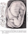
Madrid Collection

|

|
| Human Embryo CRL 10.2 mm (stage 16) | Human Embryo CRL 10.5 mm (stage 16) |
Image source: The Madrid Collection images are reproduced with the permission of Prof. Rodríguez-Vázquez, Head, Embryology Institute of Complutense University of Madrid. Images are for educational purposes only and cannot be reproduced electronically or in writing without permission.
| Madrid Collection Embryos | ||||||
|---|---|---|---|---|---|---|
| Carnegie Stage |
Embryo | Days | CRL (mm) | Section thickness |
Staining | Section plane |
| 16 | MAR 3 | 38 | 8 | 10 | (Stain - Haematoxylin Eosin)-Azan | frontal |
| 16 | BOT9 | 39 | 9 | 10 | (Stain - Haematoxylin Eosin) | transverse |
| 16 | CN 2 | 39 | 9.5 | 10 | (Stain - Haematoxylin Eosin) | sagittal |
| 16 | FAUS 4 | 40 | 9.7 | 10 | (Stain - Haematoxylin Eosin) | frontal |
| 16 | CA 2 | 40 | 10 | 10 | (Stain - Haematoxylin Eosin)-Azan | transverse |
| 16 | FE 1 | 40 | 10.5 | 10 | (Stain - Haematoxylin Eosin) | transverse |
Hill Collection
| Hill HH8 | |
|---|---|

|

|

|

|

|

|
Stereo pair animation: right lateral animation | right lateral animation | left lateral animation
| Hill HH5 | |
|---|---|

|

|

|

|

|

|

|

|
- Links: Hill Collection
Hinrichsen Collection

Hinrichsen collection Human Embryo ME34 (stage16). Left lateral view of the embryo.
Image source: The Hinrichsen Collection images are reproduced with the permission of Prof. Beate Brand-Saberi, Head, Department of Anatomy and Molecular Embryology, Ruhr-Universität Bochum. Images are for educational purposes only and cannot be reproduced electronically or in writing without permission.
Other Collection Embryos
- No. BR, 9.75 mm G. L., 8.8 mm. Summarized by Tandler (1907)[1], who, on the basis of the coital history, gave the age as the “38th day.” About stage 16.
- 9-mm embryo. The peripheral nervous system was described by Masy (1955)[2] in this embryo of stage 16.
- Huber No. 3, 10 mm. The nuclei of origin of the cranial nerves and the peripheral nervous system were described by Streeter (1908a,b).[3][4] An advanced example of stage 16.
- Wetzel (We) embryo, 10 mm. Discussed by v. Hayek (1934)[5] in regard to age, which, based on the coital history, was given as the “40th day.” Probably about stage 16.
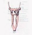
- Carnegie No. 6516, 10.5 mm (corrected). Double ureters described by Wharton (1949).[6]
- No. H60, University of Missouri, 11 mm. Described briefly by Bonnot and Severs (1906).[7] Probably belongs to stage 16.
Events
- Hearing - otic vesicle wall thickens prior to appearance of the semicircular ducts.[8] A utriculosaccular diverticulum and spiral ganglion appears. Auricular hillocks present (tragus, crus helicis, helix, and antitragus).
- Vision - (37 days) Growth of the lens body results in a D-shaped lens cavity. Perilental blood vessels (tunica vasculosa lentis) are visible. Prior to the development of the eyelids, one small sulcus or groove forms above the eye (eyelid groove) and another below it.[9]
- smell - the vomeronasal groove appears.[10] Stages Description
- Cardiovascular
- Coronary circulation plexus acquires coronary sinus connection.[11]
- Cerebral artery development of the middle cerebral artery first identified as small buds originating proximal to the anterior cerebral artery on the anterior division of the primitive internal carotid artery.[12]
- Endocrine[13]
- Hypophysis - slight indication of the infundibular recess may be seen in some embryos (O'Rahilly 1973 a).
- Epiphysis - cellular migration in an external direction occurs in the pineal body during stages 16 and 17 (Stadium 2 of Turkewitsch 1933) (O'Rahilly 1968).
- Thymus - according to Norris (1938), "not until the primordium of the parathyroid [3] has been outlined can the remaining portion of the third pouch be recognized, by exclusion, as the primordium of the endodermal thymus".
- Parathyroids - parathyrogenic zones are closely related to the third and fourth aortic arches at 9 mm (Politzer and Hann 1935, unstaged embryo). Parathyroid 3 is identifiable on the anterior wall of the third pharyngeal pouch (Weller 1933, Fig. 17) and "does not arise from a dorsal lobule" of the pouch (Norris 1937). The "sudden appearance of well-differentiated clear chief cells in the early primordia of the parathyroids" at 9 mm was emphasized by Norris (1937).
- Thyroid - has lost its continuity with the pharynx and it consists of two lobes, an isthmus, and a remnant of the pedicle (Weller 1933).
- Adrenal Cortex. Another type of cell (C3) arises from the coelomic epithelium. Both C1 and C3 cells enter the suprarenal primordium. An "enormous immigration" of C2 cells occurs.[14]
- Adrenal Medulla - cells of neural origin are migrating into the gland, separating the cortical cells into islands. Nerve fibres from the ganglia ac- company the M1 and M3 cells. The M2 cells remain in the ganglia and become sympathetic ganglion cells.[14]
- Pancreas - dorsal pancreas and the ventral pancreas are contiguous.[15]
- meninges (spinal cord) - vertebral rudiments stand out more and more distinctly from the intermediate zone. Over the dorsal surface of the spinal cord the closure membrane is separating into a peripheral and denser body-wall portion and a deeper and looser portion. The latter becomes part of the meninx primitiva, and this can be identified everywhere about the cord. The meninx primitiva has broadened, and its cells are more scattered than in the previous age group. It is deepest ventral to the neural tube and in the lateral parts between adjacent ganglia. The ganglia have migrated so that their ventral tips lie at the level of origin of the ventral nerve root. The vascular channels now surround the spinal ganglia, and small vessels penetrate into the spinal cord. A frontal section through the ganglia shows extensions of the dense lateral concentrations of the vertebral canal, passing medially between each two adjacent ganglia to become continuous with the meninx adjacent to the cord. These extensions are the first indications of the denticulate ligaments of the pia mater. These primordia of the dentate processes are also observed in transverse sections, forming a cellular concentration between the ventralateral part of the neural tube and the ventral part of the neural arch rudiments.[16]
- limb - neural the median nerve, the radial nerve, and the ulnar nerve enter into the hand plate.[17]
References
- ↑ Tandler, J. 1907. Ueber einen menschlichen Embryo vom 38. Tage. Anat. Ariz., 31. 49-56.
- ↑ Masy, S. 1955. Le systeme nerveux peripherique cranien de l'embryon humain de 9 mm. J. Embryol. Exp. Morphol, 3, 30-43.
- ↑ Streeter GL. The peripheral nervous system in the human embryo at the end of the first month (10 mm) (1908) Amer. J Anat. 8(1): 285–302.
- ↑ Streeter GL. The nuclei of origin of the cranial nerves in the 10 mm human embryo. (1908) Amer. J Anat. 2:111 - 115.
- ↑ v. Hayek, H. 1934. Ein menschlicher Embryo vom 40. Tage. Anat. Anz., 78, 315-320.
- ↑ WHARTON LR. (1949). Double ureters and associated renal anomalies in early human embryos. Contrib Embryol , 33, 103-12. PMID: 18130385
- ↑ Bonnet E. and Severs R. On the structure of a human embryo eleven millimeters in length. (1906) Anat. Anz., 29: 452-459.
- ↑ Streeter GL. Developmental horizons in human embryos. Description of age groups XV, XVI, XVII, and XVIII, being the third issue of a survey of the Carnegie collection. (1948) Contrib. Embryol., Carnegie Inst. Wash. 575, 32: 133-203.
- ↑ Pearson AA. The development of the eyelids. Part I. External features. (1980) J. Anat.: 130(1): 33-42. PMID 7364662 PDF
- ↑ Bossy J. Development of olfactory and related structures in staged human embryos. (1980) Anat. Embryol., 161(2);225-36 PMID 7469043
- ↑ Hutchins GM, Kessler-Hanna A & Moore GW. (1988). Development of the coronary arteries in the embryonic human heart. Circulation , 77, 1250-7. PMID: 3286038
- ↑ Menshawi K, Mohr JP & Gutierrez J. (2015). A Functional Perspective on the Embryology and Anatomy of the Cerebral Blood Supply. J Stroke , 17, 144-58. PMID: 26060802 DOI.
- ↑ O'Rahilly R. The timing and sequence of events in the development of the human endocrine system during the embryonic period proper. (1983) Anat. Embryol., 166: 439-451. PMID 6869855
- ↑ 14.0 14.1 Crowder RE. The development of the adrenal gland in man, with special reference to origin and ultimate location of cell types and evidence in favor of the "cell migration" theory. (1957) Contrib. Embryol., Carnegie Inst. Wash. 36, 193-210.
- ↑ Blechschmidt E. Die prdnatalen Organsysteme des Menschen. (1973) Hippokrates, Stuttgart.
- ↑ Sensenig EC. The early development of the meninges of the spinal cord in human embryos. (1951) Contrib. Embryol., Carnegie Inst. Wash. Publ. 611.
- ↑ Shinohara H. Naora H. Hashimoto R. Hatta T. and Tanaka O. Development of the innervation pattern in the upper limb of staged human embryos. (1990) Acta Anat (Basel) 138: 265-269. PMID 2389673
Additional Images
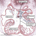
Heart four-chamber view
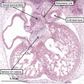
Heart four-chamber view
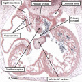
Heart four-chamber view
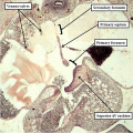
Heart septum primum
Neural tube model
Stage 16 Optical Projection Tomography
Stage16 cleft palate

External ear Stages 14-23 and adult


Historic Images
Bonnet E. and Severs R. On the structure of a human embryo eleven millimeters in length. (1906) Anat. Anz., 29: 452-459.
Model left
Model right
Model posterior
Model anterior


Streeter GL. The peripheral nervous system in the human embryo at the end of the first month (10 mm) (1908) Amer. J Anat. 8(1): 285–302.
10 mm Embryo
10 mm Embryo
10 mm Embryo
10 mm Embryo


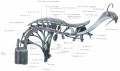
Congdon ED. Transformation of the aortic-arch system during the development of the human embryo. (1922) Contrib. Embryol., Carnegie Inst. Wash. Publ 277, 14:47-110.
Cardiovascular ventral view 11 mm embryo
Cardiovascular lateral view 11 mm embryo
Frazer embryo 10mm CRL
1921 Keith Fig. 44


- Carnegie Stages: 1 | 2 | 3 | 4 | 5 | 6 | 7 | 8 | 9 | 10 | 11 | 12 | 13 | 14 | 15 | 16 | 17 | 18 | 19 | 20 | 21 | 22 | 23 | About Stages | Timeline
Cite this page: Hill, M.A. (2026, July 6) Embryology Carnegie stage 16. Retrieved from https://embryology.med.unsw.edu.au/embryology/index.php/Carnegie_stage_16
- © Dr Mark Hill 2026, UNSW Embryology ISBN: 978 0 7334 2609 4 - UNSW CRICOS Provider Code No. 00098G