Category:Heart: Difference between revisions
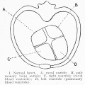
(→Notes) |
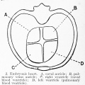
|||
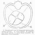
| (13 intermediate revisions by 2 users not shown) | |||
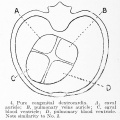
| Line 1: | Line 1: | ||
This {{Embryology}} category shows media and pages related to heart (cardiac) development. '''Pages''' section on this current page include lectures, laboratories, notes, quizzes and educational module sections that relate to cardiovascular development. | |||
{{Heart Links}} | |||
{{Heart - Historic References table}} | |||
[[Category:Muscle]][[Category:Cardiovascular]] | |||
Latest revision as of 09:14, 15 February 2017
This Embryology category shows media and pages related to heart (cardiac) development. Pages section on this current page include lectures, laboratories, notes, quizzes and educational module sections that relate to cardiovascular development.
| Heart - Historic References | ||
|---|---|---|
Tandler J. The Development of the Heart. (1912) Sect. II, chapt. 18, vol. 2, in Keibel F. and Mall FP. Manual of Human Embryology II. (1912) J. B. Lippincott Company, Philadelphia., pp. 534-570. Mall FP. On the development of the human heart. (1912) Amer. J Anat. 13: 249-298. Abbott ME. Congenital Cardiac Disease (1915) Osler & Mccrae's Modern Medicine 6, 2nd Edition. Frazer JE. The formation of the pars membranacea septi. (1916) J Anat. 51(1): 19-29. PMID 17103800 Waterston D. The development of the heart in man. (1917) Trans. Roy. Soc. Edin., 7(2): 258-302. McClure CFW. and Butler EG. The development of the vena cava inferior in man. (1925) Amer. J Anat. 35(3): 331-383. Odgers PNB. The formation of the venous valves, the foramen secundum and the septum secundum in the human heart. (1935) J. Anat., 69: 412-422. PMID 17104548 Odgers PN. The development of the pars membranacea septi in the human heart. (1938) J Anat. 72(2): 247-59. https://www.ncbi.nlm.nih.gov/pubmed/17104688 PMID 17104688] Patten BM. Developmental defects at the foramen ovale. (1938) Am J Pathol. 14(2):135-162. PMID 19970381 Odgers PNB. The development of the atrio-ventricular valves in man. (1939) J Anat. 73: 643-57. PMID 17104787 Kramer TC. The partitioning of the truncus and conus and the formation of the membranous portion of the interventricular septum in the human heart. (1942) Amer. J Anat. 71(3): 343-370. |

Subcategories
This category has the following 4 subcategories, out of 4 total.
Pages in category 'Heart'
The following 200 pages are in this category, out of 257 total.
(previous page) (next page)A
- Template:Abbott Figures
- Template:Abbott1915
- Template:Abbott1915images
- Abnormal Chick Heart Movie 1
- Abnormal Chick Heart Movie 2
- Advanced Cardiac Embryology
- ANAT2241 Cardiovascular System
- ANAT2341 Lab 11 - Heart
- ANAT2341 Lab 4 - Early Cardiovascular Development
- Template:Anderson2016 collapsetable2
- Template:Anderson2016 figures
- Template:Anderson2016 gallery1
- Template:Anderson2016 table2
- Template:Aortic stenosis
- Template:Atrial septal defects
B
- Basic - Embryonic Heart Divisions
- Basic - Primitive Heart Tube
- Basic - Vascular Heart Connections
- Basic Cardiac Embryology
- BGDA Lecture - Development of the Embryo/Fetus 2
- BGDA Practical 7 - Week 5
- Book - Congenital Cardiac Disease (1915)
- Book - Congenital Cardiac Disease - Figures
- Book - Congenital Cardiac Disease 1
- Book - Congenital Cardiac Disease 10
- Book - Congenital Cardiac Disease 11
- Book - Congenital Cardiac Disease 12
- Book - Congenital Cardiac Disease 13
- Book - Congenital Cardiac Disease 14
- Book - Congenital Cardiac Disease 15
- Book - Congenital Cardiac Disease 2
- Book - Congenital Cardiac Disease 3
- Book - Congenital Cardiac Disease 4
- Book - Congenital Cardiac Disease 5
- Book - Congenital Cardiac Disease 6
- Book - Congenital Cardiac Disease 7
- Book - Congenital Cardiac Disease 8
- Book - Congenital Cardiac Disease 9
- Book - Manual of Human Embryology 18-2
- Book - Quain's Embryology 9
- Book - Text-Book of the Embryology of Man and Mammals 17-1
C
- Template:Cardiac
- Cardiac Embryology
- Cardiac Muscle Histology
- Template:Cardiac neural crest
- Cardiovascular 3D stage 13 Movie
- Cardiovascular System - Abnormalities
- Cardiovascular System - Atrial Septal Defects
- Cardiovascular System - Circulation Development
- Cardiovascular System - Coarctation of the Aorta
- Cardiovascular System - Coronary Circulation Development
- Cardiovascular System - Developmental Shunts
- Cardiovascular System - Heart Development
- Cardiovascular System - Heart Histology
- Cardiovascular System - Heart Rate Development
- Cardiovascular System - Heart Valve Development
- Cardiovascular System - Hypoplastic Left Heart
- Cardiovascular System - Movies
- Cardiovascular System - Patent Ductus Arteriosus
- Cardiovascular System - Tetralogy of Fallot
- Cardiovascular System - Transposition of the Great Vessels
- Cardiovascular System - Tricuspid Atresia
- Cardiovascular System - Ventricular Septal Defects
- Cardiovascular System Development
- Carnegie Stage 17 Neural Movie
- Template:CHARGE syndrome
- Template:Coarctation of the aorta
- Template:Common truncus
- Template:Coronary circulation
- Template:CS11
- Template:CS12
- Template:CS13
- Template:CVS cartoons
- Template talk:CVS cartoons
E
F
H
- Template:Heart
- Template:Heart - Historic References table
- Template:Heart Abnormal
- Template:Heart abnormal cartoon gallery
- Template talk:Heart abnormal cartoon gallery
- Template:Heart abnormality timeline
- Heart Atrial Septation Movie
- Template:Heart histology
- Heart Historic Movie 1951
- Template:Heart historic movies
- Template talk:Heart historic movies
- Heart Looping Movie
- Heart Outflow Septation Movie
- Template:Heart rate
- Heart Realign Movie
- Template:Heart terms
- Template:Heart valve
- Template:Heart Volume table1
- Template:Heart Volume table2
- Template:Historic Heart 1951
- HM Practical - Cardiac Histology
- Template:Hypoplastic left heart
I
- Template:ICD-10-circulatory system Q20-Q28 table
- Intermediate - Atrial Ventricular Septation
- Intermediate - Cardiac Abnormalities
- Intermediate - Heart Tube Looping
- Intermediate - Heart Valves
- Intermediate - Outflow Tract
- Intermediate - Primordial Heart Tube
- Intermediate - Vascular Overview
- Intermediate Cardiac Embryology
- Template:Intra-embryonic coelom
L
M
P
- Paper - A contribution to the early development of the heart in mammalia, with special reference to the guinea pig
- Paper - Coarctation of the aorta 1942
- Paper - Development of the human heart from its earliest appearance to the stage found in embryos of twenty paired somites (1927)
- Paper - Development of the outflow tract and closure of the interventricular septum in the normal human heart
- Paper - Developmental Changes in the Pericardium, the Mesocardia, and the Pleural Sacs in the Human Embryo
- Paper - Developmental defects at the foramen ovale (1938)
- Paper - First contractions of the heart without cytological differentiation
- Paper - Functional limitations of the foramen ovale in the human foetal heart
- Paper - Growth allometry of the myocardium in human embryos from stages 15 to 23
- Paper - On the development of the atrial septum and the valvular apparatus in the right atrium of the pig embryo (1916)
- Paper - On the Development of the Human Heart
- Paper - On the muscular architecture of the ventricles of the human heart
- Paper - On the time of the post-natal obliteration of the fetal blood-passages (1918)
- Paper - Six specimens of abnormal heart (1912)
- Paper - Teratogenecity in the setting of cardiac development and maldevelopment
- Paper - The course of the blood flow through the fetal mammalian heart
- Paper - The Development of the Atrio-Ventricular Valves in Man
- Paper - The development of the cardiac-coronary circulatory system
- Paper - The development of the heart in man
- Paper - The Development of the Pars Membranacea Septi in the Human Heart
- Paper - The ductus arteriosus in the human fetus and newborn infant
- Paper - The early stages of the development of the pericardium
- Paper - The effect of the heart-beat upon the development of the vascular system in the chick (1918)
- Paper - The first contractions of the heart in rat embryos
- Paper - The formation of the cardiac loop in the chick
- Paper - The Formation of the Pars Membranacea Septi (1916)
- Paper - The formation of the venous valves, the foramen secundum and the septum secundum in the human heart
- Paper - The fusion of the cardiac anlages and the formation of the cardiac loop in the cat (1916)
- Paper - The human embryonic heart in the ninth week
- Paper - The human embryonic heart in the ninth week (1954)
- Paper - The human embryonic heart in the seventh week (1962)
- Paper - The origin of the heart and blood vessels in felis domestica (1924)
- Paper - The partitioning of the truncus and conus and the formation of the membranous portion of the interventricular septum in the human heart (1942)
- Paper - The physiology of the embryonic mammalian heart before circulation
- Paper - Transposition of the ventricles and the arterial stems (1931)
- Paper- The primary divisions of the myocardium in the human embryo
- Template:Patent ductus arteriosus
R
- Template:Ref-Anderson2016
- Template talk:Ref-Anderson2016
- Template:Ref-Beattie1939
- Template:Ref-Chapman1918
- Template:Ref-Chase1916
- Template:Ref-Davis1925
- Template:Ref-Davison1935
- Template:Ref-deVries1962
- Template:Ref-Fawcett1900
- Template:Ref-Field1946
- Template:Ref-Frazer1916
- Template:Ref-Goldsmith1937
- Template:Ref-Goss1938
- Template:Ref-Goss1940
- Template:Ref-Goss1942
- Template:Ref-Irvine1942
- Template:Ref-Keen1942
- Template:Ref-Keith1912
- Template:Ref-Kellogg1928
- Template:Ref-Licata1954
- Template:Ref-Magovern1986
- Template:Ref-Mall1893heart
- Template:Ref-Mall1902
- Template:Ref-Mall1911-heart
Media in category 'Heart'
The following 200 files are in this category, out of 434 total.
(previous page) (next page) 'Overriding' Aorta.PNG 840 × 575; 427 KB
 Abbott 16-18.jpg 771 × 1,000; 164 KB
 Abbott 19.jpg 880 × 791; 171 KB
 Abbott 191.jpg 1,034 × 1,000; 234 KB
 Abbott 1915.jpg 554 × 776; 38 KB
 Abbott 20.jpg 854 × 800; 129 KB
 Abbott 201.jpg 1,150 × 1,000; 203 KB
 Abbott 21.jpg 619 × 1,000; 151 KB
 Abbott 211.jpg 600 × 600; 51 KB
 Abbott 212.jpg 600 × 600; 55 KB
 Abbott 213.jpg 600 × 600; 51 KB
 Abbott 214.jpg 600 × 600; 54 KB
 Abbott 215.jpg 600 × 600; 50 KB
 Abbott 216.jpg 600 × 600; 78 KB
 Abbott 22.jpg 947 × 800; 220 KB
 Abbott 23.jpg 884 × 800; 167 KB
 Abbott 231.jpg 914 × 800; 163 KB
 Abbott 24.jpg 588 × 800; 129 KB
 Abbott 241.jpg 827 × 800; 148 KB
 Abbott 25.jpg 585 × 810; 126 KB
 Abbott 251.jpg 908 × 1,000; 167 KB
 Abbott 26.jpg 933 × 728; 140 KB
 Abbott 261.jpg 953 × 823; 162 KB
 Abbott 27.jpg 628 × 796; 132 KB
 Abbott 271.jpg 979 × 901; 161 KB
 Abbott 28.jpg 607 × 754; 118 KB
 Abbott 281.jpg 681 × 1,000; 151 KB
 Abbott 29.jpg 606 × 597; 111 KB
 Abbott 291.jpg 1,030 × 800; 188 KB
 Abbott 30.jpg 500 × 674; 85 KB
 Abbott 301.jpg 779 × 800; 118 KB
 Abbott 31.jpg 684 × 535; 81 KB
 Abbott 311.jpg 1,134 × 800; 162 KB
 Abbott 32-34.jpg 846 × 800; 84 KB
 Abbott 32.jpg 504 × 374; 21 KB
 Abbott 33.jpg 504 × 374; 19 KB
 Abbott 34.jpg 504 × 374; 22 KB
 Abbott plate 05.jpg 671 × 1,000; 132 KB
 Abbott plate 51.jpg 618 × 800; 108 KB
 Adult heart CT01.jpg 957 × 951; 212 KB
 Adult heart outflow tract CT01.jpg 747 × 747; 58 KB
 Adult heart outflow tract CT02.jpg 747 × 747; 65 KB
 Adult Heart Valves.jpg 1,475 × 1,070; 113 KB
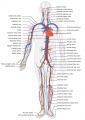 Adult human cardiovascular system.jpg 707 × 1,000; 151 KB
 Advanced Heart Development Timeline.jpg 1,772 × 769; 158 KB
 Anderson2016-fig01.jpg 800 × 800; 93 KB
 Anderson2016-fig02.jpg 800 × 788; 75 KB
 Anderson2016-fig03.jpg 800 × 800; 130 KB
 Anderson2016-fig04.jpg 800 × 800; 99 KB
 Anderson2016-fig05.jpg 796 × 573; 66 KB
 Anderson2016-fig06.jpg 800 × 800; 123 KB
 Anderson2016-fig07a.jpg 800 × 800; 193 KB
 Anderson2016-fig07b.jpg 800 × 800; 304 KB
 Anderson2016-fig08a.jpg 800 × 800; 112 KB
 Anderson2016-fig08b.jpg 800 × 800; 107 KB
 Anderson2016-fig09a.jpg 800 × 800; 106 KB
 Anderson2016-fig09b.jpg 800 × 800; 90 KB
 Anderson2016-fig10.jpg 800 × 800; 109 KB
 Anderson2016-fig11a.jpg 800 × 800; 108 KB
 Anderson2016-fig11b.jpg 800 × 800; 98 KB
 Anderson2016-fig12a.jpg 800 × 800; 120 KB
 Anderson2016-fig12b.jpg 800 × 800; 138 KB
 Anderson2016-fig13a.jpg 800 × 800; 115 KB
 Anderson2016-fig13b.jpg 800 × 800; 191 KB
 Anderson2016-fig14.jpg 800 × 800; 168 KB
 Anderson2016-fig15a.jpg 800 × 800; 142 KB
 Anderson2016-fig15b.jpg 800 × 800; 148 KB
 Anderson2016-fig16a.jpg 800 × 800; 193 KB
 Anderson2016-fig16b.jpg 800 × 800; 167 KB
 Anderson2016-fig17a.jpg 800 × 800; 98 KB
 Anderson2016-fig17b.jpg 800 × 800; 95 KB
 Anderson2016-fig18.jpg 800 × 800; 105 KB
 Anderson2016-fig19.jpg 800 × 800; 99 KB
 Anderson2016-fig20.jpg 800 × 800; 101 KB
 Anderson2016-fig21.jpg 762 × 800; 74 KB
 Anderson2016-fig22.jpg 800 × 779; 90 KB
 Anderson2016-fig23.jpg 783 × 800; 97 KB
 Anderson2016-fig24a.jpg 800 × 800; 107 KB
 Anderson2016-fig24b.jpg 800 × 800; 109 KB
 Anderson2016-fig25a.jpg 800 × 800; 122 KB
 Anderson2016-fig25b.jpg 800 × 800; 83 KB
 Anderson2016-fig26a.jpg 800 × 800; 126 KB
 Anderson2016-fig26b.jpg 800 × 800; 103 KB
 Anderson2016-fig27a.jpg 800 × 800; 117 KB
 Anderson2016-fig27b.jpg 800 × 800; 101 KB
 Anderson2016-fig28a.jpg 800 × 800; 117 KB
 Anderson2016-fig28b.jpg 800 × 800; 110 KB
 Anderson2016-fig29a.jpg 800 × 800; 111 KB
 Anderson2016-fig29b.jpg 800 × 800; 83 KB
 Anderson2016-fig30.jpg 647 × 800; 82 KB
 Anderson2016-fig31.jpg 800 × 800; 99 KB
 Anderson2016-fig32a.jpg 800 × 800; 108 KB
 Anderson2016-fig32b.jpg 800 × 800; 89 KB
 Anderson2016-fig33a.jpg 800 × 800; 110 KB
 Anderson2016-fig33b.jpg 800 × 800; 127 KB
 Anderson2016-fig34a.jpg 800 × 800; 105 KB
 Anderson2016-fig34b.jpg 800 × 800; 92 KB
 Anderson2016-fig35a.jpg 800 × 800; 118 KB
 Anderson2016-fig35b.jpg 800 × 800; 84 KB
 Anderson2016-fig36.jpg 800 × 800; 112 KB
 Anderson2016-fig37.jpg 800 × 800; 69 KB
 Anderson2016-fig38.jpg 800 × 800; 149 KB
 Anderson2016-fig39a.jpg 800 × 800; 86 KB
 Anderson2016-fig39b.jpg 800 × 800; 108 KB
 Anderson2016-fig40a.jpg 800 × 800; 102 KB
 Anderson2016-fig40b.jpg 800 × 800; 126 KB
 Anderson2016-fig41a.jpg 800 × 755; 92 KB
 Anderson2016-fig41b.jpg 800 × 800; 113 KB
 Anderson2016-fig42a.jpg 800 × 800; 90 KB
 Anderson2016-fig42b.jpg 800 × 800; 111 KB
 Anderson2016-fig43a.jpg 800 × 800; 60 KB
 Anderson2016-fig43b.jpg 800 × 800; 48 KB
 Anderson2016-fig44a.jpg 800 × 800; 83 KB
 Anderson2016-fig44b.jpg 800 × 800; 76 KB
 Anderson2016-fig45a.jpg 800 × 800; 62 KB
 Anderson2016-fig45b.jpg 800 × 800; 57 KB
 Anderson2016-fig46a.jpg 800 × 800; 100 KB
 Anderson2016-fig46b.jpg 800 × 800; 120 KB
 Anderson2016-fig47.jpg 800 × 800; 83 KB
 Anderson2016-fig48.jpg 800 × 800; 203 KB
 Anderson2016-fig49.jpg 800 × 800; 90 KB
 Anderson2016-fig50a.jpg 668 × 783; 139 KB
 Anderson2016-fig50b.jpg 798 × 786; 170 KB
 Aorta coarctation echocardiogram.jpg 601 × 283; 28 KB
 Aorta coarctation MRI.jpg 455 × 423; 25 KB
 Aortic arch and ductus arteriosus.jpg 600 × 720; 68 KB
 Aortic Stenosis.jpg 290 × 350; 16 KB
 Arey1924 fig371.jpg 1,000 × 626; 130 KB
 Atrial & Ventricular Septation 1.jpg 1,482 × 960; 97 KB
 Atrial & Ventricular Septation 2.jpg 1,482 × 1,075; 113 KB
 Atrial Septal Defect.jpg 287 × 350; 16 KB
 Atrial Septation.jpg 1,482 × 960; 90 KB
 Australia Congenital heart disease 2016–17.png 1,296 × 989; 94 KB
 AV Canal Division (Superior View).jpg 1,487 × 489; 69 KB
 AV Canal Division.jpg 1,482 × 970; 93 KB
 AV Valves.jpg 1,183 × 1,085; 129 KB
 Bailey170.jpg 709 × 457; 67 KB
 Bailey171.jpg 954 × 507; 81 KB
 Bailey172.jpg 894 × 444; 82 KB
 Bailey173.jpg 888 × 620; 113 KB
 Bailey174.jpg 955 × 542; 88 KB
 Bailey175.jpg 885 × 306; 51 KB
 Bailey176.jpg 918 × 352; 62 KB
 Bailey177.jpg 943 × 873; 199 KB
 Bailey178.jpg 913 × 653; 125 KB
 Bailey179.jpg 892 × 794; 114 KB
 Bailey181.jpg 839 × 658; 67 KB
 Baroreceptor reflex cartoon.jpg 1,200 × 1,012; 182 KB
 Basic Heart Development Timeline.jpg 1,658 × 556; 73 KB
 Bedford01.jpg 734 × 1,000; 82 KB
 Bedford02.jpg 1,069 × 911; 266 KB
 Bremer1906 fig09.jpg 915 × 1,200; 208 KB
 Cardiac Conduction System.jpg 1,201 × 862; 81 KB
 Cardiac connexins cartoon.jpg 990 × 621; 112 KB
 Cardiac muscle EM01.jpg 1,072 × 735; 231 KB
 Cardiac muscle EM02.jpg 1,072 × 735; 224 KB
 Cardiac muscle EM03.jpg 849 × 615; 135 KB
 Cardiac muscle EM04.jpg 1,000 × 680; 191 KB
 Cardiac Muscle EM05.jpg 992 × 733; 158 KB
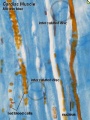 Cardiac muscle histology.jpg 300 × 400; 42 KB
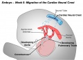 Cardiac Neural Crest Migration.jpg 1,517 × 1,116; 122 KB
 Cephalic plexus.png 600 × 557; 559 KB
 Cervical intersomitic vessels.png 600 × 462; 308 KB
 Chick Heart 002-icon.jpg 320 × 240; 5 KB
 Chicken heart 3D reconstruction from sections.jpg 1,000 × 571; 104 KB
 Coarctation of the Aorta.jpg 289 × 350; 16 KB
 Cockle01.jpg 881 × 1,000; 158 KB
 Cockle02.jpg 1,459 × 929; 266 KB
 Cockle03.jpg 810 × 1,062; 158 KB
 Cockle04.jpg 740 × 1,065; 135 KB
 Complete atrioventricular canal.jpg 320 × 240; 22 KB
 Congenital Heart Disease.jpg 475 × 421; 106 KB
 Corner1929 fig10-11.jpg 1,200 × 1,438; 730 KB
 Coronary arteries.png 800 × 472; 183 KB
 Davis1927 plate01.jpg 1,357 × 1,500; 501 KB
 Davis1927 plate02.jpg 1,357 × 1,500; 795 KB
 Dextrocardia heart position.jpg 400 × 533; 49 KB
 Dextrocardia.jpg 400 × 533; 30 KB
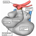
 Divisions of Early Heart Tube.jpg 1,105 × 978; 85 KB
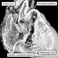
 Double Outlet Right Ventricle.jpg 289 × 350; 16 KB
 Early Development of Heart Tube.jpg 1,475 × 1,099; 132 KB
 Early Heart Tube (Dorsal).jpg 1,282 × 1,124; 111 KB
 Early Heart Tube (Lateral).jpg 1,504 × 972; 110 KB
 Ectopia cordis.jpg 800 × 603; 37 KB
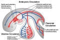 Embryonic Circulations.jpg 1,552 × 1,028; 171 KB
 Fetal blood flow 04.jpg 506 × 599; 50 KB
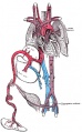 Fetal circulation1.jpg 558 × 900; 76 KB
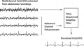 Fetal Electrocardiogram Enhancement 01.jpg 657 × 370; 40 KB
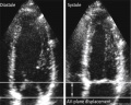 Fetal heart atrioventricular plane displacement 01.jpg 915 × 733; 55 KB
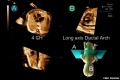 Fetal ultrasound ductal arch 01.jpg 800 × 533; 27 KB
 Folding animation 001.mov ; 1.32 MB
- Folding animation 002.mov ; 503 KB
 Frazer1916 fig01.jpg 878 × 879; 211 KB
 Frazer1916 fig02.jpg 904 × 892; 233 KB
 Frazer1916 fig03.jpg 1,469 × 828; 317 KB
 Frazer1916 fig04.jpg 1,221 × 1,020; 208 KB
 Functional Hypoplastic Left Heart.jpg 302 × 350; 18 KB
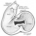
 Gray0458.gif 442 × 600; 34 KB
 Gray0458.jpg 693 × 911; 83 KB
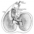
 Gray0459.jpg 1,195 × 961; 258 KB


.jpg)
.jpg)


.jpg)