Lecture - 2016 Course Introduction: Difference between revisions
mNo edit summary |
mNo edit summary |
||
| (15 intermediate revisions by the same user not shown) | |||
| Line 15: | Line 15: | ||
I like my lectures to be interactive, so ask me questions and I will also be asking you questions! | I like my lectures to be interactive, so ask me questions and I will also be asking you questions! | ||
[[Media: | [[Media:Lecture - 2016 Course Introduction - Embryology.pdf|Lecture - Print PDF]] | ||
|} | |} | ||
| Line 27: | Line 26: | ||
# Broad overview of human development. | # Broad overview of human development. | ||
<html5media height="384" width="352">File:Human development 001.mp4</html5media> | {| | ||
| {{OneMinuteClock}} | |||
[[One_Minute_Embryology#Human_Development|'''1 Minute Embryology''']] | |||
| <html5media height="384" width="352">File:Human development 001.mp4</html5media> | |||
Here is the whole course in One Minute. | Here is the whole course in One Minute. | ||
|} | |||
{| class="wikitable mw-collapsible mw-collapsed" | {| class="wikitable mw-collapsible mw-collapsed" | ||
! Introduction Movies | ! colspan=3|Introduction Movies | ||
|- | |- | ||
| valign="bottom"|{{Human fertilization movie 1}} | | valign="bottom"|{{Human fertilization movie 1}} | ||
| Line 42: | Line 45: | ||
| [[Birth]] (week 37) | | [[Birth]] (week 37) | ||
|} | |} | ||
==Understand the Course== | |||
* [[Media:2016-ANAT2341-course-outline.pdf|2016 Course Outline]] | |||
* [[ANAT2341 Course Timetable 2016]] | |||
* [[Embryology Textbooks - UNSW]] | |||
{{UNSW textbook - The Developing Human}} | |||
{{UNSW textbook - Larsen's Human Embryology}} | |||
{{2016ANAT2341textbooktable}} | |||
[[Science Student Projects]] | |||
==History== | |||
[[History - Embryologists]] | [[Embryology History]] | [[Human Embryo Collections]] | |||
[[File:BrauneB1.jpg|400px|alt=The Position of the Uterus and Fetus at Term (1872)]] | |||
[[Embryology_History_-_17th_and_18th_Century_Anatomies|17-18C]] Braune - The Position of the Uterus and Fetus at Term (1872) | |||
{| class="wikitable mw-collapsible mw-collapsed" | |||
! colspan="3"| [[Human Embryo Collections]] | |||
|- | |||
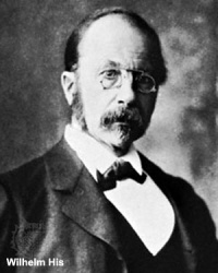
| [[File:Wilhelm_His.jpg|link=Embryology History - Wilhelm His|200px]] | |||
[[Embryology History - Wilhelm His|Wilhelm His]] (1831-1904) | |||
His's Normentafel (Normal Table) | |||
[[Book - Anatomy Of Human Embryos|Anatomie menschlicher Embryonen]] (1882) | |||
| [[File:Keibel_Mall_034a.jpg|200px]] | |||
| [[File:Keibel_Mall_034b.jpg|300px]] | |||
|- | |||
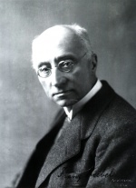
| [[File:Franz Keibel.jpg|link=Embryology History - Franz Keibel|150px]] | |||
[[Embryology History - Franz Keibel|Franz Keibel]] (1861 - 1929) | |||
Franz Keibel and Curt Elze (1908) Normal Plates of the Development of the Human Embryo | |||
| [[File:Keibel1908_plate01.jpg|200px]] | |||
| [[File:Keibel1908_plate02.jpg|200px]] | |||
|- | |||
| [[File:Franklin Mall 03.jpg|150px]] | |||
[[Embryology History - Franklin Mall|Franklin Mall]] (1862-1917) | |||
[[Carnegie Collection]] | |||
| colspan=2|[[File:Human Carnegie stage 10-23.jpg|400px]] | |||
|- | |||
| Begun by [[:File:Hideo Nishimura.jpg|Dr. Hideo Nishimura]] (1912–1995) | |||
[[File:Hideo Nishimura.jpg|150px]] | |||
Developed by Kohei Shiota and currently curated by Shigehito Yamada. | |||
[[File:Shiota_Hill_Yamada.jpg|200px]] | |||
[[Kyoto Collection]] | |||
| colspan=2|[[File:Human_Carnegie_stage_1-23.jpg|400px]] | |||
|} | |||
{| class="wikitable mw-collapsible mw-collapsed" | |||
! col span=2|[[Animal_Development|Animal Models]] | |||
|- | |||
| [[File:Frog-icon.png|right|80px|link=Frog Development]] | |||
| {{Frog links}} | |||
* The frog was used by many of the early embryology investigators and currently there are many different molecular mechanisms concerning development of the frog. | |||
* The eggs develop independently, in relative synchrony and are relatively see-through making staging and observation fairly easy. | |||
* The frog was a key model for the study of the process of gastrulation. | |||
|- | |||
| [[File:Chick icon.jpg|80px|link=Chicken Development]] | |||
| | |||
{{Chicken}} | |||
* The chicken embryo develops and hatches in 20-21 days and historically these were one of the first embryos to be studied. Cutting a window in the egg shell allows direct observation of the embryo. The Hamburger & Hamilton chicken development staging allowed researchers to develop this model as a key embryological tool. | |||
* Key research involved the transplanting of quail cells into chick embryos, to later identify their contribution to different embryonic structures, particularly for somite, neural tube and neural crest development. | |||
|- | |||
| [[File:Mouse.jpg|right|80px|link=Mouse Development]] | |||
| {{Mouse}} | |||
* The mouse has always been a good embryological model, easy to generate (litters 8-20) and quick (21d). | |||
* Mouse embryology really expanded when molecular biologists used mice for gene knockouts. | |||
|- | |||
| [[File:Fly-icon.png|right|80px|link=Fly Development]] | |||
| [[Fly Development|Fly Development]] - The fruitfly (drosophila) was and is the traditional geneticist's tool. It has been transformed to an magnificent embryologist's tool, with developmental mechanisms being uncovered in this system combined with homolgy gene searches in other species. The fly genome was one of the first to be been completely sequenced. In early development nurse cells ''sacrifice'' their cytoplasmic contents to allow egg growth and early pattern formation is through the localization of maternal messenger RNAs (mRNAs). | |||
|- | |||
| | [[File:C elegans.jpg|right|80px|link=Worm Development]] | |||
| [[Worm Development|Worm Development]] - Early embryological studies of the worm ''Caenorhabditis elegans'' (C.Elegans, so called because of its "elegant" curving movement) characterized the fate of each and every cell in the worm through all stages of development. This worm has recently had its entire genome sequenced. | |||
|- | |||
| [[File:Zebrafish-icon.png|right|80px|link=Zebrafish Development]] | |||
| [[Zebrafish Development|Zebrafish Development]] - Zebrafish are seen as the latest and greatest "model' for embryological development studies. They can be easily genetically altered and develop as practically "see through" embryos, all internal development can be clearly observed from the outside in the living embryo. | |||
|} | |||
{| | |||
! 1978 | |||
! 1981 | |||
! 1996 | |||
! 2006 | |||
! Ongoing | |||
|- | |||
| [[Assisted_Reproductive_Technology|In Vitro Fertilization]] | |||
| [[Stem Cells]] | |||
| [[Somatic Cell Nuclear Transfer]] | |||
| [[Embryology History - Shinya Yamanaka|Induced Stem Cells]] | |||
| [[Molecular Development]] | |||
|- | |||
| [[File:Intracytoplasmic_sperm_insemination.jpg|150px]] | |||
| [[File:Hematopoietic_and_stromal_cell_differentiation.jpg|150px]] | |||
| [[File:Dolly the sheep.jpg|150px]] | |||
| [[File:Mouse- embryonic stem cell signaling regulation.jpg|150px]] | |||
| [[File:Hedgehog signaling pathway.jpg|150px]] | |||
|} | |||
==Australian Data== | |||
21 July 2016 the resident population of Australia was projected to be: 24,135,202. | |||
[[File:Australian-births_2011.jpg|600px]] | |||
{| | |||
! [[Australian Statistics]] | |||
|- | |||
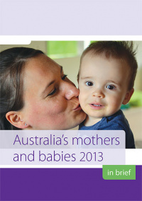
| width=250px|[[File:Australia mothers and babies 2013.jpg|link=Australia’s_mothers_and_babies_2013|200px]] | |||
| width=250px|[[File:Assisted reproductive technology in Australia and New Zealand 2010.jpg|200px]] | |||
|- | |||
| Australia’s mothers and babies (2013) | |||
| Assisted reproductive technology in Australia and New Zealand (2010) | |||
|- | |||
| Average maternal age in 2013 was [[Genetic_risk_maternal_age|'''30.1''']] years, the same as 2009 but still more than the earlier years (2000, 29.0 years; 2002, 29.4 years). Birth number was 309,489 babies in 2013, an increase of 20% from 256,925 in 2003. | |||
| [[Assisted Reproductive Technology]] (ART) was used by '''3.8%''' (2009, 3.6%) of women who gave birth. | |||
|} | |||
{{Victoria abnormal data table 2004}} | |||
==Human Development== | |||
[[File:Human development timeline graph 02.jpg|800px]] | |||
{{2016ANAT2341 footer}} | {{2016ANAT2341 footer}} | ||
Latest revision as of 13:33, 2 August 2016
| Embryology - 7 Jul 2026 |
|---|
| Google Translate - select your language from the list shown below (this will open a new external page) |
|
العربية | català | 中文 | 中國傳統的 | français | Deutsche | עִברִית | हिंदी | bahasa Indonesia | italiano | 日本語 | 한국어 | မြန်မာ | Pilipino | Polskie | português | ਪੰਜਾਬੀ ਦੇ | Română | русский | Español | Swahili | Svensk | ไทย | Türkçe | اردو | ייִדיש | Tiếng Việt These external translations are automated and may not be accurate. (More? About Translations) |
Course Introduction

Course coordinator |
This first lecture will be a general introduction to the course and the subject of Embryology.
|
Lecture Objectives

- Understand the course objectives and assessment.
- Brief understanding of the historic background of embryology.
- Brief understanding of Australian data.
- Broad overview of human development.
 |
<html5media height="384" width="352">File:Human development 001.mp4</html5media>
Here is the whole course in One Minute. |
| Introduction Movies | |||||||||||
|---|---|---|---|---|---|---|---|---|---|---|---|
|
|
| |||||||||
| Fertilization | Embryonic Development (week 1 - 8) | Birth (week 37) | |||||||||
Understand the Course
| The Developing Human: Clinically Oriented Embryology (10th edn) |
|---|
 UNSW Students have online access to the current 10th edn. through the UNSW Library subscription (with student Zpass log-in).
|
|

History
History - Embryologists | Embryology History | Human Embryo Collections

17-18C Braune - The Position of the Uterus and Fetus at Term (1872)
| Human Embryo Collections | ||
|---|---|---|
Wilhelm His (1831-1904) His's Normentafel (Normal Table) |

|

|
Franz Keibel (1861 - 1929) Franz Keibel and Curt Elze (1908) Normal Plates of the Development of the Human Embryo |

|

|

Franklin Mall (1862-1917) |

| |
| Begun by Dr. Hideo Nishimura (1912–1995)
Developed by Kohei Shiota and currently curated by Shigehito Yamada.
|

| |


| Animal Models | ||
|---|---|---|
| ||
| ||
 |
mouse
| |
| Fly Development - The fruitfly (drosophila) was and is the traditional geneticist's tool. It has been transformed to an magnificent embryologist's tool, with developmental mechanisms being uncovered in this system combined with homolgy gene searches in other species. The fly genome was one of the first to be been completely sequenced. In early development nurse cells sacrifice their cytoplasmic contents to allow egg growth and early pattern formation is through the localization of maternal messenger RNAs (mRNAs). | ||
 |
Worm Development - Early embryological studies of the worm Caenorhabditis elegans (C.Elegans, so called because of its "elegant" curving movement) characterized the fate of each and every cell in the worm through all stages of development. This worm has recently had its entire genome sequenced. | |
| Zebrafish Development - Zebrafish are seen as the latest and greatest "model' for embryological development studies. They can be easily genetically altered and develop as practically "see through" embryos, all internal development can be clearly observed from the outside in the living embryo. |
| 1978 | 1981 | 1996 | 2006 | Ongoing |
|---|---|---|---|---|
| In Vitro Fertilization | Stem Cells | Somatic Cell Nuclear Transfer | Induced Stem Cells | Molecular Development |

|

|

|

|

|
Australian Data
21 July 2016 the resident population of Australia was projected to be: 24,135,202.

| Australian Statistics | |
|---|---|
|

|
| Australia’s mothers and babies (2013) | Assisted reproductive technology in Australia and New Zealand (2010) |
| Average maternal age in 2013 was 30.1 years, the same as 2009 but still more than the earlier years (2000, 29.0 years; 2002, 29.4 years). Birth number was 309,489 babies in 2013, an increase of 20% from 256,925 in 2003. | Assisted Reproductive Technology (ART) was used by 3.8% (2009, 3.6%) of women who gave birth. |
| Victoria - 10 most reported birth anomalies | |
|---|---|
| Based upon statistics from the Victorian Perinatal Data Collection Unit in Victoria between 2003-2004. | |

|
hypospadias (More? External Genital Male Development Movie) |

|
Obstructive Defects of the Renal Pelvis (obstructive defects of the renal pelvis, uteropelvic junction obstruction, pelvo-uterero junction obstruction) Term describing a developmental renal abnormality due to partial or complete blockage of the drainage of the kidney pelvis requiring surgical correction. The blockage can also have several causes including: unusual ureter twisting or bending, ureter compression by a blood vessel, malformations of the muscular wall. The blockage leads to an accumulation of urine in the affected region, with several potential effects: nephron damage from compression (hydronephrosis); decreased urine output leading to lack of amniotic fluid (oligohydramnios); respiratory development effects due to the lack of amniotic fluid.
(More? renal abnormalities | renal) |

|
ventricular septal defect (More? ventricular septal defect)
Heart Development Timeline (see Basic Cardiac Embryology) |

|
Developmental dysplasia of the hip or Congenital Dislocated Hip
(Developmental dysplasia of the hip (DDH), congenital hip dislocation, congenital hip dysplasia) Term describes a spectrum of musculoskeletal disorders of hip instability due either to the femoral head being able to move outside the acetabulum (luxation or dislocation), or abnormally within the acetabulum (subluxation or partial dislocation). This includes presentation following a normal examination of the hips in the newborn period (Ortolani and Barlow tests). When detected can be managed with splinting (Denis-Browne splint) allows the hip joint to develop normally and does not require surgery. If undetected and left untreated, the hip joint develops abnormally and surgical reduction is required. (More? Pelvis Development) |

|
Trisomy 21 or Down syndrome - The most common genetic abnormality. (More? Trisomy 21) |

|
hydrocephalus rapid increase in head circumference or an unusually large head size due to excessive accumulation of cerebrospinal fluid in the brain.(More? hydrocephalus | Neural Abnormalities | NINDS - Hydrocephalus Fact Sheet | Hydrocephalus Support Association | USA National Hydrocephalus Foundation) |

|
cleft palate - The palate separates the nasal cavity from the oral cavity, the abnormality has many different causes, and occurs more frequently in females (57%) than in males (43%). (More? cleft palate) |

|
Trisomy 18 or Edward Syndrome - multiple abnormalities of the heart, diaphragm, lungs, kidneys, ureters and palate 86% discontinued (More? Trisomy 18) |
| Renal Agenesis/Dysgenesis - reduction in neonatal death and stillbirth since 1993 may be due to the more severe cases being identified in utero and being represented amongst the increased proportion of terminations (approximately 31%). (More? Renal Abnormalities - Renal Agenesis) | |

|
cleft lip and palate - occur with another defect in 33.7% of cases.(More? cleft lip and palate) |
| Links: Human Abnormal Development | Australian Statistics | Victoria 2004 | USA 2006 | Europe 2010 | |

Human Development

|
| |||||||||||||||||||||||||||||||||||||||||||||||||||||||||||||||||||||||||||||||||||||||||||||||||||||||||||||||||||||||||||||||||||||||||||||||||||
Glossary Links
- Glossary: A | B | C | D | E | F | G | H | I | J | K | L | M | N | O | P | Q | R | S | T | U | V | W | X | Y | Z | Numbers | Symbols | Term Link
Cite this page: Hill, M.A. (2026, July 7) Embryology Lecture - 2016 Course Introduction. Retrieved from https://embryology.med.unsw.edu.au/embryology/index.php/Lecture_-_2016_Course_Introduction
- © Dr Mark Hill 2026, UNSW Embryology ISBN: 978 0 7334 2609 4 - UNSW CRICOS Provider Code No. 00098G