M
| Embryology - 28 Apr 2024 |
|---|
| Google Translate - select your language from the list shown below (this will open a new external page) |
|
العربية | català | 中文 | 中國傳統的 | français | Deutsche | עִברִית | हिंदी | bahasa Indonesia | italiano | 日本語 | 한국어 | မြန်မာ | Pilipino | Polskie | português | ਪੰਜਾਬੀ ਦੇ | Română | русский | Español | Swahili | Svensk | ไทย | Türkçe | اردو | ייִדיש | Tiếng Việt These external translations are automated and may not be accurate. (More? About Translations) |
Glossary Links
- Glossary: A | B | C | D | E | F | G | H | I | J | K | L | M | N | O | P | Q | R | S | T | U | V | W | X | Y | Z | Numbers | Symbols | Term Link
M
MI
- (meiosis I) Acronym for meiosis I the first part of meiosis. Note, that depending on usage this acronym has been also used for metaphase I of meiosis I.
MII
- (meiosis II) Acronym for meiosis II, the second part of the meiotic nuclear division. Note, that depending on usage this acronym has been also used for metaphase II of meiosis II.
macrosomia
- (large gestational age, LGA) Term used to describe a newborn with an excessive birth weight. The definition is either a birth weight of 4000 to 4500 g (8 lb 13 oz to 9 lb 15 oz) or greater than 90% for gestational age after correcting for neonatal sex and ethnicity. There are a range of overgrowth syndromes associated with developmental delay, tumors, and other anomalies with genetic causes and syndromes (Pallister-Killian, Beckwith-Wiedemann, Sotos, Perlman, and Simpson-Golabi-Behmel) rarely diagnosed prenatally.
- (More? Macrosomia | Birth-Weight | Birth | Maternal Diabetes | Fetal Development | PMID 19609940)
main pancreatic duct
- (MPD, Wirsung's duct) The pancreas duct for exocrine secretion into the gastrointestinal tract, embryonically arises from the dorsal pancreatic bud and is present in the body and tail of the pancreas. An accessory duct (ventral) may be present as an anatomical variation due to the embryological origin of the pancreas from two pancreatic buds (dorsal and ventral).
magnesium sulfate
- (magnesium sulphate) See magnesium sulphate below.
magnesium sulphate
- (magnesium sulfate, epsom salt) A clinical tocolytic agent used to inhibit labor (labor-inhibiting), slowing or halting uterine contractions. Inhibitory mechanism not clear, but thought to; compete with calcium at the level of plasma membrane voltage-gated channels, hyperpolarize the plasma membrane, inhibit myosin light-chain kinase activity by competing with intracellular calcium. More recently recommended for neuroprotection in very preterm birth.
- (More? Birth | Preterm Birth | PMID 22227787)
magnetic resonance imaging
- (MRI, nuclear magnetic resonance imaging, NMRI) An imaging technique developed in 1973 that uses a powerful magnetic field to align the nuclear magnetization of hydrogen atoms in water in the body. This is then altered using a radio frequency (RF) field and leads to the hydrogen nuclei producing a rotating magnetic field detectable by the scanner. The technique has been recently further developed as micro-magnetic resonance imaging (MicroMRI) and diffusion tensor imaging that are particularly useful in embryological studies.
- (More? Magnetic Resonance Imaging)
magnetoencephalographic
- (MEG) A brain imaging system that measures electromagnetic changes in regions of the brain that correspond to activity.
major urinary proteins
- (MUPs) Proteins which carry volatile substances, including pheromones, and protect them during their internal passage (liver to kidneys into urine).
- (More? Mouse Estrous Cycle)
male factor
- Any cause of infertility due to deficiencies in sperm quantity, function, or motility (ability to move) that make it difficult for a sperm to fertilize an egg under normal conditions.
- (More? Week 1 | Spermatozoa Development)
malleus
- (Latin, malleus = hammer) One of the three ossicles (bones) of the middle ear (incus - malleus - stapes) that convert mechanical vibration into fluid movement within cochlea. The incus and the malleus embryonically form initially as a single structure from the first pharyngeal arch Meckel's cartilage, later formation of a joint separates the two bones.
- (More? Hearing - Middle Ear Development)
manchette
- A transient microtubule structure formed in spermatids involved in the process of assembly of the mammalian spermatozoa tail and mechanical shaping and condensation of the spermatozoa nucleus. These microtubules are also involved with specific transport, intramanchette transport, which has been likened to intraflagellar transport.
- (More? Spermatozoa Development | Testis Development | Fertilization)

mandible
- Term used to describe the lower jaw of the face, which forms from the lower part pharyngeal arch 1, the mandibular process. The smaller upper part of pharyngeal arch 1 forms the two maxillary processes, which form the upper jaw. Meckel's cartilage forms first embryonic structure that forms a template for where the mandible will form.
- (More? Head Development | Lecture - Head Development)
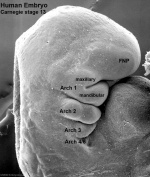
mandibular process
- (pharyngeal arch 1 mandibular process; Latin, mandibula = "jawbone") The lower portion of the first pharyngeal arch. This large prominence contributes the lower jaw. The smaller upper part of pharyngeal arch 1 is the maxillary process. In the embryo, Meckel's cartilage within the arch forms a template for the mandible location.
- (More? Head Development | Lecture - Head Development)

mammary gland
- A specialised modified secretory gland producing milk in female mammals for neonatal nutrition. Note that milk production and neonatal nutrition through milk, define us as mammals. The adult female breast contains large numbers of these secretory glands.
marginal sinus
- (subcapsular sinus) A space lying under the connective tissue capsule or covering of an organ, which receives lymph from afferent lymphatic vessels.
- (More? Lymphatics Development)
Marquette method
- (MM) A natural family planning technique used as a method of avoiding pregnancy, named after Marquette University. Couples track their fertility by self-observation of cervical mucus, by use of an electronic monitor that measures urinary levels of estrone-3-glucuronide and luteinizing hormone, and by use of basal body temperature.
- (More? Menstrual Cycle | PMID 18997569)
massive chronic intervillositis
- (chronic intervillositis, chronic histiocytic intervillositis) Rare placental abnormality and pathology defined by inflammatory placental lesions, mainly in the intervillous space (IVS), with a maternal infiltrate of mononuclear cells (monocytes, lymphocytes, histiocytes) and intervillous fibrinoid deposition.
- (More? Placenta - Abnormalities)
.jpg)
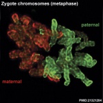
maternal
- Term used in relation to the female mother and is used genetically, biologically and legally. The term paternal relates to the male father.
maternal death
- Defined in ICD10 and WHO as the death of a woman while pregnant or within 42 days of termination of pregnancy, irrespective of the duration and site of the pregnancy, from any cause related to or aggravated by the pregnancy or its management but not from accidental or incidental causes. Pregnancy-related death
is defined as the death of a woman while pregnant or within 42 days of termination of pregnancy, irrespective of the cause of death. Late maternal death is defined as the death of a woman from direct or indirect obstetric causes, more than 42 days, but less than one year after termination of pregnancy.
- (More? Statistics - Maternal Mortality | ICD-10)
maternal passive immunity
- (maternal passive immunity) Term used to describe the transfer of maternal antibodies to the fetus (through the placenta) and the neonate (through milk).
maternal mortality rate
- Statistical term defined as the number of maternal deaths per 100,000 live births.
maternal spindle transfer
- (MST) An Assisted Reproductive Technology term. The “maternal spindle” is the group of maternal chromosomes within the oocyte, which are shaped in a spindle. The transfer technique involves removing the spindle from the mother’s oocyte before it is fertilised by the father’s spermatozoa. The spindle is then placed into a donor oocyte with healthy mitochondria, from which the donor’s spindle, and therefore her nuclear material, has been removed.
- (More? Assisted Reproductive Technology | Mitochondria)
maternal-to-zygotic transition
- (MZT) Term used to describe the genetic transition from maternal gene products to that of the zygote genome in early development.
- (More? PMID 19700615)
Math1
- (Mouse homolog of ATH1, also called Atoh1) Basic helix-loop-helix (bHLH) transcription factor both necessary and sufficient for hair cell development in the mammalian cochlea.
- (More? Inner Ear Development | Hearing Development | OMIM ATONAL)
matrix
- Cell biology and histology term used to describe the non-cellular material in which cells are embedded, as in the extracellular matrix. Epithelia have a specialized basal matrix while connective tissue (mesenchyme) cells have extensive extracellular matrix surrounding each cell. The term matrix is also used in cell biology to describe the space within mitochondria, enclosed by the two mitochondrial membranes.
MAVS
- Mitochondrial AntiViral Signaling, induces interferon expression and therefore increased antiviral defenses.
maxillary process
- (pharyngeal arch 1 maxillary process; Latin, maxilla = upper jaw) In head and face development, the upper part of pharyngeal arch 1 which forms as a pair of small lateral swellings which contributes the upper jaw and forms the palatal shelves. Larger lower part of pharyngeal arch 1 is the mandibular process. Failure of these upper processes to fuse with the frontonasal prominence leads to cleft lip and cleft palate.
- (More? Head Development)
Mayer- Rokitansky-Kuster-Hauser syndrome
- (MRKH) Abnormality of development of the female genital tract: partial or complete absence (agenesis) of the uterus; absent or hypoplastic vagina; normal fallopian tubes, ovaries, normal external genitalia and normal female chromosome pattern (46, XX). Has an incidence of approximately 1 in 4500 newborn girls and has been associated with a microdeletion at 17q12.
- (More? Uterus Development | Vagina Development | PMID 19889212)
MDCT
- Acronym for multidetector computed tomography.
- (More? Computed Tomography)
measles
- (paramyxovirus) Measles (rubeola) is mainly a respiratory viral infection, clinically different from Rubella. A single-stranded RNA virus which is highly contagious. Before measles vaccination (USA 1963) more than 90% of children had an infection before puberty and in developing countries it is still a common and often fatal childhood disease. Pregnancy effects of measles results in a higher risk of premature labor, spontaneous abortion, low-birth-weight, and possibly rare cases of birth defects with no definable pattern of malformation.
- (More? Viral Infection)
meatal plate
- An ectodermal plug that temporarily blocks the external auditory meatus (external acoustic meatus or ear canal) of the ear.
- (More? Outer Ear Development | Hearing Development)
meatus
- (Latin, meatus = a channel or way) An anatomical description of an opening or passageway (external auditory meatus, female urethral meatus).
- (More? Hearing Development)
meatoplasty
- A surgical technique allowing reconstructive surgery of a meatus or opening. The term is often used to describe the external ear canal, often used to treat external meatus stenosis. Can also be used to refer to other openings such as in the repair of a male genital hypospadia.
- (More? Ear Abnormalities)

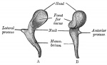
Meckel's cartilage
- (first arch cartilage) The first pharyngeal arch cartilage located within the mandibular prominence. It forms a template for the mandible and two of the middle ear ossicles (malleus and incus). This temporary cartilage first appears at Carnegie stage 16 and membranous ossification of the mandible occurs beside the template at Carnegie stage 20. Note that this is not endochondral ossification of the mandible, as the cartilage does not ossify and is lost entirely from this region. Named after Johann Friedrich Meckel, the Younger a German anatomist (1781 - 1833).
- (More? Head Development | cartoon - arch cartilages | Pharyngeal arches |

Meckel's diverticulum
- (omphalomesenteric duct malformation) Gastrointestinal tract developmental abnormality due to persistence of the early vitelline duct region. Named after Johann Friedrich Meckel (1781 - 1833) the Younger, a German anatomist.
- (More? Meckel's diverticulum | Gastrointestinal Tract Development | PMID 9138710)
meconium
- The gastrointestinal contents that accumulate in the intestines during the fetal period. This material is a mixture of liver bile and glandular secretions, amniotic fluid, and cellular debris. Meconium is also used to describe the first postnatal rectal discharge from the neonate. Fetal stress in the third trimester or at parturition can lead to premature meconium discharge, into the amniotic fluid and ingestion by the fetus (meconium aspiration syndrome) and damage to respiratory function. Damage to placental vessels meconium myonecrosis may also occur. See also meconium peritonitis and meconium periorchitis.
- (More? Birth | Meconium Aspiration Syndrome | Gastrointestinal Tract Development | Respiratory System Development)
meconium aspiration syndrome
- (MAS) Fetal stress in the third trimester, prior to/at/ or during parturition can lead to premature meconium discharge into the amniotic fluid and sunsequent ingestion by the fetus and damage to respiratory function. Damage to placental vessels meconium myonecrosis may also occur.
meconium myonecrosis
- Placental pathology resulting from prolonged meconium exposure which is toxic for myocytes of placental vessels (umbilical cord or chorionic plate).
- (More? Placenta - Abnormalities)
meconium peritonitis
- (MP) A sterile chemical peritonitis resulting from small bowel perforation leaking meconium in utero. Often detected by ultrasound and can result in a mortality rate as high as 60%.
meconium periorchitis
- A rare disorder caused following an initial fetal meconium peritonitis and subsequent leakage of meconium into the scrotal sac.
- (More? Meconium Aspiration Syndrome | PMID 19638993)
meconium plug syndrome
- (functional immaturity of the colon) Term used to describe a transient disorder of the newborn colon, which is characterized by delayed passage of meconium (more than 24 to 48 h), intestinal dilatation and yellow/green vomiting. More common in premature infants and can be determined by radiological dye study.
- (More? Gastrointestinal Tract - Abnormalities | Gastrointestinal Tract Development | Birth | PMID18485962 | U Mich - Meconium Plug Syndrome)
medial
- (Latin, medialis = middle) Anatomically towards the midline of the body or structure. The opposite anatomical term is lateral.
medial epithelial seam
- (MES) Embryonic structure formed by the fusion of the two palatal shelves, forming a two-layered medial edge epithelial seam, which is then lost with palate development.
- (More? Palate Development | Head Development | Lecture - Head Development | Medline Plus - Cleft Lip and Palate)
median section
- (Latin, medialis = middle) Histologically and anatomically a midline sagittal section or plane of the embryo or structure. The opposite anatomical term is lateral.
mediastinum testis
- (Latin, medialis = middle) A single conical mass of connective tissue within the testis (male gonad) which extends from the tunica albuginea (cortical thick capsule surrounding the testis) into the seminiferous tubule region (medullary). Embedded within this connective tissue are the rete testis component of the duct conduction system for spermatozoa
- Spermatozoa Duct Pathway: seminiferous tubule ‚ straight tubule ‚ rete testis ‚ ductuli efferentes ‚ ductus epididymidis ‚ ductus deferens)
- (More? Testis Development | Spermatozoa Development)
median eminence
- (Latin, medialis = middle) A midline pouch or recess in the floor of the third ventricle and an extension of the hypothalamus together with the neural stalk forms the infundibular stem, which in turn together with the posterior lobe forms the pituitary neurohypophysis.
medroxyprogesterone acetate
- (MPA) Synthetic progesterone used clinically in females for a range of conditions (contraceptive, hormone replacement therapy HRT, endometriosis).
medullary
- (Latin, medialis = in the middle) Term relating to the medulla; pith, marrow, inner portion of an organ. Usually combined with cortex (cortical) meaning the outer layer.
medulloblast
- An undifferentiated cell of the embryonic neural tube that can develop into either a neuroblast or spongioblast similar to a neural stem cell.
medulloblastoma
- The most common malignant brain tumor in children (leading causes of cancer-related death in children under 9 years of age) and is thought to result from the transformation of granule cell precursors in the developing cerebellum. Approximately 25% of medulloblastoma cases have mutations in components of the Sonic hedgehog - Patched signaling pathway.
- (More? Neural System - Abnormalities | Sonic hedgehog)
megalin
- A transmembrane protein acts as an endocytic receptor on the apical surface of polarised epithelial cells. It requires interaction with another protein, cubulin, for the endocytosis of ligands. In development, it has been shown that sonic hedgehog can also bind megalin and this interaction now requires further research.
megacolon
- (intestinal aganglionosis, aganglionic colon, Hirschsprung's Disease) Gastrointestinal tract abnormality due to a lack of enteric nervous system (neural ganglia) in the intestinal tract responsible for gastric motility (peristalsis). In general, its severity is dependent upon the amount of the GIT that lacks intrinsic ganglia, due to an earlier developmental lack of neural crest migration into those segments.
megaureter
- Term describing a developmental renal abnormality due to ureter blockage lower as it enters the bladder, the ureterovesicular junction (UVJ), usually involves only one kidney and the back flow enlarges the affected ureter. See also obstructive renal pelvis defect.
meiosis
- The cell division that occurs only in production of germ cells (oocyte, spermatozoa) where there is a reduction in the number of chromosomes (diploid to haploid) which is the basis of sexual reproduction. All other non-germ cells in the body divide by mitosis.
- (More? meiosis | oocyte | spermatozoa | Week 1)
meiosis I
- (MI) The first part of meiosis resulting in separation of homologous chromosomes, in humans producing two haploid cells (N chromosomes, 23), a reductional division.
- Meiosis I: Prophase I - Metaphase I - Anaphase I - Telophase I
meiosis II
- (MII) The second part of meiosis. In male human spermatogenesis, producing of four haploid cells (23 chromosomes, 1N) from the two haploid cells (23 chromosomes, 1N), each of the chromosomes consisting of two sister chromatids produced in meiosis I. In female human oogenesis, only a single haploid cell (23 chromosomes, 1N) is produced.
- Meiosis II: Prophase II - Metaphase II - Anaphase II - Telophase II
- (More? meiosis | oocyte | spermatozoa | Ovary Development)
meiotic sex chromosome inactivation
- (MSCI) The process of transcriptional silencing of the X and Y chromosomes that occurs only during male meiotic spermatogenesis. This is a specialised form of meiotic silencing of unsynapsed chromatin. Both chromosomes are transcriptionally silenced during spermatogenesis, at the primary spermatocyte stage onward. This specific silencing has also be called the second form of X chromosome inactivation, the first form occurs in all female embryo cells.
- (More? meiosis | Y Chromosome | X Chromosome | X Inactivation PMID 17329371)
meiotic silencing of unsynapsed chromatin
- (MSUC) An aneuploidy protective mechanism for subsequent generations, during meiosis where chromosomes are silenced that fail to pair with their homologous partners.
Meissner's plexus
- (submucosal plexus) Part of the enteric nervous system lying in the submucosa layer of the gastrointestinal tract. Embryologically derived from neural crest cells. Named after Georg Meissner (1829-1905) a German histologist, physiologist and anatomist.
melamine
- A commonly used compound to manufacture laminates, plastics, adhesives, and flame-resistant textiles. In the news for deliberate addition to food and animal feed to artificially boost the appearance of protein content based on nitrogen analysis. In China between 2007–2008, addition of melamine to raw milk used in powdered infant formula and other milk and dairy products caused an outbreak of kidney stones and renal failure in Chinese infants.
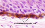
melanin
- (Greek, melanos = black) The pigment produced by melanocytes that provides photoprotection, preventing cellular DNA damage, and colouring of the cells that secret and absorb the pigment.
- (More? neural crest | integumentary)
melanoblast
- (Greek, melanos = black) The neural crest precursor cell that differentiates to form melanocytes located in the skin and other tissues that produces melanin.
- (More? neural crest | integumentary)
melanocyte
- (Greek, melanos = black) A pigmented cell, neural crest in origin, differentiating from melanoblasts located in the skin and other tissues that produces melanin. The melanocytes within the integument (skin) transfer melanin to keratinocytes to give skin colour and to the hair follicle to give hair colour. Melanocytes are also located within "non-cutaneous" tissues in the eye (for eye colour), harderian gland and inner ear. This is the cell type that proliferates in the cancer melanoma.
- (More? neural crest | integumentary)
melanosome
- (Greek, melanos = black) the large pigment filled cell organelle, about 500 nm in diameter, formed within a melanocyte and transferred to a keratinocyte within the skin. The skin colouration is established by the total number of melanosomes formed, not the number of melanocytes forming epidermal melanin units, that is about the same for all skin colours.
- (More? neural crest | integumentary)
melatonin
- An endocrine hormone secretd from the pineal gland involved with the diurnal cycle, melatoinin levels are high in dark, low in daylight.
membrana granulosa
- The granulosa cells that line the developing follicles of the ovary. These cells proliferate to form the stratum granulosa and other granulosa cells are given specific names based upon their position within the follicle. In the antral follicle, membrana granulosa sits on the follicular basal lamina and lines the antrum as a stratified epithelium. The cumulus oophorus is a column of granulosa cells that attaches the oocyte to the follicle wall. The corona radiata are the granulosa cells that directly surround the oocyte, and are released along with it at ovulation. Following ovulation the corona radiata provide physical protection to the oocyte and granulosa cells within the ovulating follicle contribute to corpus luteum.
- (More? Week 1 | Ovary Development | Oocyte Development | Menstrual Cycle)
membraneous organelle
- (MO) Term describing spermatozoa specific organelles found in the worm (C. elegans). These organelles cluster together with paternal mitochondria immediately after fertilisation and then become polyubiquitinated and associated with proteasomes.
- (More? Worm Development | Spermatozoa Development | PMID 24528894)
meninges
- (singular meninx; Greek, meninx = membrane) Anatomical term describing the three connective tissue layers that surround the entire central nervous system (brain and spinal cord). The 3 layers from the central nervous outward are: pia mater, arachnoid mater, and the dura mater. All three layers form from the meninx primitiva, a meningeal mesenchyme that is mesodermal and neural crest in origin. The space under the arachnoid layer (subarachnoid space) is filled with cerebrospinal fluid.
meningoceles
- The herniation of the meninges through a skull or spinal defect, formed from a neural tube defect. Spinal cord meningoceles have three classifications: simple meningocele, lateral meningocele and anterior sacral meningocele.
meningococcal disease
- (meningitis) Term describing the bacterial infection of cerebrospinal fluid of the spinal cord and brain. Note meningitis can also be caused by a viral or other organism infection. Treatment and outcomes differ for either viral (less severe, resolves without specific treatment) or bacterial (severe, may result in brain damage, hearing loss, or learning disability) infections.
- (More? Bacterial Infection | Postnatal Development | CDC - meningococcal disease | Medline Plus - Meningitis)
menopause
- (Greek, mene = moon, men = month, pause = end or cessation) The decrease in ovarian production of estrogen and progesterone leading to cessation of menstrual cycles, decrease in fertility, and end of female reproductive life. Clinically defined as the final menses, confirmed after 1 year without menstruation. A biological term describing the physiological changes that accompany the age related loss of fertility. Usually occurs in the mid-40's, the term was first used by the French physician, de Gardanne in 1812.
- (More? Menopause | Menstrual Cycle | Medline Plus - Menopause)
menorrhagia
- Term used to describe heavy menstrual bleeding, is common in women of reproductive age (WHO data, affects 1011 out of 5322 women).
- (More? Menstrual Cycle)
menotropin
(human menopausal gonadotropin, hMG) Originally derived from menopausal women's urine as a mixture of gonadotropins, follicle stimulating hormone (FSH) and luteinizing hormone (LH). Currently a recombinant synthetic mixture is used clinically in fertility treatments.
menstrual age
- The gestation time calculated from the first day of the last menstrual period (LMP) prior to fertilization. In humans, this differs from embryonic age by approximately two weeks.
- (More? Menstrual Cycle | Week 1)
menstrual cycle
- The human reproductive cycle, an endocrine regulated change in female anatomy and physiology that occur over 28 days (4 weeks, a lunar month) during reproductive life (between puberty and menopause). This cycle ceases during pregnancy and differs from other non-primate vertebrates (eg rats, mice, horses, pig) females that have a reproductive cycle called the estrous cycle (oestrous, British spelling).
- (More? Menstrual Cycle | Estrous Cycle)
Merkel cell
- An epidermal-derived cell in touch sensitive area of the epidermis and mediate mechanotransduction in the skin, in the epidermal basal layer of vertebrates. Merkel cells (keratin 8 positive, K8+) develop in association with primary hair follicles and are organised among columnar basal keratinocytes expressing hair follicle keratin (keratin 17 positive, K17) forming a touch dome. Previously thought to be neural crest in origin, but recently shown to arise from the embryonic epithelium. The cells are named after Friedrich Sigmund Merkel, a German anatomist who was the first to describe them in 1875.
- (More? Touch Development | integumentary | Lecture - Integumentary Development | PMID 19786578 | PMID 3782861)
merle
- The pattern of coloring observed in the coat of the domestic dog and is characterized by patches of diluted pigment. Dogs inherit trait in an autosomal, incompletely dominant fashion and heterozygous or homozygous for the merle locus exhibit a wide range of auditory and ophthalmologic abnormalities, similar to those in human Waardenburg syndrome.
- (More? Animal Development | PMID 16407134)
meroblastic
- Early development term referring to partial cleavage of the zygote generating a large yolk, occurring in birds and fish development. Alternative form of division is described as holoblastic, that refers to total or entire cleavage occurring in amphibian and mammal zygote development.
meromelia
- (Greek, melia = limb) A limb abnormality with the partial absence of a limb, as described in the original classical classification of limb deficiencies.
- (More? Limb Abnormalities)

merotelic kinetochore
- Cell division abnormality in chromosomal attachment that occurs when a single kinetochore is attached to microtubules arising from both spindle poles. Normal chromosomal attachment in early mitosis, is by only one of the two sister kinetochores attached to spindle microtubules (monotelic attachment) later sister kinetochores attach to microtubules arising from opposite spindle poles (amphitelic attachment).
- (More? Cell Division - Mitosis | PMID 21306900)
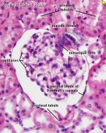
mesangial cell
- (glomerular mesangial cell) A kidney epithelial cell type located in the nephron (functional kidney unit) surrounding glomerular capillaries (blood vessels). Mesodermal in origin, there are mesangial cells within (intraglomerular) and outside (extraglomerular) the glomerulus. Have several functions including: contractile activity (smooth muscle-like) controlling blood flow and basement membrane surface area (glomerular filtration rate), structural support, phagocytosis (remove basal lamina components and immunoglobulins).
- (More? renal)
mesencephalon
- (midbrain, mes = mid, encephalon = brain) The embryonic neural tube region that will form midbrain structures of the tectum and tegmentum in the adult brain. The mesencephalon is the middle of the 5 secondary brain vesicles formed from the mesencephalon of the primary brain vesicle (there are 3 primary brain vesicles). The mesencephalon lumen (cavity of the neural tube) will form the midbrain aqueduct.
- Primary brain vesicles: prosencephalon (forebrain) - mesencephalon (midbrain) - rhombencephalon (hindbrain)
- Secondary brain vesicles: telencephalon - diencephalon - mesencephalon - metencephalon - myelencephalon
mesenchyme
- Term used to describe the cellular organisation of undifferentiated embryonic connective tissue. Mesenchymal tissue is mainly derived from mesoderm and neural crest, which will form most of the adult connective tissues. This connective tissue organization contrasts with the other main form of cellular organization, epithelial tissue.
- (More? Mesoderm | Musculoskeletal System Development | Week 3)
mesenchymal stem cell
- The cells derived from various connective tissues that form a population of stem cells with potential to differentiate for repair and replacement of connective and other tissues. Can be found mainly in bone marrow, but also in other places (dermis, lung and heart atria). The bone marrow mesenchymal stem cell (or bone marrow stromal cell) differs from the hematopoietic stem cell (which forms blood cells) and can form bone, cartilage and adipose tissue.
- (More? Stem Cells | Mesoderm)
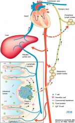
mesenteric lymph node
- (MLN) Immune system associated lymph nodes located at the base of the mesentery that collect lymph (cells and antigens) draining from the intestinal mucosa.
- (More? immune | Gastrointestinal Tract Development)
mesentery
- The tissue fold attaching gastrointestinal tract to posterior abdominal wall in which blood vessels, lymph and nerves run. Developmentally derived from lateral plate mesoderm forming splanchnic mesoderm which then forms the posterior mesogastrium.
- (More? mesentery | mesogastrium | Gastrointestinal Tract Development)
mesethmoid cartilage
- The ventral component of the nasal capsule. In the chicken embryo, it is induced by sonic hedgehog (Shh) expression from endoderm (endoderm zone I).
- (More? Head Development)
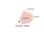
mesoderm
- (Greek, mésos = middle + derma = skin) The middle layer of the 3 germ cell layers of the trilaminar embryo (ectoderm, mesoderm, endoderm). Each region of this early layer will later form different structures, this middle layer contributes all connective tissues of the body, except in the head region where neural crest also will contribute. Mesoderm outside the embryo and covering the amnion, yolk and chorion sacs is extraembryonic mesoderm.
- Mesoderm development:
- epiblast -> mesoderm + axial mesoderm (notochord)
- lateral plate + paraxial mesoderm + axial mesoderm
- lateral plate + intermediate mesoderm + somites (body), paraxial mesoderm (head) + axial mesoderm
- somatic mesoderm + intraembryonic coelom + splanchnic mesoderm + intermediate mesoderm + somites (body), paraxial mesoderm (head) + axial mesoderm
- (More? mesoderm | somitogenesis | Musculoskeletal System Development | Week 3)
mesodiencephalic dopaminergic neurons
- (mdDA) Central nervous system neurons that control voluntary movement and reward based behaviour, which are lost in Parkinson's disease. Their embryonic origin progenitor cells are located in the caudal diencephalon and midbrain floor plate region.
- (More? neural)
mesogastrium
- The developmental term for the splanchnic mesoderm forming early mesenteries (dorsal and ventral) that support the developing gastrointestinal tract. The majority of the ventral mesentery is developmentally lost at the level of the midgut and the dorsal mesentery remains in the adult, through which blood vessels, nerves and lymph connects to the gastrointestinal wall. Note that specific visceral organs also develop within each mesogastrium.
- (More? mesentery | Gastrointestinal Tract Development)
mesometrial lymphoid aggregate of pregnancy
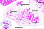
- (mesometrial lymphoid aggregate) An immune lymphoid structure that transiently forms between the uterine myometrial smooth muscle layers. Rich in uterine natural killer cells (uNK) that differentiate to form large granulated lymphocytes, initially accumulated at the site of implantation in the decidua basalis, lying also around blood vessels, and finally between the two uterine myometrial smooth muscle layers. During pregnancy uNK cells represent up to 70% of the uterine resident lymphocytes, interact with trophoblast cells, modify blood vessels (spiral arteries), and their full function is yet to be determined.
mesonephric duct
- (Wollfian duct) An early developing urogenital paired duct system that initially runs the length of the embryo, that will differentiate and form the male reproductive duct system (ductus deferens). In females, this duct degenerates occasionally some remnants may remain associated in broad ligament.
- (More? Genital - Male Development | Genital System Development | renal | Lecture - Genital Development | Caspar Friedrich Wolff)
mesonephric ridge
- Surface bulges seen on the early embryo that reflect the internal development and growth of the mesonephros stage of kidney development. These are visible on either side of the embryo in the dorsolateral region, between the upper and lower limb buds , running rostrocaudally as surface bulges on the trunk. These ridges are lost in later embryonic regression of the mesonephros and thickening of the dermis. Feature is visible at Carnegie stage 14 onward in embryonic development.
- (More? renal | Lecture - Renal Development)
mesonephros
- The second temporary stage of kidney development (pro-, meso-, meta-). The intermediate mesonephros develops and disappears with the exception of its duct, the mesonephric duct, which will form the male reproductive duct system. In males, the mesonephric tubules go on to form the ducts of the testis. In females, these degenerate. A few mesonephric tubules remain as efferent ductules in the male and vestigial remnants in the female.
- Kidney Stages: pronephros - mesonephros - metanephros
- (More? renal | Lecture - Renal Development)

mesorchium
- A peritoneal fold attaching the testes to the mesonephros during development.
mesovarium
- The mesentry of the ovary formed from a fold of the broad ligament that attaches the ovary through this structure pass the vessels and nerves to the ovary, entering at the hilus of the ovary.
- (More? Ovary Development | Week 1)
messenger RNA
- (mRNA) The form of RNA that is translated into a protein amino acid sequence by the ribosome.
metabolic syndrome
- Term used to describe the group of obesity-related metabolic abnormalities that increase an individual's risk of developing type 2 diabetes and cardiovascular disease.
meteorin
- A secreted protein expressed during gastrulation in endoderm/mesoderm development, later in in neural and astrocyte progenitors, and may also have roles in glial cell differentiation and axonal network formation.
- (More? gastrulation | neural)
metformin
- (Glucophage, N,N-Dimethylimidodicarbonimidic diamide) Drug used clinically in the treatment of diabetes (antidiabetic) for type 2 diabetes and is being considered for use in gestational diabetes. The drug is taken orally and the main effect is to increase the body sensitivity to the action of insulin, so that insulin levels fall, It does not cause low blood sugar. The drug is also used to treat polycystic ovarian syndrome (PCOS).
- (More? Maternal Diabetes | Ovary - PCOS | Genital - PCOS | MedlinePlus - Metformin | PMID 23724063)
metanephros
- The adult kidney, third stage of mammalian kidney (pro-, meso-, meta-) development within the intermediate mesoderm.
- Kidney Stages: pronephros - mesonephros - metanephros
metanephric cap
- (metanephric blastema, metanephric mesenchyme) In renal (kidney) development, the intermediate mesoderm (metanephric mesenchyme) that surrounds the outgrowing uretric bud and will later develop into the nephrons of the kidney.
metanephric mesenchyme
- (metanephric blastema) Metanephric mesenchyme caudal part of intermediate mesoderm that will develop into nephrons within the kidney. The intermediate mesoderm forms as an unsegmented strip running rostro-caudally between the somite and lateral plate mesoderm. The very caudal (tail) end of this mesoderm strip where the uteric bud forms is the metanephric mesenchyme, which induces the formation of, and surrounds the end of, the ureteric bud.
metaphase
- Cell division term referring to the third mitotic stage, mitotic spindle kinetochore microtubules align chromosomes in one midpoint plane. Metaphase ends when sister kinetochores separate. Originally based on light microscopy of living cells and electron microscopy of fixed and stained cells. A light microscope analysis called a "metaphase spread" was originally used to detect chromosomal abnormalities in cells.
- Mitosis Phases: prophase - prometaphase - metaphase - anaphase - telophase
- (More? Cell Division - Mitosis | Cell Division - Meiosis | Week 1)
metaphase spread
- A light microscope analysis technique originally used to detect chromosomal abnormalities in cells, as chromosomes are only visible during cell division.
- Mitosis Phases: prophase - prometaphase - metaphase - anaphase - telophase
metencephalon
- (Greek, encephalon = brain) The embryonic neural tube region that will form hindbrain structures of the pons and cerebellum in the adult brain. The metencephalon is the fourth of the 5 secondary brain vesicles formed from the rhombencephalon of the primary brain vesicle (there are 3 primary brain vesicles). The metencephalon lumen (cavity of the neural tube) will form the fourth ventricle.
- Secondary brain vesicles: telencephalon - diencephalon - mesencephalon - metencephalon - myelencephalon
metestrus
- In most female mammals, the third stage in the estrous cycle immediately before diestrus characterized by sexual inactivity and the formation of the corpus luteum.
- (More? Estrous Cycle | Mouse Estrous Cycle)
methionine aminopeptidase
- (MetAPs) An enzyme family of cytosolic metalloproteases which are responsible for cleavage from nascent proteins (newly translated) of the initial methionine from the N termini. Eukaryotes express two forms of MetAPs (types 1 and 2), with type 2 required for normal vascular development, deletion in mouse causes embryonic death at the midsomite stage.
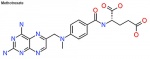
methotrexate
- (MTX, amethopterin) Drug with several different uses including the treatment of ectopic pregnancy and for the induction of medical abortions. Acts as a antimetabolite and antifolate (folic acid antagonist) drug that inhibits DNA synthesis in actively dividing cells, including trophoblasts, and therefore has other medical uses include cancer and autoimmune disease treatment. Treatment success in ectopic pregnancy relates to serum β human chorionic gonadotropin (β-hCG) concentration. Methotrexate is an antineoplastic and immunosuppressive agent also widely used in the therapy of leukemia, lymphoma, solid tumors, psoriasis and rheumatoid arthritis. Methotrexate is well known to cause serum aminotransferase elevations and long-term therapy has been linked to development of fatty liver disease, fibrosis and even cirrhosis.
- (More? Ectopic Pregnancy | Medline Plus | Methotrexate Liver Toxicity | PMID 8317518)
methyldopa
- (alpha methyldopa) A central alpha agonist used to lower blood pressure. Used as an anti-hypertensive drug to lower blood pressure in pre-eclampsia, acting by either a direct or indirect central vasodilatory mechanism. A recent study suggests this drug may have a direct effect on placenta and/or endothelial cell function in pre-eclampsia patients, altering angiogenic proteins. Drug commercial brandname (USA) "Aldomet", also available in combination with other drugs: methyldopa and chlorothiazide "Aldochlor", methyldopa and hydrochlorothiazide "Aldoril".
- (More? Placenta - Abnormalities | Medline Plus - Methyldopa | PMID 18648513)
methylenedioxypyrovalerone
- (MDPV) Illegal and frequently used psychoactive drug of abuse of cathinone family. Shown in animal models to affect the developing brain.
- (More? Illegal Drugs | PMID 25063209)
methylmercury
- (organic mercury) Toxic form, at high concentrations, of mercury found in the environment (air, water, soil, plants and animals) and is different from elemental mercury (thermometers, dental amalgams). Industrial mercury can enter water systems, which is then converted into methylmercury and can contaminate exposed fish or shellfish, entering the human food chain.
metopic suture
- A skull fibrous joint, cranial suture between adjacent developing bones of the skull. This suture begins at nose and runs superiorly to meet sagittal suture and fuses in early childhood before all other cranial sutures. Premature fusion (synostosis) of metopic suture causes trigoncephaly (wedge skull).
metritis
- Inflammation of the uterus, usually due to a bacterial or other infection, see also pelvic inflammatory disease (PID).
metrorrhagia
- Clinical term used to describe uterine bleeding at irregular intervals between the normal menstrual cycle periods (menses).
- (More? Menstrual Cycle)
Meyer-Weigert rule
- (Meyer-Weigert law) A renal clinical term for the arrangement of the ureter in a completely duplicated renal system, the ureter from the upper renal pole enters the bladder more medially and caudally than the ureter from the lower renal pole.
- (More? Renal System - Abnormalities)
MiRNA
- Acronynm for MicroRNA a small noncoding ribonucleotide-based (RNA) regulators of gene expression. They have diverse functions including regulation of cellular differentiation, proliferation and apoptosis.
microcephaly
- An abnormally small skull cranium marked by premature fusion of the skull sutures and also under-developed brain. Recently the Zika virus infection has been thought to contribute to microcephaly cases in Brazil.
- (More? Skull Development | Neural System - Abnormalities | Zika Virus |XVII Congenital Malformations)
microchimerism
- (Mc) Term used to describe when a usually small population of cells or DNA is harboured by one individual that derive from a genetically distinct individual. May occur in pregnancy when cells exchange beween fetus and mother, mother and fetus or in twinning.
micrognathia
- Clinical term for a facial malformation characterized by mandibular hypoplasia, generating a small receding chin that fails to maintain the tongue in a forward position. Several developmental causes including Pierre-Robin syndrome.
micro-magnetic resonance imaging
- (MicroMRI) A non-invasive imaging technique that allows detailed imaging of small biological structures using magnetic resonance imaging that are particularly useful in embryological studies.
- (More? Magnetic Resonance Imaging)
microphthalmia
- (microphthalmos) The abnormality of development of a small eye within the orbit. This condition has been reported in up to 11% of blind children.
microphthalmia-associated transcription factor
- (MITF) A protein, basic helix loop helix (bHLH) zipper transcription factor, key to regulating melanocyte development. Also functions as an oncogene in malignant melanoma.
- (More? OMIM - MITF | Lecture - Neural Crest Development | Lecture - Integumentary Development | Neural Crest Development)
MicroRNA
- (miRNA) a small noncoding ribonucleotide-based (RNA) regulators of gene expression. These recently discovered small RNA molecules (18-24 nucleotides) negatively regulate target mRNAs and appear to have a role in many developmental processes as well as in the adult. (See also "Dicer") There is also another class of small RNAs involved in gene expression present in cells, small interfering RNAs (siRNAs), generated from double-stranded RNA (dsRNA) precursors.
microtia
- The condition of an abnormally small external ear.
- (More? Microtia | Outer Ear Development)
microvillus inclusion disease
- (MVID) A postnatal gastrointestinal tract disease due to the shortening or absence of epithelial cell (enterocytes) apical microvilli. Infants have life-threatening watery diarrhoea and mutations in myosin VB gene (MYO5B). Other forms of congenital diarrhea include congenital secretory chloride diarrhoea, an autosomal recessive form of severe chronic diarrhoea. International Classification of Diseases - XVI Perinatal Period P78.3 Noninfective neonatal diarrhoea Neonatal diarrhoea NOS Excl.: neonatal diarrhoea NOS in countries where the condition can be presumed to be of infectious origin (A09)
- (More? Neonatal Development | Gastrointestinal Tract Development | OMIM 251850 | OMIM MYO5B | PMID 20186687 | PMID 26830108)
midbrain
- (mesencephalon) The common term used to describe the early primary brain vesicle middle subdivision of brain development at the stage when there are three primary vesicles or expansions of the early neural tube (forebrain, midbrain, hindbrain. This subdivision is the only one present at the later five secondary brain vesicle stage. Term is also used in the adult brain to describe brainstem components formed including: tectum, tegmentum, the ventricular mesocoelia, cerebral peduncles, and additional nuclei and fasciculi.
- Three primary brain vesicles: forebrain ( prosencephalon) - midbrain (mesencephalon) - hindbrain (rhombencephalon)
midbrain flexure
- (pontine flexure) pontine flexure The middle curvature that forms in the early rapidly growing neural tube.
- (More? Neural System Development)
midgut
- The middle of the three part/division (foregut - midgut - hindgut) of the early forming gastrointestinal tract. The midgut is initially connected on the ventral embryo surface to the external yolk sac by a yolk stalk, a narrow tubular connection. The midgut forms all the tract from beneath the stomach (duodenum, small intestine and large intestine) to the distral transverse colon. The midgut develops as an external loop "herniated" ventrally, until early fetal growth of the body wall recaptures this external loop, which also undergoes a rotation about the superior mesenteric artery to establish the adult anatomical position. These anatomical divisions also correspond to their 3 main vascular supply divisions of foregut coeliac artery, midgut superior mesenteric artery and hindgut inferior mesenteric artery.
mifepristone
- (RU 486) A progesterone receptor antagonist similar in structure to the natural hormone progesterone, which is used medically as a birth control drug. Commercial drug names include Mifegyne and Mifeprex. Ulipristal is new analog of mifepristone, acting as a selective progesterone receptor modulator, identified as a second generation emergency contraceptive. Progesterone binding to the progesterone receptor generates a receptor conformational change allowing it to bind to DNA and act as a transcription factor for genes. RU486 binds the same receptor, with 10-fold higher affinity, C-terminal region of the hormone-binding domain and then does not act as a transcription factor.
- (More? Mifepristone)
miRNA
- Acronym for MicroRNA
Mirror syndrome
- (Ballantyne's syndrome) An abnormality typically defined as the development of maternal edema in association with fetal hydrops with an associated high rate of fetal and maternal death. The condition has been reported associated with: rhesus isoimmunization (29%), twin-twin transfusion syndrome (18%), viral infection (16%) and fetal malformations, fetal or placental tumors (37.5%). PMID 20357423
miscarriage
- A general clinical term for the loss of embryo or fetus by spontaneous abortion. In humans, there can be a number of different specific causes and it may occur early in a pregnancy (week 1 and 2) due to conceptus genetic abnormalities.
misoprostol
- A non-steroidal anti-inflammatory drug used in medical management of first trimester miscarriage and for birth labor induction. In some countries, often in combination with mifepristone. Drug is a potential teratogen, used in non-pregnant treatment to prevent ulcers in people taking some arthritis or pain medicines. A synthetic prostaglandin E1 (PGE1) analogue marketed in the USA commercially as Cytotec.
- (More? Medline Plus - misoprostol)
missed abortion
- (blighted ovum) Term previously used to describe early fetal loss, embryo loss in first trimester.
mitochondria
- Double membraned cell organelle located in the cytoplasm, a cell may contain 100's or more mitochondria, the number can relate to the metabolic activity of that cell. Functions in cell respiration, providing energy to the cell and also has a role in the process of apoptosis (programmed cell death). Mitochondria in humans are maternally inherited and contain their own genetic material, many copies of circular DNA (mtDNA). There are a number of known mutations in this mtDNA that can cause genetic disorders, this relates also to the development of new Assisted Reproductive Technology tools.
- (More? Assisted Reproductive Technology | Mitochondria)
mitochondrial cloud
- (Balbiani body) See Balbiani body a specialised cluster region of mitochondria in oocyte cytoplasm of some species.
mitofusin
- (Mfn) Mitochondrial protein GTPases that are essential for mitochondrial fusion, the joining together of two separate mitochondria within a cell. There are two forms mitofusin 1 (Mfn1) and mitofusin 2 (Mfn2) homologous to drosophila (Fzo) that are differentially expressed in different tissues. In skeletal muscle, loss of mitofusins causes severe mitochondrial dysfunction, compensatory mitochondrial proliferation, and muscle atrophy.
- (More? PMID 14561718 | OMIM - Mfn1 | Mfn2 | Charcot-Marie-Tooth disease type 6)
mitosis
- The normal division of all cells, except germ cells, where chromosome number is maintained (diploid). In germ cell division (oocyte, spermatozoa) meiosis is a modified form of this division resulting in reduction in genetic content (haploid). Mitosis, division of the nucleus, is followed by cytokinesis the division of the cell cytoplasm and the cytoplasmic contents. cytokinesis overlaps with telophase.
- Mitosis Phases: prophase - prometaphase - metaphase - anaphase - telophase
- (More? Cell Division - Mitosis | Week 1)
mixoploidy
- (mixoploid) Genetic term used to describe an abnormal genome chromosomal set, includes mosaicism, where there are two or more genetically different cell lines in an individual.
- (More? Genetics | Abnormal Development - Genetic)
MMR
- Acronym for Measles-Mumps-Rubella vaccine.
- (More? Abnormal Development - Rubella Virus | [Postnatal - Vaccination]])
modulo
- Drosophila transcription factor which binds to the promoter of spermatid-differentiation gene Sdic and integrate meiosis and spermatid differentiation in the male germ line. (homologue of nucleolin)
- (More? PNAS paper | OMIM nucleolin)
monochorionic
- A twinning term, in a monozygotic twinning event (one fertilised egg and a single spermatazoa, form a single zygote) which occurs early (within 2 days of fertilization). Later splitting (more than 2 days after fertilization) may result in a shared placenta and only duplication of the embryonic amnionic sacs (monochorionic diamniotic twins).
- (More? Twinning)
monozygotic twin
- Twins (identical) produced from a single fertilization event (one fertilised egg and a single spermatazoa, form a single zygote), these twins therefore share the same genetic makeup. Occurs in approximately 3-5 per 1000 pregnancies, more commonly with aged mothers. Late monozygotic twins can result in conjoined twins or both a shared placenta and a shared amniotic sac (monochorionic monoamniotic twins). Note dizygotic twins (fraternal) arise from separate fertilizations of different eggs.
mongolism
- Historic term for Trisomy 21. This term has long ago been discarded as inappropriate and should not be used.
- (More? Trisomy 21 | Historic Terminology)
morbidity
- (Latin, morbidus = "sick" or "unhealthy") Clinical term refers to a diseased state, disability, or poor health due to any cause.
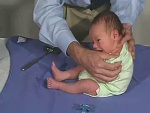
Moro reflex
- (startle reflex) Clinical term describing a primitive reflex, an involuntary response (reflex) that is present at birth and that normally disappears after 3 or 4 months. The reflex has 4 parts: startle; abduction of the upper limbs (spreading out arms); adduction of the upper limbs (unspreading the arms); crying (usually, but may be absent). Preterm birth infants (28 to 33 weeks) have an incomplete form of this reflex and postnatal persistence (beyond 4 or 5 months) occurs in infants with severe neurological defects.
- (More? Neural Exam - Newborn reflexes - Moro | Neural System Development | Neonatal Development | Medline Plus - Moro reflex)
morphogenesis
- Term used to describe the process of development involving a change in form (shape) and size or either cells or tissues.
morphogenetic field
- Term used to describe a group of cells with a similar developmental fate, forming a specific structure or organ (forelimb, eye, heart).
morula
- (Latin, morula = mulberry) An early stage (Carnegie Stage 2) in post-fertilization development when cells divide rapidly (embryonic cell cycle) producing a solid mass of cells (12-15 cells) with a "mulberry" appearance. Cell proliferation occurs still enclosed within the zone pellucida. This stage is followed by formation of a cavity in this cellular mass (blastocyst stage). In humans, morula stage of development occurs during the first week following fertilization.
- (More? Morula | Carnegie stage 2 | Week 1 | Fertilization | Week 1 | Cell Division - Mitosis)
MIS
- Acronym for Mullerian Inhibiting Substance, a hormone that regulates genital development.
MSC
- Acronym for mesenchymal stem cell, a connective tissue stem cell.
MSDS
- Acronym for Material Safety Data Sheet, a set of information about a chemical properties, risks, hazards and toxicity. This term in Australia is being replaced by Safety Data Sheet (SDS), under the United Nations Globally Harmonized System of Classification and Labelling of Chemicals (GHS) program to standardise chemical data around the world.
MSH
- Acronym for muscle specific homeobox (which is not muscle specific), a homeobox gene involved in pattern formation in several systems in development.
MSS
- Acronym for Maternal Serum Screening, used to detect potential genetic abnormalities in both mother and embryo.
- (More? Abnormal Development - Genetic)
MTOR
- Acronym for Mammalian Target Of Rapamycin a cellular protein kinase (PI3K-related kinase superfamily member) with an integrator of pathways important for cellular metabolism, proliferation, and differentiation. Studies in mouse show MTOR is expressed at all stages of oocyte development to maintain the viability of the primordial oocyte pool or recruitment into the growing oocyte cohort.
- (More? oocyte | implantation)
mucopolysaccharidosis VI
- (MPS VI) A lysosomal storage disease, due to a deficiency of the enzyme arylsulfatase B, which leads to the accumulation of dermatan sulphate (sulfate) causing a skeletal dysplasia, short stature, dysostosis multiplex and degenerative joint disease.
- (More? musculoskeletal)
mucosa
- (mucous membrane, mucosae) Histological and anatomical term used to describe lining cellular layers of tissues which function in absorption and secretion. In the gastrointestinal tract (endoderm origin) lining, the mucosa is formed by the epithelial layer, lamina propria and muscularis mucosa. The entire gastrointestinal tract along it's length has similar structural layers of: mucosa, submucosa, muscularis externa and an adventitia or serosa.
Muehrcke lines
- Clinical term for paired transverse white lines appearing in the nails secondary to edematous nail bed, resulting abnormal nail bed vasculature, forming transverse lines that disappear when the nail bed is compressed. Seen in patients with chronic hypoalbuminemia and malnutrition.
- (More? Integumentary System - Nail Development|Nail Development]])

Müllerian duct
- (paramesonephric duct) An embryonic paired duct system that will form the epithelial lining of female reproductive organs: utererine tube, uterus, upper vaginal canal. This duct system degenerate in male gonadal development. Named after Johannes Peter Müller (1801-1858) a German scientist.
Johannes Peter Müller
- Johannes Peter Müller (1801 - 1858) in 1830 was the first to describe the duct named after him, the "Müllerian duct" also called the paramesonephric duct.
- (More? Johannes Peter Müller | Uterus Development | Genital - Female Development | Menstrual Cycle | Genital System Development)
Mullerian Inhibiting Substance
- (MIS, Anti-Mullerian Hormone, AMH, Mullerian inhibiting hormone, MIH). A sertoli cell secreted glycoprotein (transforming growth factor-beta, TGF-beta superfamily) that regulates gonadal and genital tract development. The main role is to inhibit paramesonephric duct (Müllerian) development in males. Postnatally, after puberty it is also expressed in females by ovarian granulosa cells and has a role in follicle development.
- (More? Testis Development | Genital System Development | OMIM - AMH)
multidetector computed tomography
- (MDCT) A reasonably non-invasive imaging technique that uses several detectors, the multidetector can be 4- or 8-rows. Computed Tomography or computed axial tomography (CAT or CT scan) began in 1970's using x-ray and a computer to produce images either as individual slices or reconstructed to give three dimensional (3D) views of specific anatomical regions or structures. Since then technical developments have included: slip-ring technology, high-power x-ray tubes (with better cooling), and reconstruction algorithms (for nonlinear data acquisition by interpolation). Scans for a determining heart abnormalities, Coronary Coronary Computed Tomography Angiogram (CTA), require the highest possible resolution (64-slice system (64-MDCT).
multifetal pregnancy reduction
- A procedure used to decrease the number of fetuses a woman carries and improve the chances that the remaining fetuses will survive and develop into healthy infants. Multifetal reductions that occur naturally are referred to as spontaneous multifetal reductions.
multiple birth
- A pregnancy that results in the birth of more than one infant.
- (More? Abnormal Development - Twinning)
multiple hereditary exostoses
- (MHE) An genetic abnormality, occurring as an autosomal dominant disease associated with mutations in two enzymes that are required for heparan sulfate (HS) synthesis. Children with the disease form numerous benign bone tumors (osteochondromas) and also the possibility of developing chondrosarcoma.
musashi 1
- (musashi-1, MS1) A protein regulator of translation that specifically binds to the 3′-untranslated region (UTR) of some messenger RNA (mRNA) and inhibits their translation. Identified in the developing nervous system (binds m-Numb mRNA) and gastrointestinal tract.
muscarinic receptor
- (acetylcholine receptor) These are G-protein coupled receptors (GPCR) located in the cell membrane. In mammals, there are 5 different isoforms (m1-m5) with different central and peripheral nervous system and other tissue distributions.
muscle
- Term used to describe the tissue that has contractile activity, also used to describe the functional cells that contract. There are three main types of muscle: smooth muscle, cardiac muscle and skeletal muscle. Muscle is generally mesodermal in origin, each muscle type coming from a different mesoderm region.
- (More? Musculoskeletal System Development | Lecture - Mesoderm Development | Lecture - Musculoskeletal Development)
muscle atrophy
- (Latin, atrophia = ill fed) A general term applied to the wasting or decrease in muscle mass or volume. This atrophy can be due to a number of different causes (genetic, environmental and disease) and the term is mainly used in reference to skeletal muscle , but can also occur in the two other muscle types: cardiac and smooth muscle. The most common form in human development is duchenne muscular dystrophy.
mutagen
- A chemical or agent that can cause permanent damage to the deoxyribonucleic acid (DNA) in a cell. DNA damage in the human egg or sperm may lead to reduced fertility, spontaneous abortion (miscarriage), birth defects and heritable diseases.
- (More? Abnormal Development - Genetic)
myocardium
- Layer that forms the muscular wall of the heart, the thickest layer formed by spirally arranged cardiac muscle cells. The other two cardiac layers are the pericardium and endocardium. The heart embryonic origin is from the cariogenic region of prechordal splanchnic mesoderm.
- Heart layers: epicardium -> myocardium - > endocardium
- (More? Cardiovascular System Development | Cardiac Embryology tutorial | Lecture - Heart Development)
Mycoplasma genitalium
- (M. genitalium) A parasitic bacteria that can populate the epithelial layer of human uterine (Fallopian) tubes and lead to cilia damage and therefore contribute to tubal factor infertility. A recent study has shown that M. genitalium can also bind to human spermatozoa, could be carried by motile sperm, and contribute to this process of female genital disease and infertility.
- (More? Abnormal Implantation)
mycolic acid
- A family of long fatty acids normally found in the cell walls of mycoplasma bacteria (mycolata taxon).
- (More? Bacterial Infection)
myelomeningocele
- (spina bifida) A neural tube defect, a form of spina bifida (developmental abnormalities leading to failure of closure of the spinal column). Can be of two forms: spina bifida occulta (spinal column fails to close, spinal cord and meninges remain in place and defect is covered by skin) meningoceles (tissue covering the spinal cord sticks out of the spinal defect but the spinal cord remains in place).
myelencephalon
- (Greek, enkephalon = brain) The embryonic neural tube region that will form hindbrain structures of the medulla oblongata in the adult brain. The myelencephalon is the last vesicle of the 5 secondary brain vesicles before the spinal cord and formed from the rhombencephalon of the primary brain vesicle (there are 3 primary brain vesicles). The myelencephalon lumen (cavity of the neural tube) will form the lower part of the fourth ventricle.
- Secondary brain vesicles: telencephalon - diencephalon - mesencephalon - metencephalon - myelencephalon
- (More? Neural System Development)
myenteric plexus
- (Auerbach's plexus) Neural network forming part of the gastrointestinal tract enteric nervous system lying between the outer longitudinal and inner circular layers of the smooth muscle forming the muscularis externa. Its function is the motor innervation to both smooth muscle layers (peristalsis) and secretomotor innervation to the mucosa (parasympathetic and sympathetic input). The second submucosal plexus (Meissner's plexus) lies within the submucosal layer between the external smooth muscle and the mucosa of the small and large intestines from the duodenum to the internal anal sphincter. Embryologically derived from neural crest migrating into the splanchnic mesoderm, developmental abnormalities of this neural crest migration cause aganglionic colon.
myoblast
- The undifferentiated mononucleated muscle cells that will fuse together to form a multinucleated myotube, then mature into a muscle fibre.
myocardin
- A protein that acts as a transcriptional co-activator of serum response factor (Srf), which is a regulator of both smooth and cardiac muscle gene expression.
- (More? Heart Molecular | PMID 17021041)
MyoD
- Myoblast determining factor, a transcription factor involved in the determination of muscle cells in the somite. A basic helix-loop-helix factor which binds DNA.
myogenesis
- The process of muscle cell development or formation. In skeletal muscle the cellular sequence is: myoblast, myotube and myofibre.
- Development sequence: mesoderm - paraxial mesoderm - unsegmented mesoderm/somite - dermomyotome - myotome - myoblast - myotube - myofibre
myometrium
- Uterus anatomy term describing the uterus wall (uterine wall) middle smooth muscle layer. This layer lies between the lining endometrium and the covering serosa (perimetrium). During pregnancy this layer increases in thickness, mainly by smooth muscle hypertrophy, and its contraction is required for childbirth and expulsion of the placenta.
- (More? Uterus Development | Birth)
myonecrosis
- The term used to describe pathological death of myocytes. Can occur during development in placental vessels (umbilical cord or chorionic plate) following prolonged exposure to meconium (meconium myonecrosis).
- (More? Placenta Development)
myostatin
- (Mstn) A member of the transforming growth factor β (TGFβ) superfamily of secreted growth and differentiation factors. Expressed in developing and adult skeletal muscle.
myotendinous junction
- (MTJ) The specialiazed junction between a skeletal muscle and the tendon which transmit the force of the muscle contraction to the bone of the skeleton. Their role is to transmit the force of the muscle contraction. The histology of tendons is that of a dense regular connective tissue high in collagen type I. During development the MTJ has a unique microenvironment.
myotome
- The portion of the dermamyotome that generates skeletal muscle. Has 2 components epaxial (dorsal muscles) hypaxial (ventral muscles)
myotube
- The initial multinucleated cell formed by fusion of myoblasts during skeletal muscle development. Note the sequence shown below is for skeletal muscle of the body.
- Development sequence: mesoderm - paraxial mesoderm - unsegmented mesoderm/somite - dermomyotome - myotome - myoblast - myotube - myofibre
myxomatous
- The term (histology/pathology) used to describe a connective tissue embedded in mucus, for example Wharton's jelly of the placental cord.
- (More? Placenta - Histology)
Glossary Comments
Use this page to access brief definitions of specific embryology terms. Additional information can be accessed from links listed at the end of each definition. Glossary from the UNSW Embryology program compiled and written by Dr Mark Hill. Reference material used in preparing this glossary list includes: texts listed on page 1 "Reading" of each notes section, Department of Anatomy Publications, WWW resources from NCBI, NIH, OMIM, NHMRC (Australia), AMA (USA), Office of Rare Diseases (USA), PubMed Medline Dictionaries, MSDS, Merck Manual home edn. and WHO ART terminology (2009).
These notes are for Educational Purposes Only Please email Dr Mark Hill if you wish to make a comment about this current project.
Glossary Links
- Glossary: A | B | C | D | E | F | G | H | I | J | K | L | M | N | O | P | Q | R | S | T | U | V | W | X | Y | Z | Numbers | Symbols | Term Link
Cite this page: Hill, M.A. (2024, April 28) Embryology M. Retrieved from https://embryology.med.unsw.edu.au/embryology/index.php/M
- © Dr Mark Hill 2024, UNSW Embryology ISBN: 978 0 7334 2609 4 - UNSW CRICOS Provider Code No. 00098G