Lecture - Heart Development
| Embryology - 27 Apr 2024 |
|---|
| Google Translate - select your language from the list shown below (this will open a new external page) |
|
العربية | català | 中文 | 中國傳統的 | français | Deutsche | עִברִית | हिंदी | bahasa Indonesia | italiano | 日本語 | 한국어 | မြန်မာ | Pilipino | Polskie | português | ਪੰਜਾਬੀ ਦੇ | Română | русский | Español | Swahili | Svensk | ไทย | Türkçe | اردو | ייִדיש | Tiếng Việt These external translations are automated and may not be accurate. (More? About Translations) |
Introduction
Lecture Resources
| References | |||||
|---|---|---|---|---|---|
| Hill, M.A. (2020). UNSW Embryology (20th ed.) Retrieved April 27, 2024, from https://embryology.med.unsw.edu.au |
Heart Tutorial | 2017 | 2014 Lecture | 2013 Lecture Slides Anderson RH. Teratogenecity in the setting of cardiac development and maldevelopment. (2016) | ||||
| Moore, K.L., Persaud, T.V.N. & Torchia, M.G. (2015). The developing human: clinically oriented embryology (10th ed.). Philadelphia: Saunders. | The following chapter links only work with a UNSW student connection. | ||||
| Schoenwolf, G.C., Bleyl, S.B., Brauer, P.R., Francis-West, P.H. & Philippa H. (2015). Larsen's human embryology (5th ed.). New York; Edinburgh: Churchill Livingstone. | The following chapter links only work with a UNSW student connection. |
Heart Development
| Begin Intermediate: | Primordial Heart Tube | Heart Tube Looping | Atrial Ventricular Septation | Outflow Tract | Heart Valves | Cardiac Abnormalities | Vascular Overview |
The cardiovascular system is the first major system to function within the embryo, with the heart beginning to function during the fourth week. Congenital heart defects affect 8-10 of every 1000 births in the United States. Hence in both embryological and clinical contexts it is important to consider heart development.
The intermediate section of this module on cardiac embryology is directed at university level students with some previous study of embryology but minimal previous study of cardiac development specifically. This unit takes approximately 30-40 minutes to complete. The intermediate cardiac module contains 7 pages which should be worked through in order.
- Primordial Heart Tube
- Looping of the Heart Tube
- Atrial and Ventricular Septation
- Development of the Outflow Tract
- Development of Heart Valves
- Abnormalities of Cardiac Development
- Overview of Vascular Development
These pages are also listed and linked in the yellow boxes at the top of each page. When you have completed the intermediate cardiac module, you can complete the intermediate quiz to test your knowledge. For students who would like either less or more detailed information regarding each step in the developmental process, links throughout the unit will direct you to the corresponding section in the basic level or advanced level respectively. Alternatively, after completing the intermediate module you can proceed to the next advanced module or return to the simpler basic module.

|
Fetal Blood Flow - Proportions of the combined ventricular output
Numbers represent percentage of total input/output flow. Proportions of the combined ventricular output in the major vessels of the human fetal circulation by phase contrast MRI. (8 subjects, median gestational age 37 weeks, age range of 30–39 weeks)
|
Key Reference
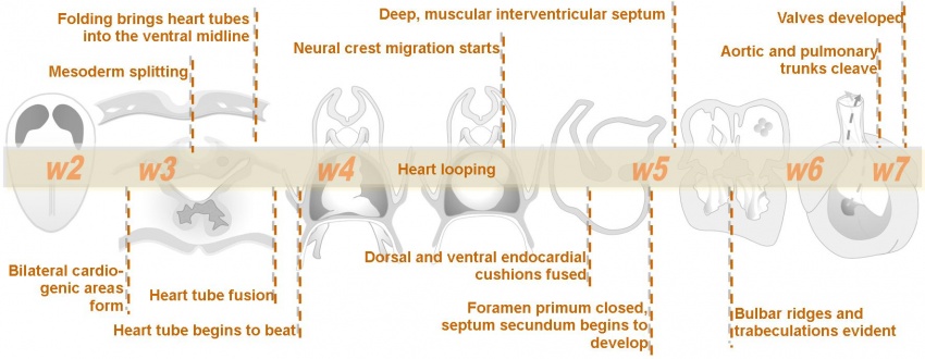
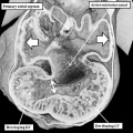
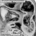
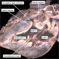
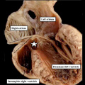
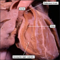
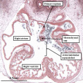
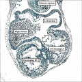
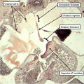
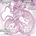
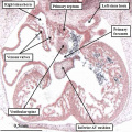
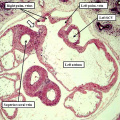
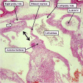
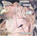
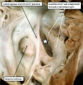
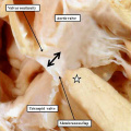
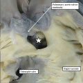
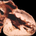
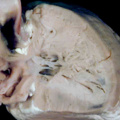
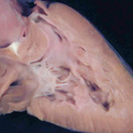
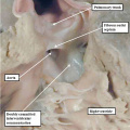
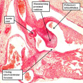
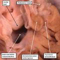
Figures from this recent paper below show human cardiac development as well as some of the the main developmental abnormalities.
| Human Heart Development |
|---|
|
| Human heart images reference[1] |
| 2018 ANAT2341 - Timetable | Course Outline | Moodle | Tutorial 1 | Tutorial 2 | Tutorial 3 |
Labs: 1 Preimplantation and Implantation | 2 Reproductive Technology Revolution | 3 Group Projects | 4 GM manipulation mouse embryos | 5 Early chicken eggs | 6 Female reproductive tract | 7 Skin regeneration | 8 Vertebral development | 9 Organogenesis Lab | 10 Cardiac development | 11 Group projects | 12 Stem Cell Journal Club |
|
Lectures: 1 Introduction | 2 Fertilization | 3 Week 1/2 | 4 Week 3 | 5 Ectoderm | 6 Placenta | 7 Mesoderm | 8 Endoderm | 9 Research Technology | 10 Cardiovascular | 11 Respiratory | 12 Neural crest | 13 Head | 14 Musculoskeletal | 15 Limb | 16 Renal | 17 Genital | 18 Endocrine | 19 Sensory | 20 Fetal | 21 Integumentary | 22 Birth | 23 Stem cells | 24 Revision |
| Student Projects: Group Projects Information Project 1 | Project 3 | Project 4 | Project 5 | 2018 Test Student | Copyright |
