Category:Heart: Difference between revisions
From Embryology
mNo edit summary |
m (→References) |
||
| Line 9: | Line 9: | ||
{{Ref-Tandler1912}} | {{Ref-Tandler1912}} | ||
{{Ref-Mall1912}} | |||
[[Category:Muscle]][[Category:Cardiovascular]] | [[Category:Muscle]][[Category:Cardiovascular]] | ||
Revision as of 09:03, 15 February 2017
This Embryology category shows media and pages related to heart (cardiac) development. Pages section on this current page include lectures, laboratories, notes, quizzes and educational module sections that relate to cardiovascular development.
References
Tandler J. The Development of the Heart. (1912) Sect. II, chapt. 18, vol. 2, in Keibel F. and Mall FP. Manual of Human Embryology II. (1912) J. B. Lippincott Company, Philadelphia., pp. 534-570. Mall FP. On the development of the human heart. (1912) Amer. J Anat. 13: 249-298.
Subcategories
This category has the following 4 subcategories, out of 4 total.
Pages in category 'Heart'
The following 200 pages are in this category, out of 257 total.
(previous page) (next page)A
- Template:Abbott Figures
- Template:Abbott1915
- Template:Abbott1915images
- Abnormal Chick Heart Movie 1
- Abnormal Chick Heart Movie 2
- Advanced Cardiac Embryology
- ANAT2241 Cardiovascular System
- ANAT2341 Lab 11 - Heart
- ANAT2341 Lab 4 - Early Cardiovascular Development
- Template:Anderson2016 collapsetable2
- Template:Anderson2016 figures
- Template:Anderson2016 gallery1
- Template:Anderson2016 table2
- Template:Aortic stenosis
- Template:Atrial septal defects
B
- Basic - Embryonic Heart Divisions
- Basic - Primitive Heart Tube
- Basic - Vascular Heart Connections
- Basic Cardiac Embryology
- BGDA Lecture - Development of the Embryo/Fetus 2
- BGDA Practical 7 - Week 5
- Book - Congenital Cardiac Disease (1915)
- Book - Congenital Cardiac Disease - Figures
- Book - Congenital Cardiac Disease 1
- Book - Congenital Cardiac Disease 10
- Book - Congenital Cardiac Disease 11
- Book - Congenital Cardiac Disease 12
- Book - Congenital Cardiac Disease 13
- Book - Congenital Cardiac Disease 14
- Book - Congenital Cardiac Disease 15
- Book - Congenital Cardiac Disease 2
- Book - Congenital Cardiac Disease 3
- Book - Congenital Cardiac Disease 4
- Book - Congenital Cardiac Disease 5
- Book - Congenital Cardiac Disease 6
- Book - Congenital Cardiac Disease 7
- Book - Congenital Cardiac Disease 8
- Book - Congenital Cardiac Disease 9
- Book - Manual of Human Embryology 18-2
- Book - Quain's Embryology 9
- Book - Text-Book of the Embryology of Man and Mammals 17-1
C
- Template:Cardiac
- Cardiac Embryology
- Cardiac Muscle Histology
- Template:Cardiac neural crest
- Cardiovascular 3D stage 13 Movie
- Cardiovascular System - Abnormalities
- Cardiovascular System - Atrial Septal Defects
- Cardiovascular System - Circulation Development
- Cardiovascular System - Coarctation of the Aorta
- Cardiovascular System - Coronary Circulation Development
- Cardiovascular System - Developmental Shunts
- Cardiovascular System - Heart Development
- Cardiovascular System - Heart Histology
- Cardiovascular System - Heart Rate Development
- Cardiovascular System - Heart Valve Development
- Cardiovascular System - Hypoplastic Left Heart
- Cardiovascular System - Movies
- Cardiovascular System - Patent Ductus Arteriosus
- Cardiovascular System - Tetralogy of Fallot
- Cardiovascular System - Transposition of the Great Vessels
- Cardiovascular System - Tricuspid Atresia
- Cardiovascular System - Ventricular Septal Defects
- Cardiovascular System Development
- Carnegie Stage 17 Neural Movie
- Template:CHARGE syndrome
- Template:Coarctation of the aorta
- Template:Common truncus
- Template:Coronary circulation
- Template:CS11
- Template:CS12
- Template:CS13
- Template:CVS cartoons
- Template talk:CVS cartoons
E
F
H
- Template:Heart
- Template:Heart - Historic References table
- Template:Heart Abnormal
- Template:Heart abnormal cartoon gallery
- Template talk:Heart abnormal cartoon gallery
- Template:Heart abnormality timeline
- Heart Atrial Septation Movie
- Template:Heart histology
- Heart Historic Movie 1951
- Template:Heart historic movies
- Template talk:Heart historic movies
- Heart Looping Movie
- Heart Outflow Septation Movie
- Template:Heart rate
- Heart Realign Movie
- Template:Heart terms
- Template:Heart valve
- Template:Heart Volume table1
- Template:Heart Volume table2
- Template:Historic Heart 1951
- HM Practical - Cardiac Histology
- Template:Hypoplastic left heart
I
- Template:ICD-10-circulatory system Q20-Q28 table
- Intermediate - Atrial Ventricular Septation
- Intermediate - Cardiac Abnormalities
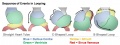
- Intermediate - Heart Tube Looping
- Intermediate - Heart Valves
- Intermediate - Outflow Tract
- Intermediate - Primordial Heart Tube
- Intermediate - Vascular Overview
- Intermediate Cardiac Embryology
- Template:Intra-embryonic coelom
L
M
P
- Paper - A contribution to the early development of the heart in mammalia, with special reference to the guinea pig
- Paper - Coarctation of the aorta 1942
- Paper - Development of the human heart from its earliest appearance to the stage found in embryos of twenty paired somites (1927)
- Paper - Development of the outflow tract and closure of the interventricular septum in the normal human heart
- Paper - Developmental Changes in the Pericardium, the Mesocardia, and the Pleural Sacs in the Human Embryo
- Paper - Developmental defects at the foramen ovale (1938)
- Paper - First contractions of the heart without cytological differentiation
- Paper - Functional limitations of the foramen ovale in the human foetal heart
- Paper - Growth allometry of the myocardium in human embryos from stages 15 to 23
- Paper - On the development of the atrial septum and the valvular apparatus in the right atrium of the pig embryo (1916)
- Paper - On the Development of the Human Heart
- Paper - On the muscular architecture of the ventricles of the human heart
- Paper - On the time of the post-natal obliteration of the fetal blood-passages (1918)
- Paper - Six specimens of abnormal heart (1912)
- Paper - Teratogenecity in the setting of cardiac development and maldevelopment
- Paper - The course of the blood flow through the fetal mammalian heart
- Paper - The Development of the Atrio-Ventricular Valves in Man
- Paper - The development of the cardiac-coronary circulatory system
- Paper - The development of the heart in man
- Paper - The Development of the Pars Membranacea Septi in the Human Heart
- Paper - The ductus arteriosus in the human fetus and newborn infant
- Paper - The early stages of the development of the pericardium
- Paper - The effect of the heart-beat upon the development of the vascular system in the chick (1918)
- Paper - The first contractions of the heart in rat embryos
- Paper - The formation of the cardiac loop in the chick
- Paper - The Formation of the Pars Membranacea Septi (1916)
- Paper - The formation of the venous valves, the foramen secundum and the septum secundum in the human heart
- Paper - The fusion of the cardiac anlages and the formation of the cardiac loop in the cat (1916)
- Paper - The human embryonic heart in the ninth week
- Paper - The human embryonic heart in the ninth week (1954)
- Paper - The human embryonic heart in the seventh week (1962)
- Paper - The origin of the heart and blood vessels in felis domestica (1924)
- Paper - The partitioning of the truncus and conus and the formation of the membranous portion of the interventricular septum in the human heart (1942)
- Paper - The physiology of the embryonic mammalian heart before circulation
- Paper - Transposition of the ventricles and the arterial stems (1931)
- Paper- The primary divisions of the myocardium in the human embryo
- Template:Patent ductus arteriosus
R
- Template:Ref-Anderson2016
- Template talk:Ref-Anderson2016
- Template:Ref-Beattie1939
- Template:Ref-Chapman1918
- Template:Ref-Chase1916
- Template:Ref-Davis1925
- Template:Ref-Davison1935
- Template:Ref-deVries1962
- Template:Ref-Fawcett1900
- Template:Ref-Field1946
- Template:Ref-Frazer1916
- Template:Ref-Goldsmith1937
- Template:Ref-Goss1938
- Template:Ref-Goss1940
- Template:Ref-Goss1942
- Template:Ref-Irvine1942
- Template:Ref-Keen1942
- Template:Ref-Keith1912
- Template:Ref-Kellogg1928
- Template:Ref-Licata1954
- Template:Ref-Magovern1986
- Template:Ref-Mall1893heart
- Template:Ref-Mall1902
- Template:Ref-Mall1911-heart
Media in category 'Heart'
The following 200 files are in this category, out of 434 total.
(previous page) (next page) Gray0462.gif 350 × 403; 49 KB
 Gray0464.gif 450 × 455; 51 KB
 Gray0467.jpg 600 × 464; 47 KB
 Gray0468.jpg 500 × 436; 43 KB
 Gray0470.jpg 800 × 371; 40 KB
 Gray0472.jpg 550 × 653; 57 KB
 Gray0474.jpg 459 × 600; 33 KB
 Gray0475.jpg 2,042 × 1,363; 350 KB
 Gray0476.jpg 600 × 609; 99 KB
 Gray0492.jpg 600 × 508; 117 KB
 Gray0498.jpg 475 × 416; 39 KB
 Gray0502.jpg 1,000 × 1,329; 215 KB
 Gray0506.jpg 375 × 464; 25 KB
 Gray0556.jpg 600 × 533; 86 KB
 Gray0971.jpg 800 × 583; 166 KB
 Head and heart muscle cartoon.jpg 874 × 800; 129 KB
 Heart chicken embryo stage 12.jpg 1,000 × 1,182; 221 KB
 Heart chicken embryo stage 16.jpg 1,000 × 1,307; 234 KB
 Heart chicken embryo stage 21.jpg 1,000 × 1,197; 207 KB
 Heart chicken embryo stage 25.jpg 1,000 × 1,154; 219 KB
 Heart conduction system-bird-monotreme-placental.jpg 1,040 × 625; 76 KB
 Heart histology 003.jpg 400 × 500; 136 KB
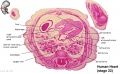
 Heart human embryo CRL10mm 01.jpg 1,000 × 1,389; 537 KB
 Heart innervation 01.jpg 1,280 × 599; 92 KB
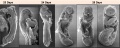 Heart Looping Sequence (SEMs).jpg 1,928 × 776; 212 KB
 Heart Looping Sequence.jpg 1,548 × 577; 85 KB
 Heart outflow tract stage 14 01.jpg 2,039 × 996; 274 KB
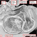 Heart outflow tract stage 14 02.jpg 996 × 996; 139 KB
 Heart outflow tract stage 14 03.jpg 989 × 996; 134 KB
 Heart Tube Fusion.jpg 1,551 × 1,139; 125 KB
 Heart Tube Segments.jpg 1,082 × 771; 63 KB
 Heart valve histology 01.jpg 1,008 × 1,280; 318 KB
 Heart valve histology 02.jpg 800 × 456; 103 KB
 Heart valve histology 03.jpg 800 × 489; 111 KB
 Heart-cartoon-001.jpg 600 × 697; 40 KB
 Heart-ventricular-septum-03.jpg 320 × 240; 14 KB
 Heart1 atrium.gif 350 × 373; 243 KB
 Heart1 ventricle.mov ; 209 KB
 HeartILP draft otcrosssection.jpg 1,433 × 1,026; 119 KB
 Historic-lungs.jpg 600 × 493; 79 KB
 Human heart developmental functional networks.jpg 833 × 614; 424 KB
 Human heart SEM1.jpg 1,200 × 330; 47 KB
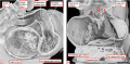
 Human stage14 heart MRI - ventricular septation.jpg 864 × 652; 39 KB
 Human stage18 heart MRI - ventricular septation.jpg 1,014 × 652; 48 KB
 Human week 10 fetus 05.jpg 1,600 × 1,200; 612 KB
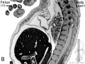
 Human- fetal week 10 heart ABCD.jpg 600 × 450; 133 KB
 Human- fetal week 10 upper body A.jpg 600 × 450; 104 KB
 Human- fetal week 10 upper body B.jpg 600 × 450; 105 KB
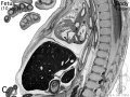 Human- fetal week 10 upper body C.jpg 600 × 450; 109 KB
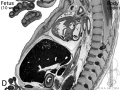 Human- fetal week 10 upper body D.jpg 600 × 450; 106 KB
 Human-heart-E3L.jpg 639 × 393; 79 KB
 Hypoplastic Left Heart.jpg 297 × 350; 17 KB
 Ingalls1920plate04.jpg 762 × 1,044; 52 KB
 Intermediate Heart Development Timeline.jpg 1,772 × 687; 133 KB
 Interrupted Aortic Arch.jpg 290 × 350; 17 KB
 Johnson1917 plate01fig02.jpg 495 × 800; 52 KB
 Johnson1917 plate03fig01.jpg 800 × 613; 57 KB
 Johnson1917 plate03fig02.jpg 800 × 620; 42 KB
 Keith1902 fig194.jpg 829 × 750; 50 KB
 Keith1902 fig212.jpg 1,123 × 750; 139 KB
 Kollmann528.jpg 744 × 587; 91 KB
 Kollmann529.jpg 737 × 593; 94 KB
 Kollmann530.jpg 733 × 524; 83 KB
 Kollmann531.jpg 688 × 570; 88 KB
 Kollmann532.jpg 702 × 409; 59 KB
 Kollmann533.jpg 717 × 594; 76 KB
 Kollmann534.jpg 684 × 599; 74 KB
 Kollmann539.jpg 736 × 696; 132 KB
 Kollmann549.jpg 689 × 573; 75 KB
 Kollmann555.jpg 605 × 585; 61 KB
 Kollmann561.jpg 739 × 591; 82 KB
 Kollmann562.jpg 670 × 581; 68 KB
 Kollmann563.jpg 754 × 848; 143 KB
 Kramer1942 fig01.jpg 1,280 × 1,108; 280 KB
 Kramer1942 fig02.jpg 1,000 × 1,526; 398 KB
 Kramer1942 fig03.jpg 1,000 × 854; 259 KB
 Kramer1942 fig04.jpg 1,000 × 654; 107 KB
 Kramer1942 fig05.jpg 1,000 × 417; 100 KB
 Kramer1942 fig06.jpg 1,000 × 482; 86 KB
 Kramer1942 fig07.jpg 1,000 × 708; 121 KB
 Kramer1942 fig08.jpg 1,000 × 457; 83 KB
 Kramer1942 fig09.jpg 1,000 × 832; 120 KB
 Le Gros Clark01.jpg 807 × 820; 159 KB
 Licata1954 fig01.jpg 1,000 × 725; 90 KB
 Licata1954 fig02.jpg 1,000 × 610; 71 KB
 Licata1954 fig03.jpg 1,000 × 708; 92 KB
 Licata1954 fig04.jpg 1,000 × 765; 84 KB
 Licata1954 fig05.jpg 1,000 × 760; 100 KB
 Licata1954 fig06.jpg 1,000 × 834; 99 KB
 Licata1954 fig07.jpg 1,000 × 931; 111 KB
 Licata1954 fig08.jpg 1,000 × 670; 82 KB
 Licata1954 fig09.jpg 1,000 × 813; 93 KB
 Licata1954 fig10.jpg 1,000 × 1,484; 367 KB
 Licata1954 fig11.jpg 1,000 × 1,502; 285 KB
 Licata1954 fig12.jpg 1,000 × 755; 82 KB
 Licata1954 fig13.jpg 1,000 × 915; 114 KB
 Licata1954 fig14.jpg 1,000 × 1,347; 177 KB
 Licata1954 fig15.jpg 1,000 × 1,324; 179 KB
 Licata1954 fig16.jpg 1,000 × 1,212; 297 KB
 Licata1954 fig16a.jpg 699 × 877; 107 KB
 Licata1954 fig16b.jpg 784 × 874; 117 KB
 Licata1954 fig16c.jpg 697 × 922; 140 KB
 Licata1954 fig16d.jpg 779 × 928; 146 KB
- Looping animation 002.mov ; 212 KB
 Mall1912-fig01.jpg 969 × 1,100; 365 KB
 Mall1912-fig02.jpg 797 × 730; 180 KB
 Mall1912-fig03.jpg 627 × 600; 95 KB
 Mall1912-fig04.jpg 832 × 650; 151 KB
 Mall1912-fig05.jpg 900 × 836; 177 KB
 Mall1912-fig06.jpg 1,000 × 991; 184 KB
 Mall1912-fig07.jpg 652 × 600; 93 KB
 Mall1912-fig08.jpg 800 × 471; 82 KB
 Mall1912-fig09.jpg 700 × 800; 158 KB
 Mall1912-fig10.jpg 800 × 550; 136 KB
 Mall1912-fig11.jpg 1,000 × 725; 124 KB
 Mall1912-fig12.jpg 816 × 800; 212 KB
 Mall1912-fig13.jpg 800 × 631; 173 KB
 Mall1912-fig14.jpg 538 × 600; 66 KB
 Mall1912-fig15.jpg 600 × 385; 33 KB
 Mall1912-fig16.jpg 735 × 650; 59 KB
 Mall1912-fig17.jpg 574 × 500; 39 KB
 Mall1912-fig18.jpg 600 × 425; 38 KB
 Mall1912-fig19.jpg 900 × 629; 77 KB
 Mall1912-fig20.jpg 400 × 494; 56 KB
 Mall1912-fig21.jpg 760 × 582; 141 KB
 Mall1912-fig22.jpg 800 × 600; 116 KB
 Mall1912-fig23.jpg 800 × 594; 159 KB
 Mall1912-fig24-25.jpg 850 × 1,000; 241 KB
 Mall1912-fig26.jpg 800 × 769; 150 KB
 Mall1912-fig27.jpg 542 × 600; 41 KB
 Mall1912-fig28.jpg 731 × 1,000; 396 KB
 Mall1912-fig29.jpg 472 × 580; 47 KB
 Mall1912-fig30.jpg 438 × 500; 58 KB
 Mall1912-fig31-33.jpg 963 × 1,000; 94 KB
 Mall1912-fig34-37.jpg 1,000 × 774; 96 KB
 Molecular & Genetic Cardiac Development Factors.jpg 1,475 × 1,541; 269 KB
 Mouse 3D Heart external E8.5-14.5.jpeg 1,280 × 559; 90 KB
 Mouse 3D Heart internal E8.5-14.5.jpeg 1,280 × 599; 106 KB
 Mouse E9 cervical intersomitic vessels A.jpg 1,714 × 1,142; 209 KB
 Mouse E9 cervical intersomitic vessels B.jpg 1,714 × 1,142; 175 KB
 Mouse E9 cervical intersomitic vessels C.jpg 1,714 × 1,142; 245 KB
 Mouse E9 cervical intersomitic vessels D.jpg 1,714 × 1,142; 147 KB
 Mouse E9 cervical intersomitic vessels.jpg 2,989 × 2,300; 777 KB
 Mouse embryo vascular.png 600 × 576; 518 KB
 Mouse heart E9.5.jpg 600 × 558; 31 KB
 Mouse heart primary cilia 01.jpg 1,200 × 908; 415 KB
 Mouse-coronary vessel formation.jpg 800 × 282; 44 KB
 Mouse-heart E17.5.jpg 353 × 1,000; 145 KB
 Nipbl heart and organ patterning.png 600 × 502; 164 KB
- Outflow tract 001.mov ; 1,020 KB
 Partial Anomalous Pulmonary Venous Drainage.jpg 290 × 350; 16 KB
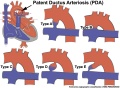 Patent ductus arteriosus classification.jpg 1,200 × 880; 138 KB
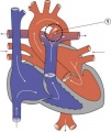 Patent Ductus Arteriosus.jpg 294 × 350; 16 KB
 Patten024.jpg 1,047 × 451; 75 KB
 Patten027.jpg 722 × 872; 123 KB
 Patten049.jpg 807 × 864; 119 KB
 Patten050.jpg 800 × 890; 109 KB
 Persistent fifth aortic arch and patent ductus arteriosus CT01.jpg 589 × 800; 69 KB
 Platypus right auricle 01.jpg 800 × 640; 124 KB
 Platypus ventricular septum 01.jpg 683 × 800; 94 KB
 Pulmonary Atresia.jpg 301 × 350; 17 KB
 Pulmonary Stenosis.jpg 289 × 350; 16 KB
 Quain595.jpg 1,200 × 803; 123 KB
 Quain596.jpg 869 × 819; 97 KB
 Robert Anderson.jpg 307 × 400; 16 KB
 Rugh 107.jpg 1,073 × 800; 188 KB
 Rugh 156.jpg 700 × 655; 86 KB
 Rugh 157.jpg 993 × 1,000; 183 KB
 Rugh 158.jpg 1,000 × 623; 158 KB
 Rugh 159.jpg 768 × 800; 87 KB
 Rugh 160.jpg 1,000 × 568; 97 KB
 Rugh 161.jpg 1,000 × 568; 104 KB
 Rugh 163.jpg 651 × 800; 75 KB
 Rugh 164.jpg 941 × 800; 108 KB
 Rugh 165.jpg 598 × 800; 132 KB
 Rugh 166.jpg 638 × 1,000; 175 KB
 Schematic ECG normal and inverted T-wave.jpg 1,001 × 384; 32 KB
 Stage 10 historic-Corner1929-1.jpg 654 × 1,000; 145 KB
 Stage 10 historic-Corner1929-1a.jpg 523 × 800; 87 KB
 Stage 11 historic-Atwell1930-1.jpg 538 × 1,000; 75 KB
 Stage 11 historic-Atwell1930-1a.jpg 430 × 800; 46 KB
 Stage 11 historic-Atwell1930-1b.jpg 323 × 600; 26 KB
 Stage 11 historic-Atwell1930-1c.jpg 215 × 400; 14 KB
 Stage 11 historic-Atwell1930-2.jpg 800 × 639; 87 KB
 Stage 11 historic-Atwell1930-2a.jpg 800 × 639; 87 KB
 Stage 11 historic-Atwell1930-2b.jpg 600 × 479; 50 KB
 Stage 11 historic-Atwell1930-2c.jpg 400 × 319; 23 KB
 Stage 11 historic-Atwell1930-4.jpg 1,000 × 1,121; 114 KB
 Stage 11 historic-Davis1923-3.jpg 1,000 × 615; 84 KB
 Stage 11 historic-Davis1923-3a.jpg 800 × 492; 59 KB
 Stage 11 historic-Davis1923-3b.jpg 600 × 369; 41 KB
 Stage 11 historic-Davis1923-3c.jpg 400 × 246; 22 KB
 Stage 13 image 022.jpg 1,000 × 473; 101 KB
 Stage 13 image 023.jpg 1,000 × 544; 110 KB
 Stage 13 image 061.jpg 1,000 × 600; 101 KB
 Stage 22 image 175.jpg 1,000 × 666; 158 KB
 Stage 22 image 178.jpg 1,000 × 658; 148 KB
 Stage 22 image 179.jpg 1,000 × 658; 131 KB
 Stage10 sem11.jpg 1,000 × 636; 76 KB
 Stage11 historic-Atwell1930-2.jpg 1,000 × 799; 131 KB


.jpg)