Category:Cardiovascular: Difference between revisions
From Embryology
No edit summary |
mNo edit summary |
||
| Line 2: | Line 2: | ||
See also the narrower categories [[:Category:Heart]] and [[:Category:Blood]]. | See also the narrower categories [[:Category:Heart]] and [[:Category:Blood]]. | ||
{{Heart Links}} | |||
[[Category:System Development]] | [[Category:System Development]] | ||
Revision as of 12:39, 13 February 2017
This is a direct link to pages, images and media related to cardiovascular system development.
See also the narrower categories Category:Heart and Category:Blood.
Subcategories
This category has the following 13 subcategories, out of 13 total.
Pages in category 'Cardiovascular'
The following 200 pages are in this category, out of 519 total.
(previous page) (next page)2
A
- Template:Abbott Figures
- Template:Abbott1915
- Template:Abbott1915images
- Abnormal Development - Hypertension
- Advanced - Abnormalities
- Advanced - Cardiac Conduction
- Advanced - Cardiac Looping
- Advanced - Cardiac Looping 2
- Advanced - Cardiac Septation
- Advanced - Cardiac Septation 2
- Advanced - Heart Fields
- Advanced - Heart Tubes
- Advanced - Molecular Development
- Advanced - Outflow Tract
- Advanced - Valve Development
- Advanced Cardiac Embryology
- Template:Advanced Cardiac menu
- ANAT2241 Cardiovascular System
- ANAT2341 Lab 4 - Early Cardiovascular Development
- ANAT3241 Lab 11
- Template:Aorta
- Template:Aortic arch
- Template:Aortic stenosis
- Template:Arachnoid mater
- Template:Artery
- Atlas of the Development of Man 2 - Cardiovascular
- Template:Atrial septal defects
- Template:Atrial septum movie links
B
- Template:Bartelmez GW.
- BGDA Lecture - Development of the Embryo/Fetus 2
- Template:BGDA Practical 14 - Maternal Decidua Interactive
- Template:BGDA Practical 14 - Placental Cord Interactive
- Template:BGDA Practical 14 - Villi Interactive
- BGDA Practical 7 - Week 5
- BGDB Lecture - Heart Development
- Template:Blood
- Template:Blood cell images
- Template:Blood vessel
- Template:Blood vessel histology
- Template:Bone marrow
- Book - A Laboratory Manual and Text-book of Embryology 9
- Book - An experimental analysis of the origin of blood and vascular endothelium (1915)
- Book - Congenital Cardiac Disease (1915)
- Book - Congenital Cardiac Disease - Figures
- Book - Congenital Cardiac Disease 1
- Book - Congenital Cardiac Disease 10
- Book - Congenital Cardiac Disease 11
- Book - Congenital Cardiac Disease 12
- Book - Congenital Cardiac Disease 13
- Book - Congenital Cardiac Disease 14
- Book - Congenital Cardiac Disease 15
- Book - Congenital Cardiac Disease 2
- Book - Congenital Cardiac Disease 3
- Book - Congenital Cardiac Disease 4
- Book - Congenital Cardiac Disease 5
- Book - Congenital Cardiac Disease 6
- Book - Congenital Cardiac Disease 7
- Book - Congenital Cardiac Disease 8
- Book - Congenital Cardiac Disease 9
- Book - Contributions to Embryology Carnegie Institution No.32
- Book - Contributions to Embryology Carnegie Institution No.36
- Book - Contributions to Embryology Carnegie Institution No.65
- Book - Contributions to Embryology Carnegie Institution No.67
- Book - Early stages of vasculogenesis in the cat with especial reference to the mesenchymal origin of endothelium (1914)
- Book - Human Embryology and Morphology 16
- Book - Quain's Embryology 9
- Book - Text-Book of the Embryology of Man and Mammals 17-1
- Template:Brain Vascular System gallery
- Template:Brain Vascular System table1
- Template:Bremer1914 figures
C
- Template:Capillary
- Template:CapillaryEM links
- Template:Cardiac
- Cardiac Embryology
- Cardiac Muscle Histology
- Template:Cardiovascular
- Cardiovascular - Arterial Development
- Cardiovascular - Detailed Cardiac Development
- Cardiovascular - Venous Development
- Cardiovascular 3D stage 13 Movie
- Cardiovascular 3D stage 22 Movie
- Template:Cardiovascular abnormalities
- Cardiovascular System - Abnormalities
- Cardiovascular System - Atrial Septal Defects
- Cardiovascular System - Blood Development
- Cardiovascular System - Blood Vessel Development
- Cardiovascular System - Bone Marrow
- Cardiovascular System - Carnegie Stage 22
- Cardiovascular System - Circulation Development
- Cardiovascular System - Coarctation of the Aorta
- Cardiovascular System - Coronary Circulation Development
- Cardiovascular System - Developmental Shunts
- Cardiovascular System - Double Outlet Right Ventricle
- Cardiovascular System - Ductus Arteriosus
- Cardiovascular System - Ductus Venosus
- Cardiovascular System - Fetal Shunts
- Cardiovascular System - Foramen Ovale
- Cardiovascular System - Heart Development
- Cardiovascular System - Heart Histology
- Cardiovascular System - Heart Rate Development
- Cardiovascular System - Heart Valve Development
- Cardiovascular System - Hypoplastic Left Heart
- Cardiovascular System - Lymphatic Development
- Cardiovascular System - Movies
- Cardiovascular System - Patent Ductus Arteriosus
- Cardiovascular System - Spleen Development
- Cardiovascular System - Tetralogy of Fallot
- Cardiovascular System - Transposition of the Great Vessels
- Cardiovascular System - Tricuspid Atresia
- Cardiovascular System - Truncus Arteriosus
- Cardiovascular System - Ventricular Septal Defects
- Cardiovascular System Development
- Template:Carotid body
- Template:Cerebral Arterial Timeline table
- Template:CHARGE syndrome
- Chicken Aortic Arches Movie
- Template:Choroid plexus
- Template:Coarctation of the aorta
- Template:Common truncus
- Computed Tomography
- Template:Congdon1922 collapse table1
- Template:Congdon1922 table1
- Template:Coronary circulation
- Template:CVS cartoons
D
- Detailed Cardiac - Arterial Roots
- Detailed Cardiac - Atrioventricular Canal
- Detailed Cardiac - Atrioventricular Conduction Axis
- Detailed Cardiac - Atrioventricular Cushions
- Detailed Cardiac - Extrapericardial Arterial Channels
- Detailed Cardiac - Interventricular Communication
- Detailed Cardiac - Intrapericardial Arterial Trunks
- Detailed Cardiac - Pulmonary Vein
- Detailed Cardiac - Sinus Node
- Detailed Cardiac - Subpulmonary Infundibulum
- Detailed Cardiac - Superior Interatrial Fold
- Detailed Cardiac - Systemic Venous Sinus
- Development Animation - Heart Atrial Septation
- Development Animation - Heart Realign
- Developmental Signals - Slit2/Robo1
- Template:Doppler ultrasound
- Template:Double outlet right ventricle
- Template:Ductus arteriosus
- Template:Ductus venosus
E
- Electrocardiogram
- Electron Microscopy Virtual Slides
- Talk:Embryo Serial Sections
- Embryology History
- Special:Badtitle/NS501:Embryology History
- History:Embryology History
- Embryology History - Douglas Reid
- Embryology History - George Bartelmez
- Embryology History - Herbert Evans
- Template:Evans HM.
- Template:Evans1909 figures
F
H
- Template:Haematopoiesis
- Template:Heart
- Template:Heart abnormal cartoon gallery
- Template talk:Heart abnormal cartoon gallery
- Heart Atrial Septation Movie
- Template:Heart histology
- Heart Historic Movie 1
- Heart Historic Movie 1951
- Heart Historic Movie 2
- Heart Historic Movie 3
- Heart Historic Movie 4
- Heart Historic Movie 5
- Heart Historic Movie 6
- Heart Historic Movie 7
- Heart Historic Movie 8
- Template:Heart Links
- Heart Looping Movie
- Heart Outflow Septation Movie
- Template:Heart rate
- Heart Realign Movie
- Template:Heart terms
- Template:Heart valve
- Historic Animation - Heart 02
- Historic Animation - Heart 04
- Template:Historic Heart
- Template talk:Historic Heart
Media in category 'Cardiovascular'
The following 200 files are in this category, out of 695 total.
(previous page) (next page) Abbott 16-18.jpg 771 × 1,000; 164 KB
 Abbott 19.jpg 880 × 791; 171 KB
 Abbott 1915.jpg 554 × 776; 38 KB
 Abbott 20.jpg 854 × 800; 129 KB
 Abbott 21.jpg 619 × 1,000; 151 KB
 Abbott 211.jpg 600 × 600; 51 KB
 Abbott 212.jpg 600 × 600; 55 KB
 Abbott 213.jpg 600 × 600; 51 KB
 Abbott 214.jpg 600 × 600; 54 KB
 Abbott 215.jpg 600 × 600; 50 KB
 Abbott 216.jpg 600 × 600; 78 KB
 Abbott 22.jpg 947 × 800; 220 KB
 Abbott 23.jpg 884 × 800; 167 KB
 Abbott 231.jpg 914 × 800; 163 KB
 Abbott 24.jpg 588 × 800; 129 KB
 Abbott 241.jpg 827 × 800; 148 KB
 Abbott 25.jpg 585 × 810; 126 KB
 Abbott 251.jpg 908 × 1,000; 167 KB
 Abbott 26.jpg 933 × 728; 140 KB
 Abbott 261.jpg 953 × 823; 162 KB
 Abbott 27.jpg 628 × 796; 132 KB
 Abbott 271.jpg 979 × 901; 161 KB
 Abbott 28.jpg 607 × 754; 118 KB
 Abbott 281.jpg 681 × 1,000; 151 KB
 Abbott 29.jpg 606 × 597; 111 KB
 Abbott 291.jpg 1,030 × 800; 188 KB
 Abbott 30.jpg 500 × 674; 85 KB
 Abbott 301.jpg 779 × 800; 118 KB
 Abbott 31.jpg 684 × 535; 81 KB
 Abbott 311.jpg 1,134 × 800; 162 KB
 Abbott 32-34.jpg 846 × 800; 84 KB
 Abbott 32.jpg 504 × 374; 21 KB
 Abbott 33.jpg 504 × 374; 19 KB
 Abbott 34.jpg 504 × 374; 22 KB
 Abbott plate 05.jpg 671 × 1,000; 132 KB
 Abbott plate 51.jpg 618 × 800; 108 KB
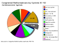 Abnormal81-92-heart.png 481 × 344; 6 KB
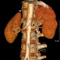 Accessory renal artery.jpg 800 × 798; 103 KB
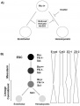 Adhesion regulation blood or vessel differentiation.jpg 600 × 788; 42 KB
 Adult heart CT01.jpg 957 × 951; 212 KB
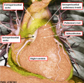
 Adult heart outflow tract CT01.jpg 747 × 747; 58 KB
 Adult heart outflow tract CT02.jpg 747 × 747; 65 KB
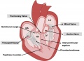 Adult Heart Valves.jpg 1,475 × 1,070; 113 KB
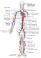 Adult human cardiovascular system.jpg 707 × 1,000; 151 KB
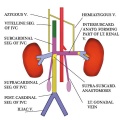 Adult renal venous cartoon.jpg 600 × 600; 62 KB
 Advanced Heart Development Timeline GA.jpg 1,000 × 434; 97 KB
 Advanced Heart Development Timeline.jpg 1,772 × 769; 158 KB
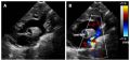 Aorta coarctation echocardiogram.jpg 601 × 283; 28 KB
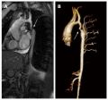 Aorta coarctation MRI.jpg 455 × 423; 25 KB
 Aortic arch and ductus arteriosus.jpg 600 × 720; 68 KB
 Aortic Stenosis.jpg 290 × 350; 16 KB
 Arey1924 fig193.jpg 1,200 × 962; 126 KB
 Arey1924 fig194.jpg 1,200 × 710; 122 KB
 Arey1924 fig195.jpg 1,583 × 950; 267 KB
 Arey1924 fig196.jpg 1,200 × 881; 196 KB
 Arey1924 fig197.jpg 1,200 × 1,615; 227 KB
 Arey1924 fig198.jpg 1,200 × 1,102; 187 KB
 Arey1924 fig199.jpg 1,200 × 956; 161 KB
 Arey1924 fig200.jpg 1,774 × 1,484; 209 KB
 Atrial & Ventricular Septation 1.jpg 1,482 × 960; 97 KB
 Atrial & Ventricular Septation 2.jpg 1,482 × 1,075; 113 KB
 Atrial Septal Defect.jpg 287 × 350; 16 KB
 Atrial Septation.jpg 1,482 × 960; 90 KB
 AV Canal Division (Superior View).jpg 1,487 × 489; 69 KB
 AV Canal Division.jpg 1,482 × 970; 93 KB
 AV Valves.jpg 1,183 × 1,085; 129 KB
 Bailey156.jpg 921 × 617; 150 KB
 Bailey157.jpg 725 × 562; 93 KB
 Bailey158.jpg 898 × 509; 101 KB
 Bailey159.jpg 933 × 896; 238 KB
 Bailey160.jpg 916 × 715; 202 KB
 Bailey161.jpg 645 × 629; 101 KB
 Bailey162.jpg 799 × 642; 102 KB
 Bailey163.jpg 757 × 691; 107 KB
 Bailey164.jpg 928 × 862; 149 KB
 Bailey165.jpg 613 × 1,045; 127 KB
 Bailey166.jpg 787 × 656; 90 KB
 Bailey167.jpg 610 × 458; 58 KB
 Bailey168.jpg 657 × 314; 43 KB
 Bailey169.jpg 504 × 264; 27 KB
 Bailey170.jpg 709 × 457; 67 KB
 Bailey171.jpg 954 × 507; 81 KB
 Bailey173.jpg 888 × 620; 113 KB
 Bailey174.jpg 955 × 542; 88 KB
 Bailey175.jpg 885 × 306; 51 KB
 Bailey176.jpg 918 × 352; 62 KB
 Bailey177.jpg 943 × 873; 199 KB
 Bailey178.jpg 913 × 653; 125 KB
 Bailey179.jpg 892 × 794; 114 KB
 Bailey180.jpg 938 × 431; 70 KB
 Bailey181.jpg 839 × 658; 67 KB
 Bailey182.jpg 913 × 718; 103 KB
 Bailey183.jpg 847 × 448; 57 KB
 Bailey184.jpg 625 × 462; 48 KB
 Bailey185.jpg 944 × 499; 98 KB
 Bailey187.jpg 817 × 732; 70 KB
 Bailey188.jpg 906 × 538; 64 KB
 Bailey189.jpg 810 × 632; 63 KB
 Bailey190.jpg 801 × 584; 69 KB
 Bailey191.jpg 863 × 509; 109 KB
 Bailey192.jpg 960 × 806; 133 KB
 Bailey193.jpg 747 × 848; 94 KB
 Bailey194.jpg 841 × 638; 80 KB
 Bailey195.jpg 924 × 781; 79 KB
 Bailey196.jpg 760 × 510; 47 KB
 Bailey197.jpg 878 × 705; 122 KB
 Bailey198.jpg 931 × 623; 83 KB
 Bailey199.jpg 924 × 451; 62 KB
 Bailey200.jpg 898 × 671; 149 KB
 Bailey201.jpg 1,059 × 1,033; 259 KB
 Bailey202.jpg 906 × 848; 138 KB
 Bailey203.jpg 406 × 614; 48 KB
 Bailey204.jpg 534 × 653; 62 KB
 Bailey205.jpg 534 × 653; 68 KB
 Bailey206.jpg 973 × 854; 162 KB
 Bailey207.jpg 896 × 951; 139 KB
 Bailey208.jpg 1,179 × 952; 165 KB
 Bailey209.jpg 1,248 × 988; 319 KB
 Bailey210.jpg 565 × 558; 63 KB
 Bailey211.jpg 767 × 820; 182 KB
 Bailey212.jpg 914 × 938; 218 KB
 Bailey213.jpg 800 × 851; 101 KB
 Bailey214.jpg 439 × 781; 81 KB
 Bailey215.jpg 944 × 958; 361 KB
 Bailey216.jpg 905 × 734; 261 KB
 Bailey217.jpg 767 × 694; 62 KB
 Bailey218.jpg 872 × 542; 84 KB
 Bailey219.jpg 911 × 575; 97 KB
 Bailey220.jpg 833 × 555; 105 KB
 Bailey221.jpg 904 × 774; 181 KB
 Bailey222.jpg 928 × 583; 145 KB
 Bailey245.jpg 499 × 733; 47 KB
 Bailey247.jpg 877 × 592; 113 KB
 Bailey248.jpg 814 × 521; 111 KB
 Baileytable03.jpg 821 × 667; 73 KB
 Bast1931 plate01.jpg 1,280 × 871; 113 KB
 Blood capillary EM 01.jpg 1,107 × 714; 260 KB
 Blood capillary EM 02.jpg 600 × 600; 99 KB
 Blood capillary EM 03.jpg 1,560 × 1,230; 441 KB
 Blood capillary EM 04.jpg 1,560 × 1,230; 462 KB
 Blood capillary EM 05.jpg 1,015 × 800; 205 KB
 Blood capillary EM 06.jpg 1,015 × 800; 216 KB
 Blood vessel wall cartoon.jpg 450 × 600; 71 KB
 Blood-brain barrier cartoon.jpg 765 × 1,000; 75 KB
 Blood-brain barrier EM01.jpg 1,656 × 810; 250 KB
 Bremer1914 plate01.jpg 684 × 1,000; 102 KB
 Bremer1914 plate02.jpg 716 × 1,000; 147 KB
 Bremer1914 plate03.jpg 706 × 1,000; 127 KB
 Bremer1914 plate04.jpg 643 × 1,000; 127 KB
 Bremer1914 plate05.jpg 643 × 1,000; 144 KB
 Buell-plate01.jpg 1,221 × 1,500; 236 KB
 Buell-plate02.jpg 1,124 × 1,500; 287 KB
 Buell01.jpg 713 × 800; 70 KB
 Buell02.jpg 977 × 800; 86 KB
 Buell03.jpg 790 × 800; 73 KB
 Buell04.jpg 913 × 800; 69 KB
 Buell05.jpg 1,176 × 800; 96 KB
 Buell06.jpg 1,037 × 800; 76 KB
 Buell07.jpg 1,200 × 878; 195 KB
 Buell08.jpg 1,037 × 1,000; 176 KB
 Buell09.jpg 724 × 1,000; 83 KB
 Buell10.jpg 665 × 1,000; 113 KB
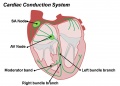 Cardiac Conduction System.jpg 1,201 × 862; 81 KB
 Cardiac muscle EM01.jpg 1,072 × 735; 231 KB
 Cardiac muscle EM02.jpg 1,072 × 735; 224 KB
 Cardiac muscle EM03.jpg 849 × 615; 135 KB
 Cardiac muscle EM04.jpg 1,000 × 680; 191 KB
 Cardiac Muscle EM05.jpg 992 × 733; 158 KB
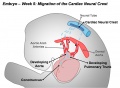 Cardiac Neural Crest Migration.jpg 1,517 × 1,116; 122 KB
 Cephalic plexus.png 600 × 557; 559 KB
 Cerebral blood supply development 01.jpg 1,200 × 460; 67 KB
 Cerebral brain artery development 01.jpg 845 × 600; 74 KB
 Cerebral brain artery development 02.jpg 996 × 400; 57 KB
 Cervical intersomitic vessels.png 600 × 462; 308 KB
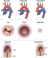
 Coarctation of the Aorta.jpg 289 × 350; 16 KB
 Complete atrioventricular canal.jpg 320 × 240; 22 KB
 Congdon-table01.jpg 764 × 1,000; 167 KB
 Congdon1922-1-16.jpg 980 × 1,000; 157 KB
 Congdon1922-17.jpg 1,000 × 411; 55 KB
 Congdon1922-18-25.jpg 1,200 × 795; 179 KB
 Congdon1922-18.jpg 494 × 506; 29 KB
 Congdon1922-19.jpg 653 × 471; 31 KB
 Congdon1922-20.jpg 794 × 446; 43 KB
 Congdon1922-21.jpg 578 × 407; 27 KB
 Congdon1922-22.jpg 511 × 489; 27 KB
 Congdon1922-23.jpg 519 × 412; 26 KB
 Congdon1922-24.jpg 790 × 482; 40 KB
 Congdon1922-25.jpg 509 × 358; 25 KB
 Congdon1922-26.jpg 746 × 726; 54 KB
 Congdon1922-27-28.jpg 997 × 612; 68 KB
 Congdon1922-29.jpg 976 × 1,000; 81 KB
 Congdon1922-30.jpg 1,133 × 1,000; 176 KB
 Congdon1922-31.jpg 1,063 × 1,000; 93 KB
 Congdon1922-32.jpg 1,133 × 1,000; 132 KB
 Congdon1922-33.jpg 920 × 1,000; 107 KB
 Congdon1922-34.jpg 920 × 1,000; 122 KB
 Congdon1922-35.jpg 920 × 1,000; 97 KB
 Congdon1922-36.jpg 920 × 1,000; 113 KB
 Congdon1922-37.jpg 1,200 × 838; 163 KB
 Congdon1922-38.jpg 1,187 × 1,000; 165 KB


.jpg)