Buccopharyngeal membrane: Difference between revisions
No edit summary |
mNo edit summary |
||
| (37 intermediate revisions by 2 users not shown) | |||
| Line 1: | Line 1: | ||
{{Header}} | |||
== Introduction == | == Introduction == | ||
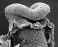
[[File:Stage11 | [[File:Stage11 sem4.jpg|thumb|300px|Human Embryo Stage {{CS11}} buccopharyngeal membrane ]] | ||
[[File:Stage11 sem2.jpg|thumb|300px|Buccopharyngeal membrane - degenerating]] | |||
[[File:Keith1902 fig015b.jpg|thumb|300px|Buccopharyngeal membrane position relative to adult anatomy]] | |||
The {{buccopharyngeal membrane}} (Latin, ''bucca'' = cheek) or {{oral membrane}}, forms the external upper membrane limit (cranial end) of the early gastrointestinal tract (GIT). This membrane region first develops in the trilaminar embryo (week 3) during gastrulation and lies above the cranial end of the {{notochord}}. The "membrane" quality comes from being composed of only ectoderm and endoderm, without a middle (intervening) layer of mesoderm. | |||
The membrane lies at the floor of the ventral depression (stomadeum) where the oral cavity will open. The membrane will break down during week 4 ({{GA}} week 6) to form the initial "oral opening" of the pharynx of the foregut. After this time the oral cavity and foregut are open to then {{amniotic cavity}}. The membrane region at the lower end of the gastrointestinal tract is the {{cloacal membrane}} that will break down at a later stage of development. | |||
{| class="wikitable mw-collapsible mw-collapsed" | |||
! Related Topics | |||
|- | |||
| | |||
{{Head Links}} | |||
<br> | |||
{{Gastrointestinal Tract Links}} | |||
|} | |||
== Some Recent Findings == | |||
{| | |||
|-bgcolor="F5FAFF" | |||
| | |||
* '''Role of JNK during buccopharyngeal membrane perforation, the last step of embryonic mouth formation'''{{#pmid:28032936|PMID28032936}} "The buccopharyngeal membrane is a thin layer of cells covering the embryonic mouth. The perforation of this structure creates an opening connecting the external and the digestive tube which is essential for oral cavity formation. In humans, persistence of the buccopharyngeal membrane can lead to orofacial defects such as choanal atresia, oral synechiaes, and cleft palate. Little is known about the causes of a persistent buccopharyngeal membrane and, importantly, how this structure ruptures. We have determined, using antisense and pharmacological approaches, that {{Xenopus}} embryos deficient c-Jun N-terminal kinase (JNK) signaling have a persistent buccopharyngeal membrane. JNK deficient embryos have decreased cell division and increased cellular stress and apoptosis. However, altering these processes independently of JNK did not affect buccopharyngeal membrane perforation. JNK deficient embryos also have increased intercellular adhesion and defects in e-cadherin localization. Conversely, embryos with overactive JNK have epidermal fragility, increased E-cadherin internalization, and increased membrane localized clathrin. In the buccopharyngeal membrane, clathrin is colocalized with active JNK. Furthermore, inhibition of endocytosis results in a persistent buccopharyngeal membrane, mimicking the JNK deficient phenotype. The results of this study suggest that JNK has a role in the disassembly adherens junctions by means of endocytosis that is required during buccopharyngeal membrane perforation." {More? JNK = [https://www.ncbi.nlm.nih.gov/gene/5599 MAPK8 mitogen-activated protein kinase 8] | [https://www.creative-diagnostics.com/JNK-Signaling-Pathway.htm JNK-Signaling-Pathway]) | |||
|} | |||
{| class="wikitable mw-collapsible mw-collapsed" | |||
! More recent papers | |||
|- | |||
| [[File:Mark_Hill.jpg|90px|left]] {{Most_Recent_Refs}} | |||
Search term: [http://www.ncbi.nlm.nih.gov/pubmed/?term=Buccopharyngeal+Membrane+Development ''Buccopharyngeal Membrane Development''] | [http://www.ncbi.nlm.nih.gov/pubmed/?term=Oral+membrane ''Oral membrane''] | [http://www.ncbi.nlm.nih.gov/pubmed/?term=Persistent+Buccopharyngeal+Membrane ''Persistent Buccopharyngeal Membrane''] | |||
|} | |||
{| class="wikitable mw-collapsible mw-collapsed" | |||
! Older papers | |||
|- | |||
| {{Older papers}} | |||
|} | |||
==Buccopharyngeal Membrane Timeline== | |||
The key developmental changes in the buccopharyngeal membrane that can be morphologically observed on the embryo surface occur during week 4 ({{GA}} week 6) of human embryonic development. | |||
{{Carnegie_stage_table_1}} | |||
<br> | |||
{{Buccopharyngeal Membrane timeline EM gallery}} | |||
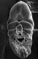
===Stage 10 Embryo === | |||
A ventral view scanning EM embryo cranial end (day 21, 4 to 5 somites) showing early cardiac tube lying beneath brain fold. Between these two structures is where the stomedeum and buccopharyngeal membrane will form. Note that the pharyngeal arches are not yet visible. | |||
[[File:Stage10 sem4.jpg|500px]] | |||
A ventral view | ===Stage 11 Embryo=== | ||
A ventral view of the embryo head region (Carnegie stage 11, week 4, 25 days, 20 somite pairs) showing the buccopharyngeal membrane breaking down and opening the gastrointestinal tract to the amnion. | |||
Note the position at the "floor" of the stomedeum and relative to the first pharyngeal arch and triangular shape. Midline crack in head is an artefact. | Note the position at the "floor" of the stomedeum and relative to the first pharyngeal arch and triangular shape. Midline crack in head is an artefact. | ||
{| | |||
! Bright Field | |||
! Scanning EM | |||
! Scanning EM | |||
|- | |||
| [[File:Stage11 bf9.jpg|300px]] | |||
| [[File:Stage11 sem3.jpg|300px]] | |||
| [[File:Stage11 sem4.jpg|300px]] | |||
|} | |||
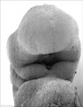
===Stage 12 Embryo=== | |||
A ventral view of the embryo head region (Carnegie stage 12, week 4, 26 days, 25 somite pairs, CRL 5 mm). Note by this stage, one day later, the buccopharyngeal membrane has been entirely lost. | |||
[[File:Stage12 sem2.jpg|500px]] | |||
===Stage 13 Embryo=== | |||
A ventral view of the embryo head region (Carnegie stage 13, week 4, CRL 5.5 mm). | |||
[[File:ME18 001.jpg|500px]] | |||
==Abnormalities== | |||
===Persistent Buccopharyngeal Membrane=== | |||
A persistent buccopharyngeal membrane is a very rare abnormality with only (2009) 23 reported cases in the literature. {{#pmid:19342107|PMID19342107}} There are a variety of clinical repair techniques.{{#pmid:9431473|PMID9431473}} | |||
Persistence of the buccopharyngeal membrane can lead to several orofacial abnormalities: | |||
* choanal atresia - narrowing of the rear opening of the nasal cavity. | |||
* oral synechiaes - fibrous bands between the mucosal surfaces of the upper and lower alveolar ridges. | |||
* {{cleft palate}} - failure of the maxillary shelves to fuse to form the palate.{{#pmid:2304741|PMID2304741}} | |||
A mouse model has shown that hedgehog mediates persistence of the buccopharyngeal membrane.{{#pmid:25300580|PMID25300580}} | |||
===1p36 Deletion Syndrome=== | |||
{| | |||
|-bgcolor="FEF9E7" | |||
| | |||
{{ICD-11}} {{ICD11weblink}}1004815242 LD44.11 Deletions of the short arm of chromosome 1] | |||
|} | |||
The {{Chr1}}p36 deletion syndrome comprises a phenotypic presentation that includes central nervous system, cardiac, craniofacial, and airway anomalies. A single patient study identified a persistent buccopharyngeal membrane and unidentifiable larynx.{{#pmid:24290305|PMID24290305}} | |||
==Animal Models== | |||
{| | |||
|+ Buccopharyngeal Membrane Animal Studies | |||
|- | |||
! Species | |||
! Reference | |||
|- | |||
| {{Chicken}} | |||
| Waterman RE & Schoenwolf GC. (1980){{#pmid:7212297|PMID7212297}} | |||
|- | |||
| {{Frog}} | |||
| Watanabe K, Sasaki F & Takahama H. (1984){{#pmid:6524693|PMID6524693}} Houssin NS. etal. (2017){{#pmid:28032936|PMID28032936}} | |||
|- | |||
| Hamster | |||
| Waterman RE. (1977){{#pmid:885288|PMID885288}} | |||
|- | |||
| {{Mouse}} | |||
| Poelmann RE. etal. (1985){{#pmid:3985368|PMID3985368}} | |||
|- | |||
| Salamander | |||
| Takahama H, Sasaki F & Watanabe K. (1988){{#pmid:3339635|PMID3339635}} | |||
|} | |||
==References== | |||
<references/> | |||
===Reviews=== | |||
{{#pmid:28514120}} | |||
===Articles=== | |||
{{#pmid:28032936}} | |||
{{#pmid:3385865}} | |||
==Additional Images== | |||
===Historic=== | |||
<gallery> | |||
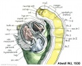
File:Low 07.jpg|Model of the Pharynx | |||
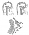
File:Low plate 03.jpg|section | |||
File:Stage 11 historic-Atwell1930-2a.jpg|17 somites stage | |||
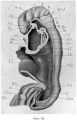
File:Keith1902 fig015b.jpg|position of the Oral Plate | |||
File:Gray1182.jpg|rabbit buccopharyngeal membrane | |||
</gallery> | |||
{{Glossary}} | |||
{{Footer}} | |||
[[Category:Gastrointestinal Tract]] | [[Category:Gastrointestinal Tract]] | ||
[[Category:Head]] | |||
Latest revision as of 13:35, 12 May 2019
| Embryology - 27 May 2024 |
|---|
| Google Translate - select your language from the list shown below (this will open a new external page) |
|
العربية | català | 中文 | 中國傳統的 | français | Deutsche | עִברִית | हिंदी | bahasa Indonesia | italiano | 日本語 | 한국어 | မြန်မာ | Pilipino | Polskie | português | ਪੰਜਾਬੀ ਦੇ | Română | русский | Español | Swahili | Svensk | ไทย | Türkçe | اردو | ייִדיש | Tiếng Việt These external translations are automated and may not be accurate. (More? About Translations) |
Introduction


The buccopharyngeal membrane (Latin, bucca = cheek) or oral membrane, forms the external upper membrane limit (cranial end) of the early gastrointestinal tract (GIT). This membrane region first develops in the trilaminar embryo (week 3) during gastrulation and lies above the cranial end of the notochord. The "membrane" quality comes from being composed of only ectoderm and endoderm, without a middle (intervening) layer of mesoderm.
The membrane lies at the floor of the ventral depression (stomadeum) where the oral cavity will open. The membrane will break down during week 4 (GA week 6) to form the initial "oral opening" of the pharynx of the foregut. After this time the oral cavity and foregut are open to then Template:Amniotic cavity. The membrane region at the lower end of the gastrointestinal tract is the cloacal membrane that will break down at a later stage of development.
Some Recent Findings
|
| More recent papers |
|---|
 This table allows an automated computer search of the external PubMed database using the listed "Search term" text link.
More? References | Discussion Page | Journal Searches | 2019 References | 2020 References Search term: Buccopharyngeal Membrane Development | Oral membrane | Persistent Buccopharyngeal Membrane |
| Older papers |
|---|
| These papers originally appeared in the Some Recent Findings table, but as that list grew in length have now been shuffled down to this collapsible table.
See also the Discussion Page for other references listed by year and References on this current page. |
Buccopharyngeal Membrane Timeline
The key developmental changes in the buccopharyngeal membrane that can be morphologically observed on the embryo surface occur during week 4 (GA week 6) of human embryonic development.
| Week: | 1 | 2 | 3 | 4 | 5 | 6 | 7 | 8 |
| Carnegie stage: | 1 2 3 4 | 5 6 | 7 8 9 | 10 11 12 13 | 14 15 | 16 17 | 18 19 | 20 21 22 23 |
- Human Buccopharyngeal Membrane Timeline Gallery (Week 4)
Stage 10

Stage 11
Stage 12
Stage 13
Stage 10 Embryo
A ventral view scanning EM embryo cranial end (day 21, 4 to 5 somites) showing early cardiac tube lying beneath brain fold. Between these two structures is where the stomedeum and buccopharyngeal membrane will form. Note that the pharyngeal arches are not yet visible.

Stage 11 Embryo
A ventral view of the embryo head region (Carnegie stage 11, week 4, 25 days, 20 somite pairs) showing the buccopharyngeal membrane breaking down and opening the gastrointestinal tract to the amnion.
Note the position at the "floor" of the stomedeum and relative to the first pharyngeal arch and triangular shape. Midline crack in head is an artefact.
| Bright Field | Scanning EM | Scanning EM |
|---|---|---|

|

|
|
Stage 12 Embryo
A ventral view of the embryo head region (Carnegie stage 12, week 4, 26 days, 25 somite pairs, CRL 5 mm). Note by this stage, one day later, the buccopharyngeal membrane has been entirely lost.

Stage 13 Embryo
A ventral view of the embryo head region (Carnegie stage 13, week 4, CRL 5.5 mm).

Abnormalities
Persistent Buccopharyngeal Membrane
A persistent buccopharyngeal membrane is a very rare abnormality with only (2009) 23 reported cases in the literature. [2] There are a variety of clinical repair techniques.[3]
Persistence of the buccopharyngeal membrane can lead to several orofacial abnormalities:
- choanal atresia - narrowing of the rear opening of the nasal cavity.
- oral synechiaes - fibrous bands between the mucosal surfaces of the upper and lower alveolar ridges.
- cleft palate - failure of the maxillary shelves to fuse to form the palate.[4]
A mouse model has shown that hedgehog mediates persistence of the buccopharyngeal membrane.[5]
1p36 Deletion Syndrome
The 1p36 deletion syndrome comprises a phenotypic presentation that includes central nervous system, cardiac, craniofacial, and airway anomalies. A single patient study identified a persistent buccopharyngeal membrane and unidentifiable larynx.[6]
Animal Models
| Species | Reference |
|---|---|
| chicken | Waterman RE & Schoenwolf GC. (1980)[7] |
| frog | Watanabe K, Sasaki F & Takahama H. (1984)[8] Houssin NS. etal. (2017)[1] |
| Hamster | Waterman RE. (1977)[9] |
| mouse | Poelmann RE. etal. (1985)[10] |
| Salamander | Takahama H, Sasaki F & Watanabe K. (1988)[11] |
References
- ↑ 1.0 1.1 Houssin NS, Bharathan NK, Turner SD & Dickinson AJ. (2017). Role of JNK during buccopharyngeal membrane perforation, the last step of embryonic mouth formation. Dev. Dyn. , 246, 100-115. PMID: 28032936 DOI.
- ↑ Verma SP & Geller K. (2009). Persistent buccopharyngeal membrane: report of a case and review of the literature. Int. J. Pediatr. Otorhinolaryngol. , 73, 877-80. PMID: 19342107 DOI.
- ↑ Bent JP, Klippert FN & Smith RJ. (1997). Management of congenital buccopharyngeal membrane. Cleft Palate Craniofac. J. , 34, 538-41. PMID: 9431473 DOI.
- ↑ Pillai KG, Kamath VV, Kumar GS & Nagamani N. (1990). Persistent buccopharyngeal membrane with cleft palate. A case report. Oral Surg. Oral Med. Oral Pathol. , 69, 164-6. PMID: 2304741
- ↑ Tabler JM, Bolger TG, Wallingford J & Liu KJ. (2014). Hedgehog activity controls opening of the primary mouth. Dev. Biol. , 396, 1-7. PMID: 25300580 DOI.
- ↑ Ferril GR, Barham HP & Prager JD. (2014). Novel airway findings in a patient with 1p36 deletion syndrome. Int. J. Pediatr. Otorhinolaryngol. , 78, 157-8. PMID: 24290305 DOI.
- ↑ Waterman RE & Schoenwolf GC. (1980). The ultrastructure of oral (buccopharyngeal) membrane formation and rupture in the chick embryo. Anat. Rec. , 197, 441-70. PMID: 7212297 DOI.
- ↑ Watanabe K, Sasaki F & Takahama H. (1984). The ultrastructure of oral (buccopharyngeal) membrane formation and rupture in the anuran embryo. Anat. Rec. , 210, 513-24. PMID: 6524693 DOI.
- ↑ Waterman RE. (1977). Ultrastructure of oral (buccopharyngeal) membrane formation and rupture in the hamster embryo. Dev. Biol. , 58, 219-29. PMID: 885288
- ↑ Poelmann RE, Dubois SV, Hermsen C, Smits-van Prooije AE & Vermeij-Keers C. (1985). Cell degeneration and mitosis in the buccopharyngeal and branchial membranes in the mouse embryo. Anat. Embryol. , 171, 187-92. PMID: 3985368
- ↑ Takahama H, Sasaki F & Watanabe K. (1988). Morphological changes in the oral (buccopharyngeal) membrane in urodelan embryos: development of the mouth opening. J. Morphol. , 195, 59-69. PMID: 3339635 DOI.
Reviews
Chen J, Jacox LA, Saldanha F & Sive H. (2017). Mouth development. Wiley Interdiscip Rev Dev Biol , 6, . PMID: 28514120 DOI.
Articles
Houssin NS, Bharathan NK, Turner SD & Dickinson AJ. (2017). Role of JNK during buccopharyngeal membrane perforation, the last step of embryonic mouth formation. Dev. Dyn. , 246, 100-115. PMID: 28032936 DOI.
Arcand P & Haikal J. (1988). Persistent buccopharyngeal membrane. J Otolaryngol , 17, 125-7. PMID: 3385865
Additional Images
Historic

Model of the Pharynx
section
17 somites stage
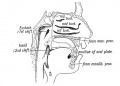
position of the Oral Plate
rabbit buccopharyngeal membrane


Glossary Links
- Glossary: A | B | C | D | E | F | G | H | I | J | K | L | M | N | O | P | Q | R | S | T | U | V | W | X | Y | Z | Numbers | Symbols | Term Link
Cite this page: Hill, M.A. (2024, May 27) Embryology Buccopharyngeal membrane. Retrieved from https://embryology.med.unsw.edu.au/embryology/index.php/Buccopharyngeal_membrane
- © Dr Mark Hill 2024, UNSW Embryology ISBN: 978 0 7334 2609 4 - UNSW CRICOS Provider Code No. 00098G