Respiratory System Development: Difference between revisions
mNo edit summary |
mNo edit summary |
||
| Line 7: | Line 7: | ||
In the head/neck region, the pharynx forms a major arched cavity within the phrayngeal arches. The lungs go through 4 distinct histological phases of development and in late fetal development thyroid hormone, respiratory motions and amniotic fliud are thought to have a role in lung maturation. | In the head/neck region, the pharynx forms a major arched cavity within the phrayngeal arches. The lungs go through 4 distinct histological phases of development and in late fetal development thyroid hormone, respiratory motions and amniotic fliud are thought to have a role in lung maturation. Branching is a key mechanism/process in lung development leading to alveolar saccules after about 23 branching generations (range of 18–30). | ||
The two main respiratory cell types, squamous alveolar type 1 and alveolar type 2 (surfactant secreting), both arise from the same bi-potetial progenitor cell.<ref name=PMID24499815><pubmed>24499815</pubmed></ref> The third main cell type are macrophages (dust cells) that arise from blood monocyte cells. | |||
Development of this system is not completed until the last weeks of Fetal development, just before birth. Therefore premature babies have difficulties associated with insufficient surfactant (end month 6 alveolar cells type 2 appear and begin to secrete surfactant). | Development of this system is not completed until the last weeks of Fetal development, just before birth. Therefore premature babies have difficulties associated with insufficient surfactant (end month 6 alveolar cells type 2 appear and begin to secrete surfactant). | ||
{{Respiratory Links}} | {{Respiratory Links}} | ||
| Line 78: | Line 78: | ||
===Mechanisms=== | ===Mechanisms=== | ||
* '''Initiation''' - Budding of foregut [[endoderm]] to generate the trachea. | |||
* '''Initiation''' - Budding of foregut endoderm to generate the trachea. | |||
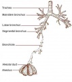
* '''Branching''' - A repeated mechanism of branching that is ongoing throughout development to form the conducting bronchioles then alveolar ducts. | * '''Branching''' - A repeated mechanism of branching that is ongoing throughout development to form the conducting bronchioles then alveolar ducts. | ||
* '''Surface area increase''' - Expansion of the surface area in late development generating eventually the thin air–blood barrier for gas exchange in the acini. | * '''Surface area increase''' - Expansion of the surface area in late development generating eventually the thin air–blood barrier for gas exchange in the acini. | ||
* '''Vascular development''' - Extension of a vascular capillary tree within the connective tissue and wall of the acini for gas exchange, and the lymphatic development for immunology of the lungs. | * '''Vascular development''' - Extension of a vascular capillary tree within the connective tissue and wall of the acini for gas exchange, and the lymphatic development for immunology of the lungs. | ||
* '''Surfactant development''' - allows lung inflation and decreases the work of breathing and also related to immunology of the lungs. | * '''Surfactant development''' - allows lung inflation and decreases the work of breathing and also related to immunology of the lungs. | ||
* '''Musculoskeletal development''' - contributes the mechanical elements of ribs, intercostals and diaphragm required for breathing. | * '''Musculoskeletal development''' - contributes the mechanical elements of ribs, intercostals and [[Diaphragm Development|diaphragm]] required for breathing. | ||
==Lung Development Stages== | ==Lung Development Stages== | ||
Revision as of 13:10, 31 August 2017
| Embryology - 15 Jun 2024 |
|---|
| Google Translate - select your language from the list shown below (this will open a new external page) |
|
العربية | català | 中文 | 中國傳統的 | français | Deutsche | עִברִית | हिंदी | bahasa Indonesia | italiano | 日本語 | 한국어 | မြန်မာ | Pilipino | Polskie | português | ਪੰਜਾਬੀ ਦੇ | Română | русский | Español | Swahili | Svensk | ไทย | Türkçe | اردو | ייִדיש | Tiếng Việt These external translations are automated and may not be accurate. (More? About Translations) |
Introduction

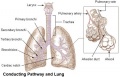
The respiratory system does not carry out its physiological function (of gas exchange) until after birth. The respiratory tract, diaphragm and lungs do form early in embryonic development. The respiratory tract is divided anatomically into 2 main parts:
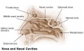
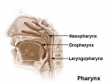
- upper respiratory tract, consisting of the nose, nasal cavity and the pharynx
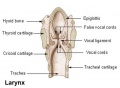
- lower respiratory tract consisting of the larynx, trachea, bronchi and the lungs.
In the head/neck region, the pharynx forms a major arched cavity within the phrayngeal arches. The lungs go through 4 distinct histological phases of development and in late fetal development thyroid hormone, respiratory motions and amniotic fliud are thought to have a role in lung maturation. Branching is a key mechanism/process in lung development leading to alveolar saccules after about 23 branching generations (range of 18–30).
The two main respiratory cell types, squamous alveolar type 1 and alveolar type 2 (surfactant secreting), both arise from the same bi-potetial progenitor cell.[1] The third main cell type are macrophages (dust cells) that arise from blood monocyte cells.
Development of this system is not completed until the last weeks of Fetal development, just before birth. Therefore premature babies have difficulties associated with insufficient surfactant (end month 6 alveolar cells type 2 appear and begin to secrete surfactant).

Some Recent Findings
|
| More recent papers |
|---|
 This table allows an automated computer search of the external PubMed database using the listed "Search term" text link.
More? References | Discussion Page | Journal Searches | 2019 References | 2020 References Search term: Lung Embryology <pubmed limit=5>Lung Embryology</pubmed> |
| Older papers |
|---|
Clinical
|
Textbooks
- Moore, K.L., Persaud, T.V.N. & Torchia, M.G. (2015). The developing human: clinically oriented embryology (10th ed.). Philadelphia: Saunders. Chapter 10 Respiratory System
- Schoenwolf, G.C., Bleyl, S.B., Brauer, P.R., Francis-West, P.H. & Philippa H. (2015). Larsen's human embryology (5th ed.). New York; Edinburgh: Churchill Livingstone. Chapter 11 Development of the Respiratory System and Body Cavities
- Before We Are Born (5th ed.) Moore and Persaud Chapter 13 p255-287
- Essentials of Human Embryology Larson Chapter 9 p123-146
- Human Embryology Fitzgerald and Fitzgerald Chapter 19,20 p119-123
- Anatomy of the Human Body 1918 Henry Gray The Respiratory Apparatus
Objectives
- Describe the development of the respiratory system from the endodermal and mesodermal components.
- Describe the main steps in the development of the lungs.
- Describe the development of the diaphragm and thoracic cavities.
- List the respiratory changes before and after birth.
- Describe the developmental aberrations responsible for the following malformations: tracheo - oesophageal fistula (T.O.F); oesphageal atresia; diaphragmatic hernia; lobar emphysema.
Development Overview
Week 4 - laryngotracheal groove forms on floor foregut.
Week 5 - left and right lung buds push into the pericardioperitoneal canals (primordia of pleural cavity)
Week 6 - descent of heart and lungs into thorax. Pleuroperitoneal foramen closes.
Week 7 - enlargement of liver stops descent of heart and lungs.
Month 3-6 - lungs appear glandular, end month 6 alveolar cells type 2 appear and begin to secrete surfactant.
Month 7 - respiratory bronchioles proliferate and end in alveolar ducts and sacs.
Mechanisms
- Initiation - Budding of foregut endoderm to generate the trachea.
- Branching - A repeated mechanism of branching that is ongoing throughout development to form the conducting bronchioles then alveolar ducts.
- Surface area increase - Expansion of the surface area in late development generating eventually the thin air–blood barrier for gas exchange in the acini.
- Vascular development - Extension of a vascular capillary tree within the connective tissue and wall of the acini for gas exchange, and the lymphatic development for immunology of the lungs.
- Surfactant development - allows lung inflation and decreases the work of breathing and also related to immunology of the lungs.
- Musculoskeletal development - contributes the mechanical elements of ribs, intercostals and diaphragm required for breathing.
Lung Development Stages

The sequence is most important rather than the actual timing, which is variable in the existing literature.
| Lung Stage | Human | Features | Vascular | |
|---|---|---|---|---|
| Embryonic | week 4 to 5 | lung buds originate as an outgrowth from the ventral wall of the foregut where lobar division occurs | extra pulmonary artery then lobular artery | |
| Pseudoglandular | week 5 to 17 | conducting epithelial tubes surrounded by thick mesenchyme are formed, extensive airway branching | Pre-acinar arteries | |
| Canalicular | week 16 to 25 | bronchioles are produced, increasing number of capillaries in close contact with cuboidal epithelium and the beginning of alveolar epithelium development | Intra-acinar arteries | |
| Saccular | week 24 to 40 | alveolar ducts and air sacs are developed | alveolar duct arteries | |
| Alveolar | late fetal to 8 years | secondary septation occurs, marked increase of the number and size of capillaries and alveoli | alveolar capillaries | |
| embryonic stage - pseudoglandular stage - canalicular stage - saccular stage - alveolar stage Links: Species Stage Comparison | respiratory | ||||
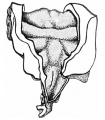
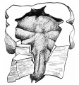
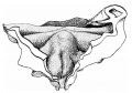
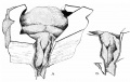
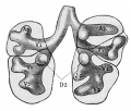
Embryonic
| Human Embryonic Lung Development | ||
|---|---|---|

|

|

|
| CRL 4.3 mm, Week 4-5, Stage 12 to 13 | CRL 8.5 mm, Week 5, Stage 15 to 16 | CRL 10.5 mm, Week 6 Stage 16 to 17 |
- Endoderm - tubular ventral growth from foregut pharynx.
- Mesoderm - mesenchyme of lung buds.
- Intraembryonic coelom - pleural cavities elongated spaces connecting pericardial and peritoneal spaces.
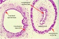
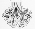
Pseudoglandular stage

|

Human lung pseudoglandular stage[10] |
Canalicular stage
- week 16 - 24
- Lung morphology changes dramatically
- differentiation of the pulmonary epithelium results in the formation of the future air-blood tissue barrier.
- Surfactant synthesis and the canalization of the lung parenchyma by capillaries begin.
- future gas exchange regions can be distinguished from the future conducting airways of the lungs.
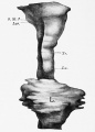
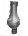
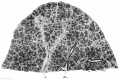
Saccular stage
|

Alveolar sac structure |
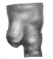
Alveolar stage
Premature babies have difficulties associated with insufficient surfactant (end month 6 alveolar cells type 2 appear and begin to secrete surfactant). |

Respiratory secondary septum[11] |
Respiratory Species Comparison

| Respiratory Stages - Species Comparison - Stages Gestational age (days) | |||||
|---|---|---|---|---|---|
| Species | Term | Embryonic | Pseudoglandular | Canalicular | Saccular |
| human | 280 | < 42 | 52 - 112 | 112 - 168 | 168 |
| primate | 168 | < 42 | 57 - 80 | 80 - 140 | 140 |
| sheep | 150 | < 40 | 40 - 80 | 80 - 120 | 120 |
| rabbit | 32 | < 18 | 21 - 24 | 24 - 27 | 27 |
| rat | 22 | < 13 | 16 - 19 | 19 - 20 | 21 |
| mouse | 20 | < 9 | 16 | 18 | 19 |
| Data modified from[13]
Links: respiratory | Respiratory Comparison | Mouse Human Respiratory | Mouse respiratory stages | mouse | rat | rabbit | Timeline Comparisons | |||||
Mouse
Note that the model mouse respiratory system differs from human in size, distribution of cell types, and the time taken to develop.

Human and mouse Sox expression[14]
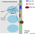
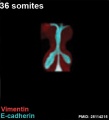
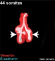
The following images are from a recent study of the development of bronchial branching in he mouse between E10 to E14.[15]
Mesenchyme (red) and epithelium (blue) the study used knockout mice to show the role of Wnt signalling in branching morphogenesis.


| Mouse lung E12.5 to E18.5 | |||
|---|---|---|---|

|

|

|

|
| E12.5 lungs | E14.5 lungs | E14.5 Sox9 | E18.5 lungs |
| Reference[16] | |||
- Links: Wnt | Mouse Development
Embryonic Respiratory Development

Pseudoglandular Respiratory Development

Pseudoglandular period identified in this paper (GA weeks 12 to 16)
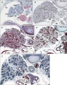
Human lung at pseudoglandular stage showing E- and N-cadherin and β-catenin localization.[17]
Endocrine Lung
| Neonatal Human | Fetal Rabbit |
|---|---|

|

|
| Pulmonary neuroendocrine cell (EM)[18] | Neuroepithelial body[18] |
Pulmonary neuroendocrine cells (PNECs)
- develop in late embryonic to early fetal period.[19][20]
- later in mid-fetal period clusters of these cells form neuroepithelial bodies (NEBs).
- first cell type to differentiate in the airway epithelium.
- differentiation regulated by proneural genes - mammalian homolog of the achaete-scute complex (Mash-1) and hairy and enhancer of split1 (Hes-1).[21]
- located in the fetal lung at bronchiole branching points.
- may stimulate mitosis to increase branching.
- secrete 2 peptides - gastrin-releasing peptide (GRP) and calcitonin gene related peptide (CGRP)
- Links: Endocrine - Other Tissues | OMIM - GRP | OMIM - CGRP
Lung Histology

|
| Fetal lung histology |
Birth Changes
At birth the lung epithelium changes from a prenatal secretory to a postnatal absorptive function. Several factors have been identified as influencing this transport change including: epinephrine, oxygen, glucocorticoids, and thyroid hormones (for review see [22])
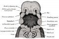
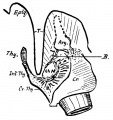
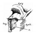
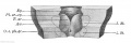
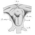
Upper Respiratory Tract

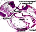
stage 11 foregut

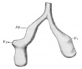
week 4 early respiratory endodermal bud
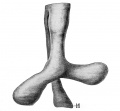
Stage 22 trachea
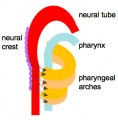
Head arches cartoon
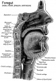
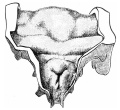
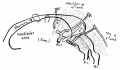
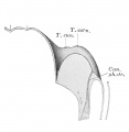
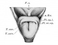
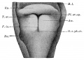
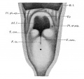
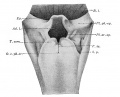
Pharynx
Nasal cavities
Pharynx
Larynx


- part of foregut development
- anatomically the nose, nasal cavity and the pharynx
- the pharynx forms a major arched cavity within the pharyngeal arches
Movies
The animations below allow a comparison of early and late embryonic lung development. Compare the size and relative position of the respiratory structures and their anatomical relationship to the developing gastrointestinal tract.
|
Early embryo (stage 13)
3 dimensional reconstruction based upon a serial reconstruction from individual Carnegie stage 13 embryo slice images. |
|
Late embryo (stage 22)
3 dimensional reconstruction based upon a serial reconstruction from individual embryo slice images Carnegie stage 22, 27 mm Human embryo, approximate day 56. |
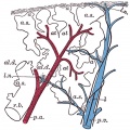
Lung Cardiovascular
Pulmonary Circulation
- pulmonary arteries and veins arise by vasculogenesis[23]
Pulmonary Veins
- vasculogenesis in the mesenchyme surrounding the terminal buds during the pseudoglandular stage.
- vasculogenesis - describes the formation of new blood vessels from pluripotent precursor cells.
- angiogenesis in the canalicular and alveolar stages.
- angiogenesis - describes the formation of new vessels from pre-existing vessels.
See also review [24]
Bronchial Circulation
Bronchial Arteries
- vascularising the walls of the airways and the large pulmonary vessels providing giving oxygen and nutrients.
- extend within the bronchial tree to the periphery of the alveolar ducts.
- not found in the lungs until around 8 weeks of gestation.
- one or two small vessels extend from the dorsal aorta and run into the lung alongside the cartilage plates of the main bronchus.
Bronchial Veins
- small bronchial veins within the airway wall drain into the pulmonary veins.
- large bronchial veins seen close to the hilum and drain into the cardinal veins and the right atrium.
See review [24]
Molecular
| Factor Links: AMH | hCG | BMP | sonic hedgehog | bHLH | HOX | FGF | FOX | Hippo | LIM | Nanog | NGF | Nodal | Notch | PAX | retinoic acid | SIX | Slit2/Robo1 | SOX | TBX | TGF-beta | VEGF | WNT | Category:Molecular |
TBX

|

|
| Mouse respiratory Tbx4 and Tbx5 model[25] | Mouse respiratory development[26]]] |
- Nkx2-1 (Titf1) - ventral wall of the anterior foregut, identifies the future trachea.
SOX
- Sox2 is essential for the initiation of lung development from the endodermal gut tube,
- Sox9 is essential for maintaining the tips and associated branching.
Several different Sox types are required for different stages of respiratory development. Wnt/β-catenin signaling does not regulate Sox9 expression in the lung.[16]
Human and mouse Sox expression[14]
FGF

- Localized Fgf10 expression not required for lung branching but prevents epithelial differentiation[27] "As the lung buds grow out, proximal epithelial cells become further and further displaced from the distal source of Fgf10 and differentiate into bronchial epithelial cells. Interestingly, our data presented here show that once epithelial cells are committed to the Sox2-positive airway epithelial cell fate, Fgf10 prevents ciliated cell differentiation and promotes basal cell differentiation."
- Opposing Fgf and Bmp activities regulate the specification of olfactory sensory and respiratory epithelial cell fates[28] " In this study, we provide evidence that in both chick and mouse, Bmp signals promote respiratory epithelial character, whereas Fgf signals are required for the generation of sensory epithelial cells. Moreover, olfactory placodal cells can switch between sensory and respiratory epithelial cell fates in response to Fgf and Bmp activity, respectively. Our results provide evidence that Fgf activity suppresses and restricts the ability of Bmp signals to induce respiratory cell fate in the nasal epithelium."
BMP
Bone morphogenic protein 4 (Bmp4) acts as in an autocrine signalling mechanism to limit bud outgrowth and is therefore involved in branching.
Other
- Heparan sulfate in lung morphogenesis[29] "Heparan sulfate (HS) is a structurally complex polysaccharide located on the cell surface and in the extracellular matrix, where it participates in numerous biological processes through interactions with a vast number of regulatory proteins such as growth factors and morphogens. ...he potential contribution of HS to abnormalities of lung development has yet to be explored to any significant extent, which is somewhat surprising given the abnormal lung phenotype exhibited by mutant mice synthesizing abnormal HS."
- Signaling via Alk5 controls the ontogeny of lung Clara cells[30] "Clara cells, together with ciliated and pulmonary neuroendocrine cells, make up the epithelium of the bronchioles along the conducting airways. Clara cells are also known as progenitor or stem cells during lung regeneration after injury. ...Using lung epithelial cells, we show that Alk5-regulated Hes1 expression is stimulated through Pten and the MEK/ERK and PI3K/AKT pathways. Thus, the signaling pathway by which TGFbeta/ALK5 regulates Clara cell differentiation may entail inhibition of Pten expression, which in turn activates ERK and AKT phosphorylation."
- Wt1 and retinoic acid signaling in the subcoelomic mesenchyme control the development of the pleuropericardial membranes and the sinus horns[31] "Pericardium and sinus horn formation are coupled and depend on the expansion and correct temporal release of pleuropericardial membranes from the underlying subcoelomic mesenchyme. Wt1 and downstream Raldh2/retinoic acid signaling are crucial regulators of this process."
References
- ↑ 1.0 1.1 <pubmed>24499815</pubmed>
- ↑ 2.0 2.1 <pubmed>26119728</pubmed>| Cell Rep.
- ↑ <pubmed>26586225</pubmed>
- ↑ <pubmed>25564622</pubmed>
- ↑ <pubmed>24058167</pubmed>
- ↑ <pubmed>20535580</pubmed>
- ↑ <pubmed>20484817</pubmed>
- ↑ <pubmed>20378729</pubmed>
- ↑ <pubmed>20371042</pubmed>
- ↑ <pubmed>24693478</pubmed>| Anat Cell Biol.
- ↑ <pubmed>25973420</pubmed>| Front Med (Lausanne).]]
- ↑ <pubmed>15005800</pubmed>| BMC Developmental Biology
- ↑ Pinkerton KE & Joad JP. (2000). The mammalian respiratory system and critical windows of exposure for children's health. Environ. Health Perspect. , 108 Suppl 3, 457-62. PMID: 10852845
- ↑ 14.0 14.1 <pubmed>28806170</pubmed>
- ↑ <pubmed>25114215</pubmed>PMC4151720 | Proc Natl Acad Sci U S A.
- ↑ 16.0 16.1 <pubmed>24191021</pubmed>
- ↑ Kaarteenaho R, Lappi-Blanco E, Lehtonen S. Epithelial N-cadherin and nuclear β-catenin are up-regulated during early development of human lung. BMC Dev Biol. 2010 Nov 16;10:113. PMID: 21080917 | PMC2995473 | BMC Dev Biol.
- ↑ 18.0 18.1 <pubmed>6376101</pubmed>
- ↑ <pubmed>6188605</pubmed>
- ↑ <pubmed>3906540</pubmed>
- ↑ <pubmed>20027181</pubmed>
- ↑ <pubmed>12235057</pubmed>
- ↑ <pubmed>11867341</pubmed>
- ↑ 24.0 24.1 <pubmed>12430957</pubmed>
- ↑ <pubmed>22876201</pubmed>| PLoS Genet.
- ↑ 26.0 26.1 Cardoso WV, Kotton DN. Specification and patterning of the respiratory system. StemBook [Internet]. Cambridge (MA): Harvard Stem Cell Institute; 2008 Jul 16. PMID20614584 | StemBook - Specification and patterning of the respiratory system
- ↑ <pubmed>23924632</pubmed>
- ↑ <pubmed>20392740</pubmed>
- ↑ <pubmed> 20301217</pubmed>
- ↑ <pubmed> 20147383</pubmed>
- ↑ <pubmed> 20185795</pubmed>
Reviews
<pubmed></pubmed> <pubmed></pubmed> <pubmed>28144783</pubmed> <pubmed>24449833</pubmed> <pubmed>20691848</pubmed> <pubmed>20152174</pubmed> <pubmed>16770071</pubmed> <pubmed>12456356</pubmed> <pubmed>6370120</pubmed>
Articles
<pubmed></pubmed> <pubmed></pubmed> <pubmed></pubmed> <pubmed>18651668</pubmed> <pubmed>16770071</pubmed> <pubmed>11867341</pubmed> <pubmed>10919986</pubmed> <pubmed>10100986</pubmed>
Search PubMed
Search April 2010
- Respiratory System Development - All (30795) Review (3706) Free Full Text (7943)
- Respiratory Development - All (28939) Review (5876) Free Full Text (7203)
Search Pubmed: Respiratory System Development | Respiratory Development
Terms
| Respiratory Terms (expand to view) |
|---|
|
| Other Terms Lists |
|---|
| Terms Lists: ART | Birth | Bone | Cardiovascular | Cell Division | Endocrine | Gastrointestinal | Genital | Genetic | Head | Hearing | Heart | Immune | Integumentary | Neonatal | Neural | Oocyte | Palate | Placenta | Radiation | Renal | Respiratory | Spermatozoa | Statistics | Tooth | Ultrasound | Vision | Historic | Drugs | Glossary |
Additional Images
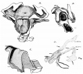
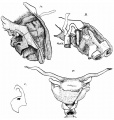
Upper Respiratory Tract
Head arches cartoon
Pharynx
Nasal cavities
Pharynx
Larynx

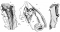
Lower Respiratory Tract
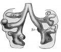
Model Sox9 lung development PMID 24274029
Human right lung 7-8 weeks PMID 24693478
Mouse 36 somites PMID 25114215
Mouse 44 somites PMID 25114215
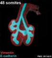
Mouse 48 somites PMID 25114215
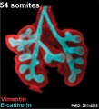
Mouse 54 somites PMID 25114215
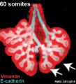
Mouse 60 somites PMID 25114215


Historic Images
| Historic Disclaimer - information about historic embryology pages |
|---|
|
week 4 early respiratory endodermal bud

week 4 later ventral endoderm growth
lower respiratory tract
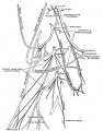
conducting system bronchi to lungs
secondary lobule from the human lung
longitudinal section of a primary lung lobule
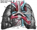
heart and lungs
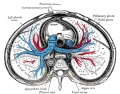
transverse section of the thorax showing the middle and posterior mediastinum
Adult diaphragm
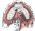
Adult cervical plexus (phrenic nerve shown lower right)


Frazer JE. Development of the larynx. (1910) J Anat. 44: 156-191. PMID 17232839
- Larynx
embryo 5 mm
embryo 6 mm
embryo 7.5 mm
embryo 8.5 mm
embryo 12 mm
embryo 6.6 mm
embryo 22 mm
embryo 35 mm
embryo 9.5 mm
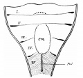
fig 10
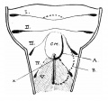
fig 11
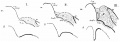
fig 12
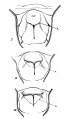
fig 13
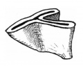
fig 14
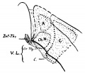
fig 15
embryo 22 mm
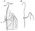
fig 17
embryo 18 mm
fig 19


Keibel F. and Mall FP. Manual of Human Embryology II. (1912) J. B. Lippincott Company, Philadelphia.
- “Respiratory
embryo 2.5 mm (Rob. Meyer No. 300)
Lung embryo 4.25 mm
Lung left embryo 4.25 mm
larynx entrance embryo 8 mm
larynx entrance embryo 8-9 mm (28-29 days)
larynx embryo 8-9 mm (28-29 days)
larynx entrance embryo 15-16 mm (40-42 days)
larynx entrance embryo 30 mm
larynx entrance embryo of 16/23 cm
larynx entrance embryo of 29/43 cm
Epithelial lung embryo 18.5 mm (Rob. Meyer No. 338)
Epithelial lung embryo 28.7 mm (Chr. 1)
Lung week 5
Lung week 5
lung embryo 10.5 mm (N)
ight lung fetus of 100 mm

mesodermal lungs embryo of 5 mm
Mesodermal lung embryo 7 mm (Chr. 1)
Mesodermal lung embryo 15 mm
Mesodermal lung embryo 15 mm
R Lung embryo 17.5 mm

L Lung embryo 17.5 mm
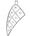
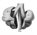
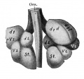
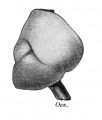
lobation of the lung


External Links
External Links Notice - The dynamic nature of the internet may mean that some of these listed links may no longer function. If the link no longer works search the web with the link text or name. Links to any external commercial sites are provided for information purposes only and should never be considered an endorsement. UNSW Embryology is provided as an educational resource with no clinical information or commercial affiliation.
| System Links: Introduction | Cardiovascular | Coelomic Cavity | Endocrine | Gastrointestinal Tract | Genital | Head | Immune | Integumentary | Musculoskeletal | Neural | Neural Crest | Placenta | Renal | Respiratory | Sensory | Birth |
Glossary Links
- Glossary: A | B | C | D | E | F | G | H | I | J | K | L | M | N | O | P | Q | R | S | T | U | V | W | X | Y | Z | Numbers | Symbols | Term Link
Cite this page: Hill, M.A. (2024, June 15) Embryology Respiratory System Development. Retrieved from https://embryology.med.unsw.edu.au/embryology/index.php/Respiratory_System_Development
- © Dr Mark Hill 2024, UNSW Embryology ISBN: 978 0 7334 2609 4 - UNSW CRICOS Provider Code No. 00098G