Category:Cardiovascular: Difference between revisions
From Embryology
mNo edit summary |
mNo edit summary |
||
| Line 1: | Line 1: | ||
This | This {{Embryology}} category shows pages, images and media related to cardiovascular system development. | ||
See also the narrower categories [[:Category:Heart]] and [[:Category:Blood]]. | See also the narrower categories [[:Category:Heart]] and [[:Category:Blood]]. | ||
{{Heart Links}} | {{Heart Links}} | ||
Latest revision as of 12:39, 13 February 2017
This Embryology category shows pages, images and media related to cardiovascular system development.
See also the narrower categories Category:Heart and Category:Blood.
Subcategories
This category has the following 13 subcategories, out of 13 total.
Pages in category 'Cardiovascular'
The following 200 pages are in this category, out of 519 total.
(previous page) (next page)2
A
- Template:Abbott Figures
- Template:Abbott1915
- Template:Abbott1915images
- Abnormal Development - Hypertension
- Advanced - Abnormalities
- Advanced - Cardiac Conduction
- Advanced - Cardiac Looping
- Advanced - Cardiac Looping 2
- Advanced - Cardiac Septation
- Advanced - Cardiac Septation 2
- Advanced - Heart Fields
- Advanced - Heart Tubes
- Advanced - Molecular Development
- Advanced - Outflow Tract
- Advanced - Valve Development
- Advanced Cardiac Embryology
- Template:Advanced Cardiac menu
- ANAT2241 Cardiovascular System
- ANAT2341 Lab 4 - Early Cardiovascular Development
- ANAT3241 Lab 11
- Template:Aorta
- Template:Aortic arch
- Template:Aortic stenosis
- Template:Arachnoid mater
- Template:Artery
- Atlas of the Development of Man 2 - Cardiovascular
- Template:Atrial septal defects
- Template:Atrial septum movie links
B
- Template:Bartelmez GW.
- BGDA Lecture - Development of the Embryo/Fetus 2
- Template:BGDA Practical 14 - Maternal Decidua Interactive
- Template:BGDA Practical 14 - Placental Cord Interactive
- Template:BGDA Practical 14 - Villi Interactive
- BGDA Practical 7 - Week 5
- BGDB Lecture - Heart Development
- Template:Blood
- Template:Blood cell images
- Template:Blood vessel
- Template:Blood vessel histology
- Template:Bone marrow
- Book - A Laboratory Manual and Text-book of Embryology 9
- Book - An experimental analysis of the origin of blood and vascular endothelium (1915)
- Book - Congenital Cardiac Disease (1915)
- Book - Congenital Cardiac Disease - Figures
- Book - Congenital Cardiac Disease 1
- Book - Congenital Cardiac Disease 10
- Book - Congenital Cardiac Disease 11
- Book - Congenital Cardiac Disease 12
- Book - Congenital Cardiac Disease 13
- Book - Congenital Cardiac Disease 14
- Book - Congenital Cardiac Disease 15
- Book - Congenital Cardiac Disease 2
- Book - Congenital Cardiac Disease 3
- Book - Congenital Cardiac Disease 4
- Book - Congenital Cardiac Disease 5
- Book - Congenital Cardiac Disease 6
- Book - Congenital Cardiac Disease 7
- Book - Congenital Cardiac Disease 8
- Book - Congenital Cardiac Disease 9
- Book - Contributions to Embryology Carnegie Institution No.32
- Book - Contributions to Embryology Carnegie Institution No.36
- Book - Contributions to Embryology Carnegie Institution No.65
- Book - Contributions to Embryology Carnegie Institution No.67
- Book - Early stages of vasculogenesis in the cat with especial reference to the mesenchymal origin of endothelium (1914)
- Book - Human Embryology and Morphology 16
- Book - Quain's Embryology 9
- Book - Text-Book of the Embryology of Man and Mammals 17-1
- Template:Brain Vascular System gallery
- Template:Brain Vascular System table1
- Template:Bremer1914 figures
C
- Template:Capillary
- Template:CapillaryEM links
- Template:Cardiac
- Cardiac Embryology
- Cardiac Muscle Histology
- Template:Cardiovascular
- Cardiovascular - Arterial Development
- Cardiovascular - Detailed Cardiac Development
- Cardiovascular - Venous Development
- Cardiovascular 3D stage 13 Movie
- Cardiovascular 3D stage 22 Movie
- Template:Cardiovascular abnormalities
- Cardiovascular System - Abnormalities
- Cardiovascular System - Atrial Septal Defects
- Cardiovascular System - Blood Development
- Cardiovascular System - Blood Vessel Development
- Cardiovascular System - Bone Marrow
- Cardiovascular System - Carnegie Stage 22
- Cardiovascular System - Circulation Development
- Cardiovascular System - Coarctation of the Aorta
- Cardiovascular System - Coronary Circulation Development
- Cardiovascular System - Developmental Shunts
- Cardiovascular System - Double Outlet Right Ventricle
- Cardiovascular System - Ductus Arteriosus
- Cardiovascular System - Ductus Venosus
- Cardiovascular System - Fetal Shunts
- Cardiovascular System - Foramen Ovale
- Cardiovascular System - Heart Development
- Cardiovascular System - Heart Histology
- Cardiovascular System - Heart Rate Development
- Cardiovascular System - Heart Valve Development
- Cardiovascular System - Hypoplastic Left Heart
- Cardiovascular System - Lymphatic Development
- Cardiovascular System - Movies
- Cardiovascular System - Patent Ductus Arteriosus
- Cardiovascular System - Spleen Development
- Cardiovascular System - Tetralogy of Fallot
- Cardiovascular System - Transposition of the Great Vessels
- Cardiovascular System - Tricuspid Atresia
- Cardiovascular System - Truncus Arteriosus
- Cardiovascular System - Ventricular Septal Defects
- Cardiovascular System Development
- Template:Carotid body
- Template:Cerebral Arterial Timeline table
- Template:CHARGE syndrome
- Chicken Aortic Arches Movie
- Template:Choroid plexus
- Template:Coarctation of the aorta
- Template:Common truncus
- Computed Tomography
- Template:Congdon1922 collapse table1
- Template:Congdon1922 table1
- Template:Coronary circulation
- Template:CVS cartoons
D
- Detailed Cardiac - Arterial Roots
- Detailed Cardiac - Atrioventricular Canal
- Detailed Cardiac - Atrioventricular Conduction Axis
- Detailed Cardiac - Atrioventricular Cushions
- Detailed Cardiac - Extrapericardial Arterial Channels
- Detailed Cardiac - Interventricular Communication
- Detailed Cardiac - Intrapericardial Arterial Trunks
- Detailed Cardiac - Pulmonary Vein
- Detailed Cardiac - Sinus Node
- Detailed Cardiac - Subpulmonary Infundibulum
- Detailed Cardiac - Superior Interatrial Fold
- Detailed Cardiac - Systemic Venous Sinus
- Development Animation - Heart Atrial Septation
- Development Animation - Heart Realign
- Developmental Signals - Slit2/Robo1
- Template:Doppler ultrasound
- Template:Double outlet right ventricle
- Template:Ductus arteriosus
- Template:Ductus venosus
E
- Electrocardiogram
- Electron Microscopy Virtual Slides
- Talk:Embryo Serial Sections
- Embryology History
- Special:Badtitle/NS501:Embryology History
- History:Embryology History
- Embryology History - Douglas Reid
- Embryology History - George Bartelmez
- Embryology History - Herbert Evans
- Template:Evans HM.
- Template:Evans1909 figures
F
H
- Template:Haematopoiesis
- Template:Heart
- Template:Heart abnormal cartoon gallery
- Template talk:Heart abnormal cartoon gallery
- Heart Atrial Septation Movie
- Template:Heart histology
- Heart Historic Movie 1
- Heart Historic Movie 1951
- Heart Historic Movie 2
- Heart Historic Movie 3
- Heart Historic Movie 4
- Heart Historic Movie 5
- Heart Historic Movie 6
- Heart Historic Movie 7
- Heart Historic Movie 8
- Template:Heart Links
- Heart Looping Movie
- Heart Outflow Septation Movie
- Template:Heart rate
- Heart Realign Movie
- Template:Heart terms
- Template:Heart valve
- Historic Animation - Heart 02
- Historic Animation - Heart 04
- Template:Historic Heart
- Template talk:Historic Heart
Media in category 'Cardiovascular'
The following 95 files are in this category, out of 695 total.
(previous page) (next page) Stage 13 image 093.jpg 1,000 × 454; 92 KB
 Stage 13 image 094.jpg 1,000 × 469; 90 KB
 Stage 13 image 095.jpg 1,000 × 452; 76 KB
 Stage 13 image 096.jpg 1,000 × 469; 75 KB
 Stage 13 image 097.jpg 1,000 × 720; 162 KB
 Stage 13 image 098.jpg 1,000 × 623; 144 KB
 Stage 13 image 101.jpg 1,000 × 649; 65 KB
 Stage 22 image 175.jpg 1,000 × 666; 158 KB
 Stage 22 image 178.jpg 1,000 × 658; 148 KB
 Stage 22 image 179.jpg 1,000 × 658; 131 KB
 Stage 22 image 192.jpg 1,000 × 658; 180 KB
 Stage11 sem9.jpg 1,203 × 1,653; 301 KB
 Stage11 sem9a.jpg 728 × 1,000; 145 KB
 Stage11 sem9b.jpg 582 × 800; 104 KB
 Stage13 bloodflow.jpg 437 × 297; 24 KB
 Streeter-plate01.jpg 1,200 × 950; 138 KB
 Streeter-plate02.jpg 1,200 × 871; 180 KB
 Streeter-plate03.jpg 1,398 × 1,000; 192 KB
 Streeter-plate04.jpg 1,301 × 1,000; 214 KB
 Streeter-plate05.jpg 833 × 1,000; 115 KB
 Streeter1915 fig17.jpg 700 × 513; 76 KB
 Streeter1921 fig01.jpg 981 × 1,000; 103 KB
 Streeter1921 fig02.jpg 1,286 × 1,000; 138 KB
 Streeter1921 fig03.jpg 1,154 × 1,000; 142 KB
 Streeter1921 fig04.jpg 1,030 × 1,000; 101 KB
 Streeter1921 fig05.jpg 600 × 342; 19 KB
 Streeter1921 fig06.jpg 1,007 × 1,000; 106 KB
 Streeter1921 fig07-09.jpg 1,200 × 494; 92 KB
 Streeter1921 fig10.jpg 1,108 × 1,000; 95 KB
 Streeter1921 fig11.jpg 908 × 751; 103 KB
 Streeter1921 fig12.jpg 960 × 745; 117 KB
 Streeter1921 fig13.jpg 800 × 600; 45 KB
 Streeter1921 fig14.jpg 800 × 600; 55 KB
 Streeter1921 fig15.jpg 800 × 600; 54 KB
 Streeter1921 fig16.jpg 800 × 600; 60 KB
 Streeter1921 fig17.jpg 800 × 600; 60 KB
 Streeter1921 fig18.jpg 800 × 600; 48 KB
 Streeter1921 fig19.jpg 800 × 600; 48 KB
 Streeter1921 fig20.jpg 800 × 600; 40 KB
 Streeter1921 fig21.jpg 800 × 600; 40 KB
 Streeter1921 fig22.jpg 917 × 1,000; 157 KB
 Streeter1921 fig23.jpg 917 × 1,000; 152 KB
 Streeter1921 fig24.jpg 921 × 1,000; 130 KB
 Streeter1921 fig25.jpg 921 × 1,000; 118 KB
 Streeter1921 fig26.jpg 1,328 × 1,000; 228 KB
 Streeter1921 fig27.jpg 949 × 1,000; 138 KB
 Streeter1921 table01.jpg 800 × 347; 44 KB
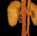
 Supernumerary renal vein 01.jpg 800 × 798; 72 KB
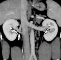
 Supernumerary renal vein 02.jpg 800 × 795; 89 KB
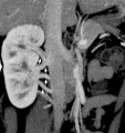
 Supernumerary renal vein 03.jpg 800 × 794; 80 KB
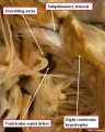
 Supernumerary renal vein 04.jpg 800 × 850; 76 KB
 Tetralogy of Fallot 01.jpg 700 × 873; 113 KB
 Tetralogy of Fallot 02.jpg 800 × 796; 63 KB
 Tetralogy of Fallot.jpg 300 × 350; 17 KB
 Thalidomide - CPS49 vascular effect.jpg 1,000 × 1,002; 197 KB
 Thalidomide - limb signaling.jpg 628 × 540; 88 KB
 Total Anomalous Pulmonary Venous Connection.jpg 295 × 350; 17 KB
 Transposition of the Great Vessels.jpg 299 × 350; 18 KB
 Tricuspid Atresia.jpg 303 × 350; 16 KB
 Trigeminal artery 01.jpg 947 × 800; 102 KB
 Trigeminal artery 02.jpg 520 × 490; 35 KB
 Ultrasound - Hypoplastic left heart syndrome 01.jpg 800 × 600; 53 KB
 Ultrasound - Hypoplastic left heart syndrome 02.jpg 890 × 626; 54 KB
 Ultrasound - Hypoplastic left heart syndrome 03.jpg 874 × 612; 51 KB
 Ultrasound - Hypoplastic left heart syndrome 04.jpg 919 × 618; 68 KB
 Uterine and placental vasculature.jpg 614 × 472; 143 KB
 Uterine arterial vessel cartoon.jpg 600 × 483; 44 KB
 Uterine vascular anastomoses.jpg 1,200 × 370; 48 KB
 Vasculature development 01 cartoon.jpg 599 × 900; 106 KB
 Vasculature development 02 cartoon.jpg 601 × 1,002; 93 KB
 Vein valve animation.gif 300 × 200; 54 KB
 Ventricular septal defect 01.jpg 1,024 × 692; 84 KB
 Ventricular Septal Defect.jpg 289 × 350; 16 KB
 Venule microvessel EM.jpg 600 × 626; 91 KB
 Waterston13.jpg 429 × 681; 63 KB
 Waterston14.jpg 438 × 680; 66 KB
 Waterston16.jpg 593 × 675; 73 KB
 Waterston17.jpg 500 × 710; 74 KB
 Waterston18.jpg 500 × 662; 71 KB
 Waterston19.jpg 500 × 642; 66 KB
 Week17 fetal heart rate.mp4 ; 398 KB
 West11.jpg 367 × 963; 27 KB
 Woollard-plate01.jpg 744 × 1,000; 153 KB
 Woollard-plate02.jpg 788 × 1,000; 196 KB
 Woollard001.jpg 739 × 858; 142 KB
 Woollard002.jpg 699 × 873; 125 KB
 Woollard003.jpg 1,107 × 848; 159 KB
 Woollard004.jpg 1,037 × 717; 148 KB
 Woollard005.jpg 1,034 × 655; 144 KB
 Ziegler model 12.jpg 475 × 700; 39 KB
 Ziegler model 18.jpg 700 × 465; 42 KB
 Ziegler model 19.jpg 700 × 465; 23 KB
 ZPulmonary Atresia.jpg 653 × 618; 85 KB
 ZUltrasound Image of Fetal Aorta.jpg 640 × 480; 47 KB


