Embryonic Development: Difference between revisions
mNo edit summary |
mNo edit summary |
||
| (One intermediate revision by the same user not shown) | |||
| Line 2: | Line 2: | ||
== Introduction == | == Introduction == | ||
{| class="wikitable mw-collapsible mw-collapsed" | {| class="wikitable mw-collapsible mw-collapsed" | ||
! Author Comments | ! Author Comments | ||
|- | |- | ||
| [[File:Mark_Hill.jpg|50px]] '''Start here''' by looking at the external appearance of embryos in sequence from [[#Carnegie_Stages|1 to 23]]. It is not so important to memorise the dates, as they are only approximate, but more important to understand '''growth''' (size changes) and the '''development''' (overall sequence of events) during this period. | | [[File:Mark_Hill.jpg|50px]] '''Start here''' by looking at the external appearance of embryos in sequence from [[#Carnegie_Stages|1 to 23]]. It is not so important to memorise the dates, as they are only approximate, but more important to understand '''growth''' (size changes) and the '''development''' (overall sequence of events) during this period. | ||
| Line 32: | Line 32: | ||
! Carnegie Collection | ! Carnegie Collection | ||
|- | |- | ||
| [[File:Human Carnegie stage 1-23.jpg|550px]] | | [[File:Human Carnegie stage 1-23.jpg|alt=Kyoto Collection Embryos|link=Kyoto Collection|550px]] | ||
| | | | ||
| [[File:Human Carnegie stage 10-23.jpg|420px]] | | [[File:Human Carnegie stage 10-23.jpg|alt=Carnegie Collection Embryos|link=Carnegie Collection|420px]] | ||
|- | |- | ||
| Human Embryo, Carnegie stages 1-23 | | Human Embryo, Carnegie stages 1-23 | ||
Latest revision as of 06:46, 13 December 2018
Embryology - 13 Aug 2026    Expand to Translate Expand to Translate
|
|---|
| Google Translate - select your language from the list shown below (this will open a new external page) |
|
العربية | català | 中文 | 中國傳統的 | français | Deutsche | עִברִית | हिंदी | bahasa Indonesia | italiano | 日本語 | 한국어 | မြန်မာ | Pilipino | Polskie | português | ਪੰਜਾਬੀ ਦੇ | Română | русский | Español | Swahili | Svensk | ไทย | Türkçe | اردو | ייִדיש | Tiếng Việt These external translations are automated and may not be accurate. (More? About Translations) |
Introduction
| Author Comments |
|---|
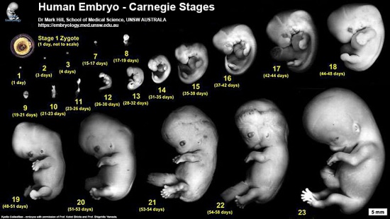
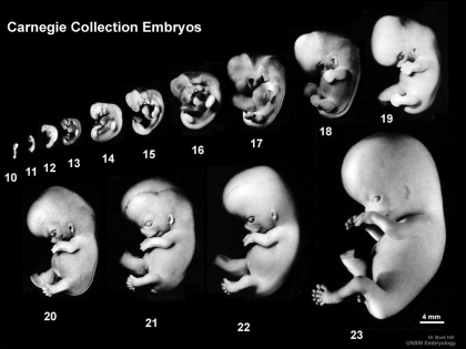
 Start here by looking at the external appearance of embryos in sequence from 1 to 23. It is not so important to memorise the dates, as they are only approximate, but more important to understand growth (size changes) and the development (overall sequence of events) during this period. Start here by looking at the external appearance of embryos in sequence from 1 to 23. It is not so important to memorise the dates, as they are only approximate, but more important to understand growth (size changes) and the development (overall sequence of events) during this period.
Clicking the Carnegie stage numbers opens a page dedicated to describing that single stage and the associated developmental events. |
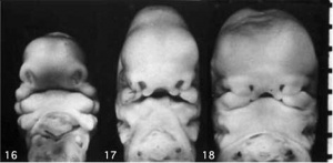
| This page shows some key events of human development during the embryonic period of the first eight weeks (weeks 1 - 8) following fertilization. This period is also considered the organogenic period, when most organs within the embryo have begun to form.
|

|
| Kyoto Collection | Carnegie Collection | |
|---|---|---|
|
| |
| Human Embryo, Carnegie stages 1-23 | Human Embryo, Carnegie stages 10-23 |
| <html5media height="530" width="375">File:Embryo stages 003.mp4</html5media> | Use the stage number links to images and information about each specific stage of human development over the first 8 weeks. The links below give a broad overview of developmental events during each week.
Embryo sizes - stage 14 compared to 23
Embryo Week: Week 1 | Week 2 | Week 3 | Week 4 | Week 5 | Week 6 | Week 7 | Week 8 | Week 9 |

Week 1
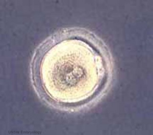
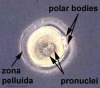
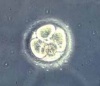
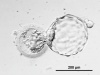
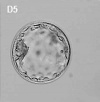
- Week 1 Carnegie stage - 1 | 2 | 3 | 4
- oocyte | spermatozoa | fertilization
- zygote
- morula
- blastocyst
| Week 1 Links: stage 1 | stage 2 | stage 3 | menstrual cycle | fertilization | zygote | morula | blastocyst | Lecture - Fertilization | meiosis | mitosis | Lecture - Week 1 and 2 | menstrual cycle | oocyte | spermatozoa | twinning | Genetic risk maternal age | Trisomy 21 | Trisomy 18 | Trisomy 13 | hydatidiform mole | GA week 3 |
| Carnegie stages | |||
|---|---|---|---|
|
|
|
|
| stage 1 | stage 2 | stage 3 | stage 4 |
Week 2
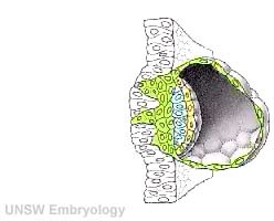
- Week 2 Carnegie stage - 5 | 6
- trophoblast - (trophectoderm) outer cell layer
- Embryoblast - inner cell mass
- implantation
- Bilaminar embryo
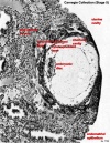
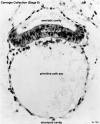
| Week 2 Links: stage 4 | stage 5 | stage 6 | Lecture - Week 1 and 2 | implantation | trophoblast | human chorionic gonadotropin | pregnancy test | twinning | Category:Week 2 | GA week 4 |
| Carnegie stages | ||
|---|---|---|
|
|
|
| stage 4 | stage 5 | stage 6 |
Week 3
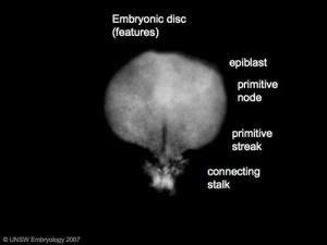
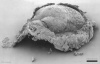
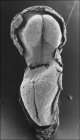
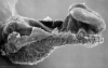
| Week 3 Links: stage 7 | stage 8 | stage 9 | Gastrulation | Endoderm | Mesoderm | Ectoderm | Placenta | Coelomic Cavity | Lecture | Category:Week 3 | GA week 5 |
| Carnegie stages | ||
|---|---|---|
|
|
|
| stage 7 | stage 8 | stage 9 |
Week 4
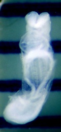
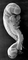
| Week 4 Links: stage 10 | stage 11 | stage 12 | 13 | Placodes | Pharyngeal arches | Lecture - Mesoderm | Lecture - Ectoderm | Lecture - Early Vascular | Category:Week 4 | GA week 6 |
| Carnegie stages | |||
|---|---|---|---|
|
|
|
|
| stage 10 | stage 11 | stage 12 | stage 13 |
Week 5
| Carnegie stages | |
|---|---|
|
|
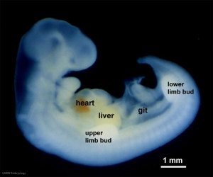
| stage 14 | stage 15 |
Week 6
| Carnegie stages | |
|---|---|
|
|
| stage 16 | stage 17 |
Week 7
| Carnegie stages | |
|---|---|
|
|
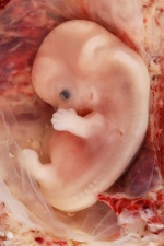
| stage 18 | stage 19 |
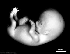
Week 8
| Carnegie stages | |||
|---|---|---|---|
|
|
|
|
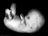
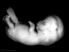
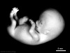
| stage 20 | stage 21 | stage 22 | stage 23 |
- Carnegie Stages: 1 | 2 | 3 | 4 | 5 | 6 | 7 | 8 | 9 | 10 | 11 | 12 | 13 | 14 | 15 | 16 | 17 | 18 | 19 | 20 | 21 | 22 | 23 | About Stages | Timeline
Carnegie Stage Table
Weeks shown in the table below are embryonic post ovulation age, for clinical Gestational Age (GA) measured from last menstrual period, add 2 weeks.
(not to scale) |
||||
 |
fertilized oocyte, zygote, pronuclei | |||
 |
morula cell division with reduction in cytoplasmic volume, blastocyst formation of inner and outer cell mass | |||
 |
loss of zona pellucida, free blastocyst | |||
 |
attaching blastocyst | |||
(week 2) |
 |
implantation | ||
 |
extraembryonic mesoderm, primitive streak, gastrulation | |||
 |
gastrulation, notochordal process | |||
 |
primitive pit, notochordal canal | |||
 |
Somitogenesis Somite Number 1 - 3 neural folds, cardiac primordium, head fold | |||
 |
Somite Number 4 - 12 neural fold fuses | |||
 |
Somite Number 13 - 20 rostral neuropore closes | |||
 |
Somite Number 21 - 29 caudal neuropore closes | |||
 |
Somite Number 30 leg buds, lens placode, pharyngeal arches | |||
 |
lens pit, optic cup | |||
 |
lens vesicle, nasal pit, hand plate | |||
 |
nasal pits moved ventrally, auricular hillocks, foot plate | |||
 |
finger rays | |||
 |
ossification commences | |||
 |
straightening of trunk | |||
 |
upper limbs longer and bent at elbow | |||
 |
hands and feet turned inward | |||
 |
eyelids, external ears | |||
 |
rounded head, body and limbs | |||
The embryos shown in the table are from the Kyoto and Carnegie collection and other sources.
References
- ↑ Findlay JK, Gear ML, Illingworth PJ, Junk SM, Kay G, Mackerras AH, Pope A, Rothenfluh HS & Wilton L. (2007). Human embryo: a biological definition. Hum. Reprod. , 22, 905-11. PMID: 17178746 DOI.
- ↑ O'Rahilly R. 1979. Early human development and the chief sources of information on staged human embryos. Europ. J. Obstet. Gynec. Reprod. Biol., 9, 273-280. PMID 400868
- ↑ O'Rahilly R. and Müller F. Developmental Stages in Human Embryos. Contrib. Embryol., Carnegie Inst. Wash. 637 (1987).
Glossary Links
- Glossary: A | B | C | D | E | F | G | H | I | J | K | L | M | N | O | P | Q | R | S | T | U | V | W | X | Y | Z | Numbers | Symbols | Term Link
Cite this page: Hill, M.A. (2026, August 13) Embryology Embryonic Development. Retrieved from https://embryology.med.unsw.edu.au/embryology/index.php/Embryonic_Development
- © Dr Mark Hill 2026, UNSW Embryology ISBN: 978 0 7334 2609 4 - UNSW CRICOS Provider Code No. 00098G


