|
|
| (32 intermediate revisions by the same user not shown) |
| Line 1: |
Line 1: |
| | {{Header}} |
| | [[File:Carnegie_Institute_of_Washington_logo.jpg|thumb|150px|Carnegie Institute of Washington]] |
| | |
| | ==Introduction== |
| | |
| | Carnegie stages are named after the famous US Institute which began collecting and classifying embryos in the early 1900's. Stages are based on the external and/or internal morphological development of the embryo, and are not directly dependent on either age or size. The human embryonic period proper is divided into 23 Carnegie stages covering the first 8 weeks post-ovulation. |
| | |
| | See also the historic early work of Mall (1910)<ref name=Mallchapter8-1910>{{Ref-Mallchapter8-1910}}</ref> on embryonic and fetal growth.This staging system can also be applied to other species, see [[Carnegie Stage Comparison]]. |
| | |
| | |
| | {{Carnegie_stage_table_1}} |
| | |
| {{Carnegie stages}} | | {{Carnegie stages}} |
|
| |
|
| {| border="0" | | :{{Week}} |
| |-bgcolor="CEDFF2"
| | |
| | width="40" |<center>'''Stage'''</center>
| | :'''Links:''' [[Embryo Virtual Slides]] | [[Human Embryo Collections]] |
| | width="65" |<center>'''Days''' (approx)</center>
| | |
| | width="101" |<center>'''Size'''</center> <center>(mm)</center>
| | {{Carnegie_stage_table}} |
| | width="100" |<center>'''Images<br />'''(not to scale)</center>
| | |
| | width="400" |<center>'''Events'''</center>
| | |
| |-
| | ==References== |
| | width="40" |<center>[[Carnegie_stage_1|'''1''']]</center>
| | |
| | width="65" |<center> 1 </center>([[week 1|'''week 1''']]) | | ===Stage 11-12=== |
| | width="101" |<center>0.1 - 0.15</center>
| | {{Ref-Streeter1942}} |
| | width="100" bgcolor="#000000" |<center>[[File:Human_zygote_two_pronuclei_02.jpg|60px|Link=Carnegie_stage_1]]</center>
| | |
| | width="400" | fertilized oocyte, [[zygote]], pronuclei
| | ===Stage 13-14=== |
| |-bgcolor="F5FAFF"
| | {{Ref-Streeter1945}} |
| | width="40" |<center>[[Carnegie_stage_2|'''2''']]</center>
| | |
| | width="65" |<center> 2 - 3</center>
| | ===Stage 15-18=== |
| | width="101" |<center>0.1 - 0.2</center>
| | {{Ref-Streeter1948}} |
| | width="100" bgcolor="#000000" |<center>[[File:Human_embryo_day_3.jpg|60px|Link=Carnegie_stage_2]]</center>
| | |
| | width="400" | [[morula]] cell division with reduction in cytoplasmic volume, [[blastocyst]] formation of inner and outer cell mass
| | ===Stage 11-23=== |
| |-
| | {{Ref-Streeter1951a}} |
| | width="40" |<center>[[Carnegie_stage_3|'''3''']]</center>
| | |
| | width="65" |<center> 4 - 5</center>
| | ===Stage 19-23=== |
| | width="101" |<center>0.1 - 0.2</center>
| | {{Ref-Streeter1957}} |
| | width="100" bgcolor="#000000" |<center>[[File:Human_embryo_day_5.jpg|60px|Link=Carnegie_stage_3]]</center>
| | |
| | width="400" | loss of zona pellucida, free [[blastocyst]]
| | |
| |-bgcolor="F5FAFF"
| | ==Historic Stages== |
| | width="40" |<center>[[Carnegie_stage_4|'''4''']]</center>
| | {{Historic Disclaimer}} |
| | width="65" |<center> 5 - 6</center>
| | His's Normentafel |
| | width="101" |<center>0.1 - 0.2</center>
| | <gallery> |
| | width="100" bgcolor="#000000" |<center>[[File:Week2_001_icon.jpg|60px|Link=Carnegie_stage_4]]</center>
| | File:Keibel_Mall_034a.jpg|His's Normentafel (Early Stages) |
| | width="400" | attaching [[blastocyst]]
| | File:Keibel_Mall_034b.jpg|His's Normentafel (Late Stages) |
| |-
| | File:Keibel1908_fig01.jpg| |
| | width="40" |<center>[[Carnegie_stage_5|'''5''']]</center>
| | File:Prentiss_090.jpg| |
| | width="65" |<center> 7 - 12<br /> ([[week 2|'''week 2''']])</center>
| | File:Bailey091.jpg| |
| | width="101" |<center> 0.1 - 0.2</center>
| | </gallery> |
| | width="100" bgcolor="#000000" |<center>[[File:Stage5_bf11L.jpg|60px|Link=Carnegie_stage_5]]</center>
| | |
| | width="400" | [[implantation]]
| | Keibel and Elze Normentafel (1908) |
| |-bgcolor="F5FAFF"
| | <gallery> |
| | width="40" |<center>[[Carnegie_stage_6|'''6''']]</center>
| | File:Keibel1908_plate01.jpg|link=Book_-_Normal_Plates_of_the_Development_of_Vertebrates_8|Normal Plates of the Development of the Human Embryo |
| | width="65" |<center> 13 - 15</center>
| | File:Keibel1908_plate02.jpg| |
| | width="101" |<center> 0.2</center>
| | File:Keibel1908_plate03.jpg| |
| | width="100" bgcolor="#000000" |<center>[[File:Human_embryo_16-18_days_02.jpg|60px|Link=Carnegie_stage_6]]</center>
| | File:Keibel1908_plate04.jpg| |
| | width="400" | extraembryonic mesoderm, primitive streak, [[gastrulation]]
| | </gallery> |
| |-
| | |
| | width="40" |<center>[[Carnegie_stage_7|'''7''']]</center>
| | ==Growth== |
| | width="65" |<center> 15 - 17 </center><center>([[week 3|'''week 3''']])</center>
| | The following human growth data is from Mall (1910)<ref name=Mallchapter8-1910>{{Ref-Mallchapter8-1910}}</ref> |
| | width="101" |<center> 0.4</center>
| | |
| | width="100" bgcolor="#000000" |<center>[[File:Stage7_features.jpg|60px|Link=Carnegie_stage_7]]</center>
| | <gallery> |
| | width="400" | [[gastrulation]], [[notochord|notochordal process]]
| | File:Keibel Mall 145.jpg|Fig. 143. Human Embryo Length |
| |-bgcolor="F5FAFF"
| | File:Keibel Mall 146.jpg|Fig. 146. Human Embryo and Fetal Lengths |
| | width="40" |<center>[[Carnegie_stage_8|'''8''']]</center>
| | File:Keibel Mall Table-embryo and fetal age.jpg|Table Human Embryo and Fetal Ages |
| | width="65" |<center> 17 - 19</center>
| | </gallery> |
| | width="101" |<center> 1.0 - 1.5</center>
| | |
| | width="100" bgcolor="#000000" |<center>[[File:Stage8_bf4.jpg|60px|Link=Carnegie_stage_8]]</center>
| | ==References== |
| | width="400" |primitive pit, notochordal canal
| | <references/> |
| |-
| | |
| | width="40" |<center>[[Carnegie_stage_9|'''9''']]</center>
| |
| | width="65" |<center> 19 - 21</center>
| |
| | width="101" |<center> 1.5 - 2.5</center>
| |
| | width="100" bgcolor="#000000" |<center>[[File:Stage9_dorsal.jpg|60px|Link=Carnegie_stage_9]]</center>
| |
| | width="400" |[[Somitogenesis]] '''Somite Number 1 - 3''' neural folds, cardiac primordium, head fold
| |
| |-bgcolor="F5FAFF"
| |
| | width="40" |<center>[[Carnegie_stage_10|'''10''']]</center>
| |
| | width="65" |<center> 22 - 23 </center><center>([[week 4|'''week 4''']])</center>
| |
| | width="101" |<center> 2 - 3.5</center>
| |
| | width="100" bgcolor="#000000" |<center>[[File:Stage10_bf4b.jpg|60px|Link=Carnegie_stage_10]]</center>
| |
| | width="400" |'''Somite Number 4 - 12''' neural fold fuses
| |
| |-
| |
| | width="40" |<center>[[Carnegie_stage_11|'''11''']]</center>
| |
| | width="65" |<center> 23 - 26</center>
| |
| | width="101" |<center> 2.5 - 4.5</center>
| |
| | width="100" bgcolor="#000000" |<center>[[File:Stage11 bf7b.jpg|60px|Link=Carnegie_stage_11]]</center>
| |
| | width="400" |'''Somite Number 13 - 20''' rostral neuropore closes
| |
| |-bgcolor="F5FAFF"
| |
| | width="40" |<center>[[Carnegie_stage_12|'''12''']]</center>
| |
| | width="65" |<center> 26 - 30</center>
| |
| | width="101" |<center> 3 - 5</center>
| |
| | width="100" bgcolor="#000000" |<center>[[File:Stage12 bf5b.jpg|60px|Link=Carnegie_stage_12]]</center>
| |
| | width="400" |'''Somite Number 21 - 29''' caudal neuropore closes
| |
| |-
| |
| | width="40" |<center>[[Carnegie_stage_13|'''13''']]</center>
| |
| | width="65" |<center> 28 - 32 </center>([[week 5|'''week 5''']])
| |
| | width="101" |<center> 4 - 6</center>
| |
| | width="100" bgcolor="#000000" |<center>[[File:Stage13 bf2c.jpg|60px|Link=Carnegie_stage_13]]</center>
| |
| | width="400" |'''Somite Number 30''' leg buds, lens placode, pharyngeal arches
| |
| |- bgcolor="#CCFFCC"
| |
| | colspan="5" width="376" height="18" |<center> [[Carnegie_stage_13_-_serial_sections|Stage 13/14 shown in serial embryo sections]] series of Embryology Program</center> | |
| |-bgcolor="F5FAFF"
| |
| | width="40" |<center>[[Carnegie_stage_14|'''14''']]</center>
| |
| | width="65" |<center> 31 - 35</center>
| |
| | width="101" |<center> 5 - 7</center>
| |
| | width="100" bgcolor="#000000" |<center>[[File:Stage14_bf2c.jpg|60px|Link=Carnegie_stage_14]]</center>
| |
| | width="400" |lens pit, optic cup
| |
| |-
| |
| | width="40" |<center>[[Carnegie_stage_15|'''15''']]</center>
| |
| | width="65" |<center> 35 - 38</center>
| |
| | width="101" |<center> 7 - 9</center>
| |
| | width="100" bgcolor="#000000" |<center>[[File:Stage15 bf1c.jpg|60px|Link=Carnegie_stage_15]]</center>
| |
| | width="400" |lens vesicle, nasal pit, hand plate
| |
| |-bgcolor="F5FAFF"
| |
| | width="40" |<center>[[Carnegie_stage_16|'''16''']]</center>
| |
| | width="65" |<center> 37 - 42 </center>([[week 6|'''week 6''']])
| |
| | width="101" |<center> 8 - 11</center>
| |
| | width="100" bgcolor="#000000" |<center>[[File:Stage16 bf1c.jpg|60px|Link=Carnegie_stage_16]]</center>
| |
| | width="400" |nasal pits moved ventrally, auricular hillocks, foot plate
| |
| |-
| |
| | width="40" |<center>[[Carnegie_stage_17|'''17''']]</center>
| |
| | width="65" |<center> 42 - 44</center>
| |
| | width="101" |<center> 11 - 14</center>
| |
| | width="100" bgcolor="#000000" |<center>[[File:Stage17 bf1c.jpg|60px|Link=Carnegie_stage_17]]</center>
| |
| | width="400" |finger rays
| |
| |-bgcolor="F5FAFF"
| |
| | width="40" |<center>[[Carnegie_stage_18|'''18''']]</center>
| |
| | width="65" |<center> 44 - 48 </center>([[week 7|'''week 7''']])
| |
| | width="101" |<center> 13 - 17</center>
| |
| | width="100" bgcolor="#000000" |<center>[[File:Stage18 bf1c.jpg|60px|Link=Carnegie_stage_18]]</center>
| |
| | width="400" |ossification commences
| |
| |-
| |
| | width="40" |<center>[[Carnegie_stage_19|'''19''']]</center>
| |
| | width="65" |<center> 48 - 51</center>
| |
| | width="101" |<center> 16 - 18</center>
| |
| | width="100" bgcolor="#000000" |<center>[[File:Stage19 bf1c.jpg|60px|Link=Carnegie_stage_19]]</center>
| |
| | width="400" |straightening of trunk
| |
| |-bgcolor="F5FAFF"
| |
| | width="40" |<center>[[Carnegie_stage_20|'''20''']]</center>
| |
| | width="65" |<center> 51 - 53 </center>([[week 8|'''week 8''']])
| |
| | width="101" |<center> 18 - 22</center>
| |
| | width="100" bgcolor="#000000" |<center>[[File:Stage20 bf1c.jpg|60px|Link=Carnegie_stage_20]]</center>
| |
| | width="400" |upper limbs longer and bent at elbow
| |
| |-
| |
| | width="40" |<center>[[Carnegie_stage_21|'''21''']]</center>
| |
| | width="65" |<center>53 - 54</center>
| |
| | width="101" |<center>22 - 24</center>
| |
| | width="100" bgcolor="#000000" |<center>[[File:Stage21 bf1c.jpg|60px|Link=Carnegie_stage_21]]</center>
| |
| | width="400" |hands and feet turned inward
| |
| |- bgcolor="#FFCCCC"
| |
| | colspan="5" width="376" |<center> [[Carnegie_stage_22_-_serial_sections|Stage 22 shown in serial embryo sections series]] of Embryology Program</center>
| |
| |-bgcolor="F5FAFF"
| |
| | width="40" |<center>[[Carnegie_stage_22|'''22''']]</center>
| |
| | width="65" |<center> 54 - 56</center>
| |
| | width="101" |<center> 23 - 28</center>
| |
| | width="100" bgcolor="#000000" |<center>[[File:Stage22 bf1c.jpg|60px|Link=Carnegie_stage_22]]</center>
| |
| | width="400" |eyelids, external ears
| |
| |-
| |
| | width="40" |<center>[[Carnegie_stage_23|'''23''']]</center>
| |
| | width="65" |<center> 56 - 60</center>
| |
| | width="101" |<center>27 - 31</center>
| |
| | width="100" bgcolor="#000000" |<center>[[File:Stage23 bf1c.jpg|60px|Link=Carnegie_stage_23]]</center>
| |
| | width="400" |rounded head, body and limbs
| |
| |-bgcolor="F5FAFF"
| |
| | colspan="5" width="376" |<center>Following this stage [[Fetal Development]] occurs until birth (approx 40 weeks)</center>
| |
| |}
| |
|
| |
|
|
| |
|
| {{Glossary}} | | {{Glossary}} |
| | |
| | |
| {{Footer}} | | {{Footer}} |

Carnegie Institute of Washington
Introduction
Carnegie stages are named after the famous US Institute which began collecting and classifying embryos in the early 1900's. Stages are based on the external and/or internal morphological development of the embryo, and are not directly dependent on either age or size. The human embryonic period proper is divided into 23 Carnegie stages covering the first 8 weeks post-ovulation.
See also the historic early work of Mall (1910)[1] on embryonic and fetal growth.This staging system can also be applied to other species, see Carnegie Stage Comparison.
- Carnegie Stages: 1 | 2 | 3 | 4 | 5 | 6 | 7 | 8 | 9 | 10 | 11 | 12 | 13 | 14 | 15 | 16 | 17 | 18 | 19 | 20 | 21 | 22 | 23 | About Stages | Timeline
- Embryo Week: Week 1 | Week 2 | Week 3 | Week 4 | Week 5 | Week 6 | Week 7 | Week 8 | Week 9
- Links: Embryo Virtual Slides | Human Embryo Collections
Carnegie Stage Table
Weeks shown in the table below are embryonic post ovulation age, for clinical Gestational Age (GA) measured from last menstrual period, add 2 weeks.
| Stage
|
Days (approx)
|
Size (mm)
|
Images
(not to scale)
|
Events
|
| 1
|
1 (week 1)
|
0.1 - 0.15
|

|
fertilized oocyte, zygote, pronuclei
|
| 2
|
2 - 3
|
0.1 - 0.2
|

|
morula cell division with reduction in cytoplasmic volume, blastocyst formation of inner and outer cell mass
|
| 3
|
4 - 5
|
0.1 - 0.2
|

|
loss of zona pellucida, free blastocyst
|
| 4
|
5 - 6
|
0.1 - 0.2
|

|
attaching blastocyst
|
| 5
|
7 - 12
(week 2)
|
0.1 - 0.2
|

|
implantation
|
| 6
|
13 - 15
|
0.2
|

|
extraembryonic mesoderm, primitive streak, gastrulation
|
| 7
|
15 - 17 (week 3)
|
0.4
|

|
gastrulation, notochordal process
|
| 8
|
17 - 19
|
1.0 - 1.5
|

|
primitive pit, notochordal canal
|
| 9
|
19 - 21
|
1.5 - 2.5
|

|
Somitogenesis Somite Number 1 - 3 neural folds, cardiac primordium, head fold
|
| 10
|
22 - 23 (week 4)
|
2 - 3.5
|

|
Somite Number 4 - 12 neural fold fuses
|
| 11
|
23 - 26
|
2.5 - 4.5
|

|
Somite Number 13 - 20 rostral neuropore closes
|
| 12
|
26 - 30
|
3 - 5
|

|
Somite Number 21 - 29 caudal neuropore closes
|
| 13
|
28 - 32 (week 5)
|
4 - 6
|

|
Somite Number 30 leg buds, lens placode, pharyngeal arches
|
| Stage 13/14 shown in serial embryo sections series of Embryology Program
|
| 14
|
31 - 35
|
5 - 7
|

|
lens pit, optic cup
|
| 15
|
35 - 38
|
7 - 9
|

|
lens vesicle, nasal pit, hand plate
|
| 16
|
37 - 42 (week 6)
|
8 - 11
|

|
nasal pits moved ventrally, auricular hillocks, foot plate
|
| 17
|
42 - 44
|
11 - 14
|

|
finger rays
|
| 18
|
44 - 48 (week 7)
|
13 - 17
|

|
ossification commences
|
| 19
|
48 - 51
|
16 - 18
|

|
straightening of trunk
|
| 20
|
51 - 53 (week 8)
|
18 - 22
|

|
upper limbs longer and bent at elbow
|
| 21
|
53 - 54
|
22 - 24
|

|
hands and feet turned inward
|
| Stage 22 shown in serial embryo sections series of Embryology Program
|
| 22
|
54 - 56
|
23 - 28
|

|
eyelids, external ears
|
| 23
|
56 - 60
|
27 - 31
|

|
rounded head, body and limbs
|
| Following this stage Fetal Development occurs until birth (approx 37 weeks)
|
The embryos shown in the table are from the Kyoto and Carnegie collection and other sources.
References
Stage 11-12
Streeter GL. Developmental horizons in human embryos. Description of age group XI, 13 to 20 somites, and age group XII, 21 to 29 somites. (1942) Contrib. Embryol., Carnegie Inst. Wash. Publ. 541, 30: 211-245.
Stage 13-14
Streeter GL. Developmental horizons in human embryos. Description of age group XIII, embryos about 4 or 5 millimeters long, and age group XIV, period of indentation of the lens vesicle. (1945) Carnegie Instn. Wash. Publ. 557, Contrib. Embryol., Carnegie Inst. Wash., 31: 27-63.
Stage 15-18
Streeter GL. Developmental horizons in human embryos. Description of age groups XV, XVI, XVII, and XVIII, being the third issue of a survey of the Carnegie collection. (1948) Contrib. Embryol., Carnegie Inst. Wash. 575, 32: 133-203.
Stage 11-23
Streeter GL. Developmental horizons in human embryos. Age groups XI to XXIII. (1951) Carnegie Institution of Washington, Washington, D. C.
Stage 19-23
Streeter GL. Developmental Horizons In Human Embryos Description Or Age Groups XIX, XX, XXI, XXII, And XXIII, Being The Fifth Issue Of A Survey Of The Carnegie Collection. (1957) Carnegie Instn. Wash. Publ. 611, Contrib. Embryol., 36: 167-196.
Historic Stages
| Historic Disclaimer - information about historic embryology pages
|
| Pages where the terms "Historic" (textbooks, papers, people, recommendations) appear on this site, and sections within pages where this disclaimer appears, indicate that the content and scientific understanding are specific to the time of publication. This means that while some scientific descriptions are still accurate, the terminology and interpretation of the developmental mechanisms reflect the understanding at the time of original publication and those of the preceding periods, these terms, interpretations and recommendations may not reflect our current scientific understanding. (More? Embryology History | Historic Embryology Papers)
|
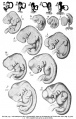
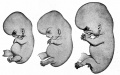
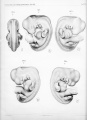
His's Normentafel
His's Normentafel (Early Stages)
His's Normentafel (Late Stages)
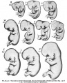
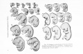
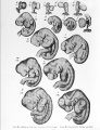
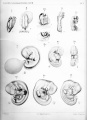
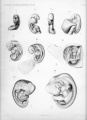
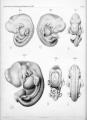
Keibel and Elze Normentafel (1908)
Normal Plates of the Development of the Human Embryo
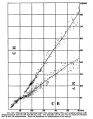
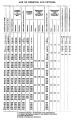
Growth
The following human growth data is from Mall (1910)[1]
Fig. 143. Human Embryo Length
Fig. 146. Human Embryo and Fetal Lengths
Table Human Embryo and Fetal Ages
References
Glossary Links
- Glossary: A | B | C | D | E | F | G | H | I | J | K | L | M | N | O | P | Q | R | S | T | U | V | W | X | Y | Z | Numbers | Symbols | Term Link
Cite this page: Hill, M.A. (2024, June 20) Embryology Carnegie stage table. Retrieved from https://embryology.med.unsw.edu.au/embryology/index.php/Carnegie_stage_table
- What Links Here?
- © Dr Mark Hill 2024, UNSW Embryology ISBN: 978 0 7334 2609 4 - UNSW CRICOS Provider Code No. 00098G


