Category:Cardiovascular: Difference between revisions
From Embryology
mNo edit summary |
mNo edit summary |
||
| Line 1: | Line 1: | ||
This | This {{Embryology}} category shows pages, images and media related to cardiovascular system development. | ||
See also the narrower categories [[:Category:Heart]] and [[:Category:Blood]]. | See also the narrower categories [[:Category:Heart]] and [[:Category:Blood]]. | ||
{{Heart Links}} | {{Heart Links}} | ||
Latest revision as of 12:39, 13 February 2017
This Embryology category shows pages, images and media related to cardiovascular system development.
See also the narrower categories Category:Heart and Category:Blood.
Subcategories
This category has the following 13 subcategories, out of 13 total.
Pages in category 'Cardiovascular'
The following 200 pages are in this category, out of 519 total.
(previous page) (next page)2
A
- Template:Abbott Figures
- Template:Abbott1915
- Template:Abbott1915images
- Abnormal Development - Hypertension
- Advanced - Abnormalities
- Advanced - Cardiac Conduction
- Advanced - Cardiac Looping
- Advanced - Cardiac Looping 2
- Advanced - Cardiac Septation
- Advanced - Cardiac Septation 2
- Advanced - Heart Fields
- Advanced - Heart Tubes
- Advanced - Molecular Development
- Advanced - Outflow Tract
- Advanced - Valve Development
- Advanced Cardiac Embryology
- Template:Advanced Cardiac menu
- ANAT2241 Cardiovascular System
- ANAT2341 Lab 4 - Early Cardiovascular Development
- ANAT3241 Lab 11
- Template:Aorta
- Template:Aortic arch
- Template:Aortic stenosis
- Template:Arachnoid mater
- Template:Artery
- Atlas of the Development of Man 2 - Cardiovascular
- Template:Atrial septal defects
- Template:Atrial septum movie links
B
- Template:Bartelmez GW.
- BGDA Lecture - Development of the Embryo/Fetus 2
- Template:BGDA Practical 14 - Maternal Decidua Interactive
- Template:BGDA Practical 14 - Placental Cord Interactive
- Template:BGDA Practical 14 - Villi Interactive
- BGDA Practical 7 - Week 5
- BGDB Lecture - Heart Development
- Template:Blood
- Template:Blood cell images
- Template:Blood vessel
- Template:Blood vessel histology
- Template:Bone marrow
- Book - A Laboratory Manual and Text-book of Embryology 9
- Book - An experimental analysis of the origin of blood and vascular endothelium (1915)
- Book - Congenital Cardiac Disease (1915)
- Book - Congenital Cardiac Disease - Figures
- Book - Congenital Cardiac Disease 1
- Book - Congenital Cardiac Disease 10
- Book - Congenital Cardiac Disease 11
- Book - Congenital Cardiac Disease 12
- Book - Congenital Cardiac Disease 13
- Book - Congenital Cardiac Disease 14
- Book - Congenital Cardiac Disease 15
- Book - Congenital Cardiac Disease 2
- Book - Congenital Cardiac Disease 3
- Book - Congenital Cardiac Disease 4
- Book - Congenital Cardiac Disease 5
- Book - Congenital Cardiac Disease 6
- Book - Congenital Cardiac Disease 7
- Book - Congenital Cardiac Disease 8
- Book - Congenital Cardiac Disease 9
- Book - Contributions to Embryology Carnegie Institution No.32
- Book - Contributions to Embryology Carnegie Institution No.36
- Book - Contributions to Embryology Carnegie Institution No.65
- Book - Contributions to Embryology Carnegie Institution No.67
- Book - Early stages of vasculogenesis in the cat with especial reference to the mesenchymal origin of endothelium (1914)
- Book - Human Embryology and Morphology 16
- Book - Quain's Embryology 9
- Book - Text-Book of the Embryology of Man and Mammals 17-1
- Template:Brain Vascular System gallery
- Template:Brain Vascular System table1
- Template:Bremer1914 figures
C
- Template:Capillary
- Template:CapillaryEM links
- Template:Cardiac
- Cardiac Embryology
- Cardiac Muscle Histology
- Template:Cardiovascular
- Cardiovascular - Arterial Development
- Cardiovascular - Detailed Cardiac Development
- Cardiovascular - Venous Development
- Cardiovascular 3D stage 13 Movie
- Cardiovascular 3D stage 22 Movie
- Template:Cardiovascular abnormalities
- Cardiovascular System - Abnormalities
- Cardiovascular System - Atrial Septal Defects
- Cardiovascular System - Blood Development
- Cardiovascular System - Blood Vessel Development
- Cardiovascular System - Bone Marrow
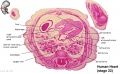
- Cardiovascular System - Carnegie Stage 22
- Cardiovascular System - Circulation Development
- Cardiovascular System - Coarctation of the Aorta
- Cardiovascular System - Coronary Circulation Development
- Cardiovascular System - Developmental Shunts
- Cardiovascular System - Double Outlet Right Ventricle
- Cardiovascular System - Ductus Arteriosus
- Cardiovascular System - Ductus Venosus
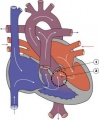
- Cardiovascular System - Fetal Shunts
- Cardiovascular System - Foramen Ovale
- Cardiovascular System - Heart Development
- Cardiovascular System - Heart Histology
- Cardiovascular System - Heart Rate Development
- Cardiovascular System - Heart Valve Development
- Cardiovascular System - Hypoplastic Left Heart
- Cardiovascular System - Lymphatic Development
- Cardiovascular System - Movies
- Cardiovascular System - Patent Ductus Arteriosus
- Cardiovascular System - Spleen Development
- Cardiovascular System - Tetralogy of Fallot
- Cardiovascular System - Transposition of the Great Vessels
- Cardiovascular System - Tricuspid Atresia
- Cardiovascular System - Truncus Arteriosus
- Cardiovascular System - Ventricular Septal Defects
- Cardiovascular System Development
- Template:Carotid body
- Template:Cerebral Arterial Timeline table
- Template:CHARGE syndrome
- Chicken Aortic Arches Movie
- Template:Choroid plexus
- Template:Coarctation of the aorta
- Template:Common truncus
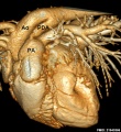
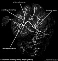
- Computed Tomography
- Template:Congdon1922 collapse table1
- Template:Congdon1922 table1
- Template:Coronary circulation
- Template:CVS cartoons
D
- Detailed Cardiac - Arterial Roots
- Detailed Cardiac - Atrioventricular Canal
- Detailed Cardiac - Atrioventricular Conduction Axis
- Detailed Cardiac - Atrioventricular Cushions
- Detailed Cardiac - Extrapericardial Arterial Channels
- Detailed Cardiac - Interventricular Communication
- Detailed Cardiac - Intrapericardial Arterial Trunks
- Detailed Cardiac - Pulmonary Vein
- Detailed Cardiac - Sinus Node
- Detailed Cardiac - Subpulmonary Infundibulum
- Detailed Cardiac - Superior Interatrial Fold
- Detailed Cardiac - Systemic Venous Sinus
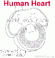
- Development Animation - Heart Atrial Septation
- Development Animation - Heart Realign
- Developmental Signals - Slit2/Robo1
- Template:Doppler ultrasound
- Template:Double outlet right ventricle
- Template:Ductus arteriosus
- Template:Ductus venosus
E
- Electrocardiogram
- Electron Microscopy Virtual Slides
- Talk:Embryo Serial Sections
- Embryology History
- Special:Badtitle/NS501:Embryology History
- History:Embryology History
- Embryology History - Douglas Reid
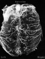
- Embryology History - George Bartelmez
- Embryology History - Herbert Evans
- Template:Evans HM.
- Template:Evans1909 figures
F
H
- Template:Haematopoiesis
- Template:Heart
- Template:Heart abnormal cartoon gallery
- Template talk:Heart abnormal cartoon gallery
- Heart Atrial Septation Movie
- Template:Heart histology
- Heart Historic Movie 1
- Heart Historic Movie 1951
- Heart Historic Movie 2
- Heart Historic Movie 3
- Heart Historic Movie 4
- Heart Historic Movie 5
- Heart Historic Movie 6
- Heart Historic Movie 7
- Heart Historic Movie 8
- Template:Heart Links
- Heart Looping Movie
- Heart Outflow Septation Movie
- Template:Heart rate
- Heart Realign Movie
- Template:Heart terms
- Template:Heart valve
- Historic Animation - Heart 02
- Historic Animation - Heart 04
- Template:Historic Heart
- Template talk:Historic Heart
Media in category 'Cardiovascular'
The following 200 files are in this category, out of 695 total.
(previous page) (next page) Congdon1922-39.jpg 1,200 × 756; 138 KB
 Congdon1922-40.jpg 1,013 × 1,000; 110 KB
 Congdon1922-plate01.jpg 877 × 1,200; 145 KB
 Congdon1922-plate02.jpg 877 × 1,200; 191 KB
 Congdon1922-plate03.jpg 1,200 × 885; 182 KB
 Coronary arteries.png 800 × 472; 183 KB
 Cullen1916 fig33.jpg 1,154 × 2,052; 422 KB
 Dextrocardia heart position.jpg 400 × 533; 49 KB
 Dextrocardia.jpg 400 × 533; 30 KB
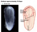
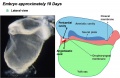
 Divisions of Early Heart Tube.jpg 1,105 × 978; 85 KB
 Doan-plate01.jpg 924 × 1,170; 207 KB
 Doan01.jpg 800 × 369; 79 KB
 Doan02.jpg 800 × 637; 82 KB
 Doan03.jpg 800 × 312; 31 KB
 Dog liver portosystemic shunts.jpg 800 × 264; 19 KB
 Dog patent ductus arteriosus computed tomography.jpg 600 × 654; 101 KB
 Double Outlet Right Ventricle.jpg 289 × 350; 16 KB
 Early Heart Tube (Dorsal).jpg 1,282 × 1,124; 111 KB
 Early Heart Tube (Lateral).jpg 1,504 × 972; 110 KB
 Ectopia cordis.jpg 800 × 603; 37 KB
 Embryo renal venous cartoon.jpg 600 × 600; 68 KB
 Embryonic Circulations.jpg 1,552 × 1,028; 171 KB
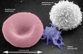
 Erythrocyte and lymphocyte SEM01.jpg 800 × 522; 74 KB
 Erythrocyte and lymphocyte SEM02.jpg 800 × 522; 78 KB
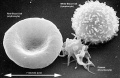 Erythrocyte and lymphocyte SEM03.jpg 800 × 522; 80 KB
 Evans1909 fig01-03.jpg 2,874 × 1,421; 443 KB
 Evans1909 fig01.jpg 596 × 1,000; 88 KB
 Evans1909 fig02.jpg 596 × 1,000; 97 KB
 Evans1909 fig03.jpg 596 × 1,000; 92 KB
 Evans1909 fig03b-06.jpg 3,000 × 2,100; 457 KB
 Evans1909 fig06.jpg 822 × 1,000; 82 KB
 Evans1909 fig07-08.jpg 1,265 × 1,116; 92 KB
 Evans1909 fig09-12.jpg 3,089 × 1,606; 290 KB
 Evans1909 fig09.jpg 800 × 1,181; 41 KB
 Evans1909 fig10.jpg 800 × 1,298; 47 KB
 Evans1909a fig01.jpg 653 × 1,280; 128 KB
 Fawcett1905 fig01.jpg 800 × 1,114; 145 KB
 Fetal blood flow 01.jpg 1,000 × 599; 75 KB
 Fetal blood flow 02.jpg 491 × 599; 38 KB
 Fetal blood flow 03.jpg 500 × 599; 43 KB
 Fetal blood flow 04.jpg 506 × 599; 50 KB
 Fetal blood flow liver and brain.jpg 677 × 790; 69 KB
 Fetal cardiac state diagram 01.jpg 1,200 × 321; 29 KB
 Fetal cardiac state diagram 02.jpg 1,200 × 458; 27 KB
 Fetal cardiac ultrasound 01.jpg 900 × 600; 88 KB
 Fetal Circulation Pathway.jpg 1,371 × 1,069; 112 KB
 Fetal circulation1.jpg 558 × 900; 76 KB
 Fetal ductus venosus cartoon.jpg 1,000 × 1,329; 255 KB
 Fetal ductus venosus pressure wave 01.jpg 706 × 755; 52 KB
 Fetal ductus venosus ultrasound 01.jpg 783 × 1,000; 68 KB
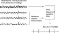 Fetal Electrocardiogram Enhancement 01.jpg 657 × 370; 40 KB
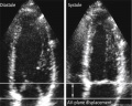 Fetal heart atrioventricular plane displacement 01.jpg 915 × 733; 55 KB
 Fetal heartbeat 01.mp3 ; 225 KB
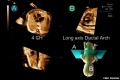 Fetal ultrasound ductal arch 01.jpg 800 × 533; 27 KB
 Fetalblood.jpg 400 × 307; 24 KB
 Foster137.jpg 786 × 471; 69 KB
 Foster138.jpg 755 × 540; 51 KB
 Foster139.jpg 412 × 684; 39 KB
 Functional Hypoplastic Left Heart.jpg 302 × 350; 18 KB
 Gillilan1959-fig01.jpg 1,000 × 1,313; 155 KB
 Gillilan1959-fig02.jpg 905 × 1,000; 159 KB
 Gillilan1959-fig03.jpg 1,042 × 1,366; 317 KB
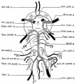
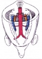
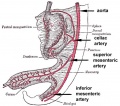 GIT blood supply.jpg 568 × 500; 47 KB
 Gonad blood 01 icon.jpg 296 × 413; 28 KB
 Gray0448.jpg 356 × 600; 70 KB
 Gray0458.jpg 693 × 911; 83 KB
 Gray0460.jpg 818 × 551; 106 KB
 Gray0461.jpg 859 × 841; 82 KB
 Gray0462.gif 350 × 403; 49 KB
 Gray0462.jpg 822 × 800; 157 KB
 Gray0463.jpg 1,099 × 755; 134 KB
 Gray0464.gif 450 × 455; 51 KB
 Gray0464.jpg 993 × 961; 258 KB
 Gray0465.jpg 954 × 938; 207 KB
 Gray0467.jpg 600 × 464; 47 KB
 Gray0468.jpg 500 × 436; 43 KB
 Gray0470.jpg 800 × 371; 40 KB
 Gray0472.jpg 550 × 653; 57 KB
 Gray0473.gif 500 × 418; 32 KB
 Gray0473.jpg 499 × 418; 0 bytes
 Gray0474.jpg 459 × 600; 33 KB
 Gray0475.jpg 2,042 × 1,363; 350 KB
 Gray0476.jpg 600 × 609; 99 KB
 Gray0477.jpg 597 × 560; 22 KB
 Gray0478.jpg 597 × 560; 21 KB
 Gray0479.jpg 597 × 560; 26 KB
 Gray0480.jpg 597 × 560; 31 KB
 Gray0492.jpg 600 × 508; 117 KB
 Gray0498.jpg 475 × 416; 39 KB
 Gray0502.jpg 1,000 × 1,329; 215 KB
 Gray0506.jpg 375 × 464; 25 KB
 Gray0532.jpg 680 × 700; 155 KB
 Gray0533.jpg 705 × 750; 164 KB
 Gray0540.jpg 1,355 × 1,000; 261 KB
 Gray0556.jpg 600 × 533; 86 KB
 Gray0585.jpg 752 × 800; 188 KB
 Gray0589.jpg 900 × 534; 134 KB
 Gray0769.jpg 600 × 454; 101 KB
 Gray0872.jpg 499 × 600; 93 KB
 Gray0873.jpg 919 × 545; 124 KB
 Gray0874.jpg 600 × 545; 120 KB
 Gray0877.jpg 500 × 794; 101 KB
 Gray0878.jpg 580 × 550; 120 KB
 Gray0879.jpg 600 × 522; 66 KB
 Gray0880.jpg 800 × 496; 147 KB
 Gray0985.jpg 682 × 600; 93 KB
 Gray1120.jpg 683 × 900; 297 KB
 Gray1121.jpg 600 × 669; 154 KB
 Gray1122.jpg 520 × 404; 57 KB
 Gray1123.jpg 564 × 450; 66 KB
 Gray1124.jpg 714 × 500; 138 KB
 Gray1129.jpg 396 × 500; 55 KB
 Gray1130.jpg 301 × 400; 25 KB
 Gray1132.jpg 625 × 500; 86 KB
 Gray1170.jpg 1,000 × 718; 170 KB
 Gray1174.jpg 782 × 800; 165 KB
 Gray1192.jpg 800 × 415; 48 KB
 Haemoglobin comparison oxygen saturation curve.png 639 × 541; 15 KB
 Heart conduction system-bird-monotreme-placental.jpg 1,040 × 625; 76 KB
 Heart fields 001 icon.jpg 720 × 540; 31 KB
 Heart folding 001 icon.jpg 720 × 540; 39 KB
 Heart folding 002 icon.jpg 720 × 540; 39 KB
 Heart human embryo CRL10mm 01.jpg 1,000 × 1,389; 537 KB
 Heart innervation 01.jpg 1,280 × 599; 92 KB
 Heart looping 006 icon.jpg 720 × 540; 51 KB
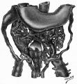
 Heart Looping Sequence (SEMs).jpg 1,928 × 776; 212 KB
 Heart Looping Sequence.jpg 1,548 × 577; 85 KB
 Heart outflow tract stage 14 01.jpg 2,039 × 996; 274 KB
 Heart outflow tract stage 14 02.jpg 996 × 996; 139 KB
 Heart outflow tract stage 14 03.jpg 989 × 996; 134 KB
 Heart septation 001 icon.jpg 720 × 540; 26 KB
 Heart Tube Fusion.jpg 1,551 × 1,139; 125 KB
 Heart Tube Segments.jpg 1,082 × 771; 63 KB
 Heart valve histology 01.jpg 1,008 × 1,280; 318 KB
 Heart valve histology 02.jpg 800 × 456; 103 KB
 Heart valve histology 03.jpg 800 × 489; 111 KB
 Heart-cartoon-001.jpg 600 × 697; 40 KB
 Heart-ventricular-septum-03.jpg 320 × 240; 14 KB
 Heart1 atrium.gif 350 × 373; 243 KB
 Heart1 ventricle.mov ; 209 KB
 Human heart developmental functional networks.jpg 833 × 614; 424 KB
 Human heart SEM1.jpg 1,200 × 330; 47 KB
 Human placenta vascular 01.jpg 1,200 × 644; 125 KB
 Human placenta vascular CT 01.jpg 938 × 1,000; 126 KB
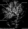 Human placenta vascular MRI 01.jpg 938 × 1,000; 151 KB
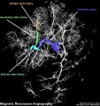 Human placenta vascular MRI 02.jpg 938 × 1,000; 156 KB
 Human placenta vasohibin 1 expression.jpg 1,206 × 908; 168 KB
 Human placenta vasohibin 2 expression.jpg 1,198 × 904; 165 KB
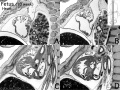 Human- fetal week 10 heart ABCD.jpg 600 × 450; 133 KB
 Human-heart-E3L.jpg 639 × 393; 79 KB
 Hypoplastic Left Heart.jpg 297 × 350; 17 KB
 Ingalls1908 plate01.jpg 927 × 1,000; 204 KB
 Ingalls1920plate04.jpg 762 × 1,044; 52 KB
 Interrupted Aortic Arch.jpg 290 × 350; 17 KB
 Johnson1917 plate05.jpg 1,214 × 1,000; 170 KB
 Keibel Mall 2 293.jpg 1,280 × 766; 304 KB
 Keibel Mall 2 297-299.jpg 1,280 × 786; 135 KB
 Keibel Mall 2 297.jpg 354 × 359; 13 KB
 Keibel Mall 2 298.jpg 599 × 605; 40 KB
 Keibel Mall 2 299.jpg 733 × 1,034; 120 KB
 Keibel Mall 2 447.jpg 1,280 × 1,701; 250 KB
 Keibel Mall 2 539.jpg 1,280 × 1,414; 185 KB
 Keibel Mall 2 540.jpg 1,280 × 938; 182 KB
 Keibel Mall 2 541.jpg 1,280 × 1,683; 313 KB
 Keibel Mall 2 542.jpg 1,280 × 1,670; 276 KB
 Keibel Mall 2 543.jpg 1,280 × 1,193; 241 KB
 Keibel Mall 2 544.jpg 971 × 1,093; 117 KB
 Keibel Mall 2 545.jpg 761 × 1,093; 104 KB
 Keibel Mall 2 546.jpg 1,000 × 777; 67 KB
 Keibel Mall 2 547.jpg 648 × 483; 36 KB
 Keibel Mall 2 548.jpg 672 × 483; 53 KB
 Keibel Mall 2 572.jpg 1,280 × 969; 219 KB
 Keibel Mall 2 573.jpg 1,280 × 886; 165 KB
 Keibel Mall 2 574.jpg 1,280 × 1,902; 327 KB
 Keibel Mall 2 575.jpg 1,000 × 1,288; 120 KB
 Keith1902 fig028.jpg 707 × 800; 77 KB
 Keith1902 fig029.jpg 872 × 600; 81 KB
 Keith1902 fig181.jpg 648 × 601; 44 KB
 Keith1902 fig182.jpg 637 × 601; 38 KB
 Keith1902 fig183.jpg 879 × 800; 113 KB
 Keith1902 fig184.jpg 814 × 828; 90 KB
 Keith1902 fig185.jpg 588 × 828; 48 KB
 Keith1902 fig186.jpg 746 × 800; 91 KB
 Keith1902 fig187.jpg 916 × 800; 138 KB
 Keith1902 fig188.jpg 951 × 800; 115 KB
 Keith1902 fig189.jpg 807 × 750; 70 KB
 Keith1902 fig190.jpg 780 × 800; 83 KB
 Keith1902 fig191.jpg 1,000 × 411; 74 KB
 Keith1902 fig192.jpg 1,000 × 643; 119 KB
 Keith1902 fig193.jpg 970 × 800; 97 KB
 Keith1902 fig194.jpg 829 × 750; 50 KB
 Keith1902 fig195.jpg 881 × 800; 115 KB
 Keith1902 fig196.jpg 1,065 × 700; 106 KB
 Keith1902 fig197.jpg 1,116 × 800; 191 KB
 Keith1902 fig198.jpg 1,026 × 800; 170 KB
 Keith1902 fig199.jpg 993 × 600; 127 KB
 Keith1902 fig200.jpg 800 × 420; 61 KB
 Keith1902 fig201.jpg 826 × 800; 87 KB
 Keith1902 fig202.jpg 1,176 × 800; 149 KB
 Keith1902 fig203.jpg 1,000 × 633; 91 KB


.jpg)
.jpg)


.jpg)