Embryology History - Osborne Heard
| Embryology - 26 Apr 2024 |
|---|
| Google Translate - select your language from the list shown below (this will open a new external page) |
|
العربية | català | 中文 | 中國傳統的 | français | Deutsche | עִברִית | हिंदी | bahasa Indonesia | italiano | 日本語 | 한국어 | မြန်မာ | Pilipino | Polskie | português | ਪੰਜਾਬੀ ਦੇ | Română | русский | Español | Swahili | Svensk | ไทย | Türkçe | اردو | ייִדיש | Tiếng Việt These external translations are automated and may not be accurate. (More? About Translations) |
Introduction

Osborne O. Heard (1891 – 1983) was one of the first to be hired, to the newly formed Carnegie Institute of Embryology in 1913, he was modeller who spent 42 years at the department and made over 700 wax-based reconstructions.
The results of this team effort still stand as the international standard by which human embryos are described and classified.
The models were mainly made by the lost-wax casting process and his models were also much more detailed than the earlier (1880's) Ziegler embryo models.
- Links: Carnegie Collection | Carnegie Stages | Ziegler Models
| Embryologists: William Hunter | Wilhelm Roux | Caspar Wolff | Wilhelm His | Oscar Hertwig | Julius Kollmann | Hans Spemann | Francis Balfour | Charles Minot | Ambrosius Hubrecht | Charles Bardeen | Franz Keibel | Franklin Mall | Florence Sabin | George Streeter | George Corner | James Hill | Jan Florian | Thomas Bryce | Thomas Morgan | Ernest Frazer | Francisco Orts-Llorca | José Doménech Mateu | Frederic Lewis | Arthur Meyer | Robert Meyer | Erich Blechschmidt | Klaus Hinrichsen | Hideo Nishimura | Arthur Hertig | John Rock | Viktor Hamburger | Mary Lyon | Nicole Le Douarin | Robert Winston | Fabiola Müller | Ronan O'Rahilly | Robert Edwards | John Gurdon | Shinya Yamanaka | Embryology History | Category:People | ||
|
- Carnegie Stages: 1 | 2 | 3 | 4 | 5 | 6 | 7 | 8 | 9 | 10 | 11 | 12 | 13 | 14 | 15 | 16 | 17 | 18 | 19 | 20 | 21 | 22 | 23 | About Stages | Timeline
1957
Heard OO. Methods used by C. H. Heuser in preparing and sectioning early embryos. (1957.) Carnegie Instn. Wash. Publ. 611, Contrib. Embryol., 36, 1-18.
1921
Reference: Mall,, F.P and Arthur William Meyer, A.W. Studies on Abortuses: A Survey of Pathologic Ova in the Carnegie Embryological Collection. (1921) Carnegie Institution No.68. Vol. 12 No.56.
Chapter 2. Care and Utilization of the Collection - Modelling Methodology


(Text except below from reference above)
In nearly all studies on embryo-anatomy it is necessary to resort to methods which will enable one to see the structure and form of organs in their serial sections. As far as possible this is always done by observing the whole embryo or parts of one dissected under the dissecting microscope. A valuable specimen, however, can not be treated in this way. Such a specimen is cut into sections and from the sections an effort is made to reconstruct the anatomy. In the first publication the anatomy of the embryo was worked out by Bern's (1883) method of reconstruction, but unfortunately at that time this method gave little more than the external form, the finer structures being lost in the model. The plan of dissecting the model was then tried, and it soon became clear that success in reconstruction depended very largely upon one's power to visualize the structures from the serial sections, and to some extent upon inventive ability in eliminating a part of the model, as deeper structures can not be shown without removing the superficial ones. Gradually we evolved what we term dissectible wax models, which, however, are somewhat clumsy and usually warp in warm water. Finally, Dr. Bardeen (1901), who is skilled in work of this kind, discovered that excellent results could be obtained by making a foundation model in wax and then projecting and elaborating it as a drawing by the graphic method of His (1892), thus combining the good features of two very valuable methods.
At first our record of sections was made by drawing them with the camera lucida or, better still, in a dark room by the aid of a magic lantern. This, however, is a very laborious and time-consuming task, and has a tendency to check the mental activity of the investigator, for unfortunately the work can not be done successfully by a technical assistant. When the collection was transferred to the Carnegie Institution we availed ourselves of the opportunity to install an accurate projection apparatus patterned somewhat after the one used by His (1892). With this apparatus sections of embryo No. 460, previously referred to, were photographed on glass negatives with an enlargement of 50 diameters. Thus it was possible to make several prints. As the photographs were taken with a 50-mm. planar lens, they show all the details wonderfully well.
As everything which we wished to reconstruct had to be transferred to wax, it was highly desirable that the final model should be composed of some temperature resisting substance. A method has therefore been devised whereby all the structures desired are eliminated from the wax. Thus, in a block of wax the model is represented in outline by a hole or a series of holes, according to the number of structures to be reproduced, and these holes are subsequently filled with plaster of pans. When this hardens and the wax is removed, the finished plaster model is liberated.
This process of modeling depends solely upon absolute accuracy in superposing the wax plates to correspond to the original sections of the embryo. Hence a perpendicular line must be established, and this is done as follows: The wax plates of the sections are placed one upon another until a complete model of the embryo is built up (fig. 3), the construction being guided by photographs of the embryo made before it was cut. Thus it is possible to duplicate accurately the external form of an embryo on an enlarged scale. After this is done it is a simple matter to mark the wax plates in a perpendicular direction that is, by rightangle lines drawn upon every plate through its axis. These constitute what we call guide-lines, the instrument for marking which was invented by Dr. W. H.
Lewis (1915) and is known as the Lewis guide liner (fig. 4). After the guide-lines have been drawn on each wax plate they are transferred to the photographs or drawings. This is done by superposing each wax plate upon its photograph or drawing and marking the end of the guide-lines. When the wax plate is removed, these points are connected by lines similar to those on the plates. After the two principal guide-lines have been established, it has been found convenient to use secondary guide-lines, 5 cm. apart, which run parallel to the primary ones over the entire surface of the photograph.
In order to make a reconstruction of any portion of an embryo it is necessary first to transfer the outlines of that structure from the photograph to the wax plates by means of carbon paper and a glass point used as a pencil. These outlines are then cut and removed from the wax and the plates squared off along the secondary guide-lines sufficiently far outside of the proposed model not to conflict with its casting.
In order that these finished plates, which we call mold plates, may be kept in exact apposition, they are piled in a rectangular corner made of plate glass (fig. 5). While this is being done it is necessary to cut certain artificial channels as vents, so that the air will escape from the openings when the plaster is poured in.
It is also well to bridge loosely attached parts with copper wire, which is done by heating the wire and laying upon the wax plates as they are being piled up. The entire mass forms a mold which, to distinguish it from the others, is called a plate mold. This is cast with plaster of paris, and after the plaster has set the wax is removed by heating. Sometimes it is necessary to proceed farther and make a break mold over the first cast, after which it is possible to make as many duplicate casts as desired. In the above description the main outlines of this valuable method of reconstruction, as now practised in this laboratory, have been given.
Numerous general studies have also been made in embryometrics, in which linear, gravity, and time values are considered. Furthermore, the technique of gross anatomy has been applied to differentiate, by means of injection, the arteries, veins, and lymphatics of the whole clarified embryo. Similar specimens can be prepared to show the entire cartilaginous and bony systems. Thus many embryos frequently are considered in a single publication, especially those illustrating causes of abortion and the production of moles and monsters. Attention is therefore called to the numbers following the references in the appended list of publications. These represent the catalogue numbers of the specimens studied.
It will be noted that certain specimens are referred to frequently, showing that these have been of more general use than others. While these numbers may seem somewhat confusing as they here appear, references to a given embryo are found together in the Key, and this makes it quite easy to find the published description of that embryo. Where the description is accompanied by illustrations, the specimen number is printed in bold type. A comprehensive view of the entire bibliography shows that the papers are sometimes general but often special in character. Whenever the line of thought is general, it is necessary to consider the topic in many different embryos, and one topic is frequently represented by several collaborators. In other words, our department is a large cooperative effort on the part of many contributors and collaborators.
Our first papers were published in the Journal of Morphology and the Johns Hopkins Hospital Bulletin and Reports, as well as in the foreign journals. We had hoped at that time that the excellent Journal of Morphology would continue, and that it would publish all of our best papers; but unfortunately it could not be financed, and thus for a time we had no adequate outlet for our work. At the Christmas (1900) meeting of the American Association of Anatomists, held in Baltimore, a committee was named to consider the advisability of founding a new journal for the publication of serious anatomical studies. In May following, three trustees (Huntington, Mall, and Minot) were appointed to launch this enterprise. In November of that year the first number of the American Journal of Anatomy appeared, and at the next meeting of the Anatomists, held in Chicago, it was voted to substitute the new journal for the Proceedings of the Association, without additional expense to the members. In this way national support was at once assured. A glance over the first ten volumes of the Journal discloses the fact that about 30 per cent of the papers emanated from the Anatomical Laboratory of the Johns Hopkins Medical School. In other words, our collection was one of the chief incentives for its publication. In rapid succession other journals were established and published in Baltimore; first, the Journal of Experimental Zoology in 1904, then the Anatomical Record in 1906. The stimulus given by these national efforts brought about the revival of the Journal of Morphology by the Wistar Institute in 1908, the opening volume consisting of a monograph entitled "A study of the causes underlying the origin of human monsters" (Mall). These serial publications mark the conversion of our studies of embryology from a local to a national enterprise.
1922
Reference: Congdon, E. D. (1922) 47-110 Transformation of the aortic-arch system during the development of the human embryo. Carnegie Institution No.68.
- "Microscopic study was supplemented in each case by models made by the wax-plate method. Several of these reconstructions were already in the laboratory, having been prepared in connection with other studies, notably those of Ingalls, Bartelmez, Davis, Evans, and Streeter. Plaster casts were made from some of the plates by Mr. O. O. Heard, whose skilful aid is greatly appreciated. The colored figures were the work of Mr. J. F. Didusch and were drawn from models. I am much indebted to him for their excellent rendering and for further assistance in reconstructing some parts."
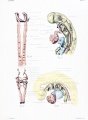
Plate 1
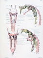
Plate 2

Plate 3


Glossary Links
- Glossary: A | B | C | D | E | F | G | H | I | J | K | L | M | N | O | P | Q | R | S | T | U | V | W | X | Y | Z | Numbers | Symbols | Term Link
Cite this page: Hill, M.A. (2024, April 26) Embryology Embryology History - Osborne Heard. Retrieved from https://embryology.med.unsw.edu.au/embryology/index.php/Embryology_History_-_Osborne_Heard
- © Dr Mark Hill 2024, UNSW Embryology ISBN: 978 0 7334 2609 4 - UNSW CRICOS Provider Code No. 00098G