Embryology History - Robert Meyer
| Embryology - 18 Apr 2026 |
|---|
| Google Translate - select your language from the list shown below (this will open a new external page) |
|
العربية | català | 中文 | 中國傳統的 | français | Deutsche | עִברִית | हिंदी | bahasa Indonesia | italiano | 日本語 | 한국어 | မြန်မာ | Pilipino | Polskie | português | ਪੰਜਾਬੀ ਦੇ | Română | русский | Español | Swahili | Svensk | ไทย | Türkçe | اردو | ייִדיש | Tiếng Việt These external translations are automated and may not be accurate. (More? About Translations) |
Introduction


Robert Meyer (1864 – 1947) was a German embryologist and pathologist.
He studied medicine at the universities of Leipzig, Heidelberg and Strassburg, receiving his doctorate in 1889 (Strassburg). From 1890 to 1894 he was a medical practitioner in the community of Dedeleben, and afterwards worked as assistant to gynecologist Johann Veit in Berlin.
Head of the laboratory in the women's clinic at the Berlin Charité (1909 to 1911), and in 1912 succeeded Carl Arnold Ruge as chief of the pathological institute of the university women's clinic. In 1932 he became an honorary professor to the faculty of medicine at the university. Because of his Jewish ancestry, he was removed from his position at Berlin in 1935 and emigrated to the United States, settling in Minneapolis in 1939.
In 1897 he published in Veit’s "Handbuch der Gynäkologie" 1st edition (1897) from his studies of 125 fetuses reviews on gynatresia, hematocolpos, and duplex uterus.
In 1932 he published studies (Archiv für Gynäkologie) of the development of the vagina based on his serial sections of 112 fetuses.
The "Weigert-Meyer law" is a rule concerning the anatomical relationship of the two ureters, named in conjunction with pathologist Carl Weigert.
| Online Editor |
|---|
| The following templates within the site text point to this current page.
Professor R. Meyer | Dr. Robert Meyer | Meyer R. Do not confuse Robert Meyer with the American embryologist Arthur William Meyer (1873 – 1966). |
| Embryologists: William Hunter | Wilhelm Roux | Caspar Wolff | Wilhelm His | Oscar Hertwig | Julius Kollmann | Hans Spemann | Francis Balfour | Charles Minot | Ambrosius Hubrecht | Charles Bardeen | Franz Keibel | Franklin Mall | Florence Sabin | George Streeter | George Corner | James Hill | Jan Florian | Thomas Bryce | Thomas Morgan | Ernest Frazer | Francisco Orts-Llorca | José Doménech Mateu | Frederic Lewis | Arthur Meyer | Robert Meyer | Erich Blechschmidt | Klaus Hinrichsen | Hideo Nishimura | Arthur Hertig | John Rock | Viktor Hamburger | Mary Lyon | Nicole Le Douarin | Robert Winston | Fabiola Müller | Ronan O'Rahilly | Robert Edwards | John Gurdon | Shinya Yamanaka | Embryology History | Category:People | ||
|
Meyer Embryo Collection
Robert Meyer's original Berlin collection was purchased by the late Hedwig Frey and bequeathed by him to the Anatomisches Institut, University of Zurich. References to specific collection embryos are made in a number of historic publications.
| Hedwig Frey (1877-1938) |
|---|
| Hedwig Frey (1877-1938) graduated in 1897 from her teacher training in Zurich and in 1905 attended lectures at the local university. She was denied a medical degree, instead she began in 1908 a degree in anthropology and anatomy. She then became an assistant at the Anatomical Institute of Zurich and in 1918 became a Privatdozentin for anatomy and development history, after she had submitted her habilitation thesis on the transformation process of the thorax. From 1920 to 1938 she was employed as a prosector. In 1924 Frey received the honorary professorship from the University of Zurich and was the first professor of anatomy in Switzerland.(modified text) |
Meyer 249

Meyer 250
Keibel F. The Interdependence of the Various Developmental Processes in Keibel F. and Mall FP. Manual of Human Embryology II. (1912) J. B. Lippincott Company, Philadelphia.
Online Editor - Table Meyer 250 to be formatted.
Meyer 264

|
Fig. 657. Part of a transverse section through a human embryo of 60 mm. head-foot length. (Embryo R. Meyer 264; slide 44, row 2, section 2.)
By the union of the mesonephric fold and testis to the anterior abdominal wall, formed by the chorda gubernaculi, both have been carried quite away from the posterior abdominal wall. Between the urogenital fold and the posterior wall a layer of loose mesenchyme has formed. Thereby the portion of the abdominal cavity which contains the urogenital fold is separated off from the rest of the cavity as the saccus vaginalis. The separation between the two is completed by a peritoneal fold formed by the a. umbilicalis. |
Meyer 267

|
Fig. 643. Transverse section of a male embryo of 70 mm head-foot length (embryo R. Meyer 267; slide 67, row 1, section 3), below the symphysis pubis. X ca. 23.
From behind forwards the following parts are cut: rectum, pars pelvina of the urogenital sinus and scrotum. From the pars pelvina Cowper's glands have developed and between A the mesenchyme of the urogenital sinus and the epithelium of the scrotum there is stretched the septum scroti. |

|
Fig. 655. Transverse section of the urogenital sinus of a male embryo of 70 mm head-foot length. (Embryo R. Meyer 267; slide 60, row 2, section 3.) X47.
The section shows Mailer's tubercle projecting into the lumen of the sinus and around the sinus the anlagen of the prostatic glands. |
Meyer 268

|
Fig. 653 a and b. Two sections through the clitoris of an embryo of 60 mm head-foot length. (Embryo R. Meyer 268; slide 66, row 2, section 1 and slide 67, row 1, section 2.)
The urethral plate in section a extends from the anal surface of the clitoris only to the centre. In section 6, which passes through the tip, the clitoris is completely divided by the urethral plate. |

|
Fig. 654. Longitudinal section of the clitoris of an embryo of 60 mm head-foot length. (Embryo H. Meyer 268; slide 64, row 2, section 1.) X 50.
The section cuts the sinus urogenitalis lengthwise from the crista urethralis int. to the ostium urogenitale. The lumen of the sinus is enlarged at its end to a triangular basin, on whose distal wall is the urethral plate. To the right and left are the genital swellings, the anlagen of the labia majora, which are becoming separated from the clitoris. |
Meyer 300
| Human Embryo No. 300 | ||||||||||||||||||||||
|---|---|---|---|---|---|---|---|---|---|---|---|---|---|---|---|---|---|---|---|---|---|---|
| Collection of Professor R. Meyer, Berlin. Carnegie stage 12 | ||||||||||||||||||||||
| Desig. | Size | Age | Body form | Primitive streak | Primitive segments | Chorda | Nervous system | Eye | Ear | Nose | Hypophysis | Mouth | Digestive trac, liver and pancreas | Branchial pouches, threoid thymus, trachea and lungs | Urogenital system | Heart and vessels | Integument | Skeleton | Extremities | Amnion | Allantois | Remarks |
| 7 N.T. Fig. VI 1, VI r ind VI v. |
Gr. L. 2.5 mm. | Remains of primitive streak. Cloacal membrane. | 23 pairs of primitive segments. | Chorda separated from entoderm, | Anterior neuropore closed. but its position still recognizable. Medullary canal stili wide open for a stretch caudally. Roof of 4th vent, beginning to thin out. Neuromeres. Trigeminus and acustico-facialis ganglia distinct. | Optic vesicles. Mesoderm between ectoderm and optic vesicle, | Auditory vesicle almost closed (open through 4 or 5 sections of 5μ). Ductus endolymphaticus not yet visible. | Anlage doubtful. | Pharyngeal membrane just torn, still abundant remains of it. | Intestine still communicates widely with the yolk sack. Liver a thick-walled sack from which the trabeculse are beginning to bud. | The 3 anterior branchial pouches reacts the ectoderm; the 4th, though formed, does not. Thyreoidea mediana formed. Pulmotracheal groove. The paired condition of the pulmonary anlage already indicated. | Rudimentary "pronephric anlage" | Heart S-shaped. | Allantoic duct | The embryo was obtained by operation. | |||||||
| Remarks
Abundant mitoses in the embryo. Fixation: ? Stain: Borax carmine. Sections: 5 μ. The embryo was modelled by Dr. Peter Thompson (1907) Also see Meyer (1904) | ||||||||||||||||||||||
| Reference: Keibel F. and Elze C. Normal Plates of the Development of the Human Embryo (Homo sapiens). (1908) Vol. 8 in series by Keibel F. Normal plates of the development of vertebrates (Normentafeln zur Entwicklungsgeschichte der Wirbelthiere) Fisher, Jena., Germany. Cited in: 1912 Human Embryology and Thompson P. Description of a human embryo of twenty-three paired somites. (1907) J Anat Physiol, 41(3):159-71. PMID 17232726; Meyer, Rob., Ueber die Beziehung der Umierenkanalchen zum Coelomepithel, etc., Anat. Anz., Vol. 25, 1904 | ||||||||||||||||||||||
| 1908 Embryo Tables: Klb (stage 10) | Pfannenstiel III (stage 11) | Meyer 300 (stage 12) | Strahl 4mm (stage 13) | Hertwig G31 (stage 14) | ||||||||||||||||||||||

|
Fig. 317. Pharynx of the embryo. Pharynx of the embryo Meyer 300 (Normentafel Xo. 7, 23 pairs of primitive segments, 2.5 mm vertex-breech length) seen from the ventral surface. The closing membranes of the gillclefts are outlined in black.
At. mes., the somewhat depressed oral end of the area mesobranchialis ; Hw., heart swelling; Lar., laryngotracheal groove; V.V., ventral prolongations of the pharyngeal 'pouches. Other lettering as in Fig. 314. x 150. |

|
Fig. 318. Pharynx of the embryo Rob. Meyer No. 300 lateral surface. The model of embryo No. Meyer 300 shown in Fig. 317 from the lateral surface.
Ka., doubtful branchial anlage; L., liver; Lar., laryngotracheal groove; Lu., lung anlage; Mw., angle of mouth. |

|
Fig. 331. Anlage of the respiratory tract of an embryo of 23 primitive segments. (Rob. Meyer No. Meyer 300, 2.5 mm.; compare Figs. 317 and 318). L., liver; Lar„ laryngeal groove; Tr., tracheal groove; Lu., lung anlage; 5. St. f, doubtful anlage of the fifth pharyngeal pouch. X 150. |

|
Fig. 317. Pharynx of the embryo Meyer 300 (Normentafel Xo. 7, 23 pairs of primitive segments, 2.5 mm vertex-breech length) seen from the ventral surface. The closing membranes of the gillclefts are outlined in black. |

|
Fig. 373. Model of the heart of a human embryo 2.5 mm. greatest length. No. Meyer 300 of Rob. Meyer's collection (Normentafel Xo. 7), 2.5 mm. greatest length.
Modelled by P. Thompson. (After Thompson.) A., atrium; Au., region of the atrial canal; B., bulbus cordis; S., sinus venosus; T ., truncus arteriosus; U., vena umbilicalis sinistra; V., ventricular limb. X 50. |
Meyer 300 References
Waterston D. A human embryo of twenty-seven pairs of somites, embedded in decidua. (1914) J Anat Physiol., 49(1): 90-118 PMID 17233016
Meyer 302

|
Fig. 57. Embryo shows a distinct depression between the root of the nose and the forehead. The pinna, tragus, and antitragus are evident; and while the concha is still widely open it shows a tendency to deepen. Early indications of the eyelids are to be seen. The nape flexure is still very evident, but it forms an obtuse angle, and the depression of the dorsal line just below it is less marked. The nose and mouth have both separated from contact with the pericardium ; the mouth is slightly open and the mandible lies close upon the breast. The shoulder is distinctly marked off from the body, the upper arm makes an angle with the forearm, and the elbow projects strongly. The hand plate is beginning to turn ventrally, and the finger anlagen are very distinct; their tips are becoming free and the thumb is strongly abducted. In the lower limb the knee is very distinct and anlagen of the toes are appearing on the foot plate. The dorsum of the foot is not yet marked off from the crus. |
Meyer 307

Meyer 321
Keibel F. The Interdependence of the Various Developmental Processes in Keibel F. and Mall FP. Manual of Human Embryology II. (1912) J. B. Lippincott Company, Philadelphia.
Online Editor - Table Meyer 321 to be formatted.
Meyer 335
9–10 somites, Embryo R. Meyer 335. Listed by Bartelmez and Evans (1926), and cited by Felix (1912).
Meyer 338
 Fig. 341. Epithelial lung anlage of the embryo Rob. Meyer 338 (Normentafel No. 18.5 mm), from the ventral surface. M, stomach. X 100.
Meyer 399

Embryo R.Meyer 399 of the Zurich Anatomical Institute (Stage I of Blisnianskaja). X 150.
Manual of Human Embryology II - Meyer Embryos
Felix W. The development of the urinogenital organs. In Keibel F. and Mall FP. Manual of Human Embryology II. (1912) J. B. Lippincott Company, Philadelphia. pp 752-979.
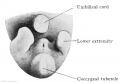
Fig. 637. Caudal end of the body of an embryo of 18 mm GL. Between the umbilicus, the coccygeal tubercle, and the right and left lower limbs is the cloacal tubercle; on its anal slope are the ostium urogenitale and the anal groove.
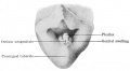
Fig. 640. The indifferent external genitalia of an embryo of 28 mm GL. The cloacal tubercle is divided into the phallus and the genital tubercles or swellings. The ostium urogenitale and anus are still close together.
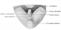
Fig. 641. Female external genitalia of an embryo of 32.5 mm GL. The phallus has become the clitoris, the glans and coronary sulcus are recognizable. The ostium urogenitale has preserved its relation to the anal opening, but no longer reaches the sulcus coronarius glandis.
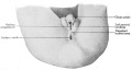
Fig. 642. Male external genitalia of an embryo of 3.5 months. The phallus has become the glans penis, the scrotal swellings are fully developed and the unpaired scrotal area is indicated. The ostium urogenitale and ostium anale have separated, the ostium urogenitale extends to the sulcus coronarius glandis.
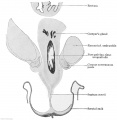
Fig. 643. Transverse section of a male embryo of 70 mm head-foot length (embryo R. Meyer 267; slide 67, row 1, section 3), below the symphysis pubis. X ca. 23. From behind forwards the following parts are cut: rectum, pars pelvina of the urogenital sinus and scrotum. From the pars pelvina Cowper's glands have developed and between A the mesenchyme of the urogenital sinus and the epithelium of the scrotum there is stretched the septum scroti.
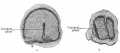
Fig. 653 a and b. Two sections through the clitoris of an embryo of 60 mm head-foot length. (Embryo R. Meyer 268; slide 66, row 2, section 1 and slide 67, row 1, section 2.) The urethral plate in section a extends from the anal surface of the clitoris only to the centre. In section 6, which passes through the tip, the clitoris is completely divided by the urethral plate.
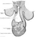
Fig. 654. Longitudinal section of the clitoris of an embryo of 60 mm head-foot length. (Embryo H. Meyer 268; slide 64, row 2, section 1.) X 50. The section cuts the sinus urogenitalis lengthwise from the crista urethralis int. to the ostium urogenitale. The lumen of the sinus is enlarged at its end to a triangular basin, on whose distal wall is the urethral plate. To the right and left are the genital swellings, the anlagen of the labia majora, which are becoming separated from the clitoris.
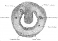
Fig. 655. Transverse section of the urogenital sinus of a male embryo of 70 mm head-foot length. (Embryo R. Meyer 267; slide 60, row 2, section 3.) X47. The section shows Mailer's tubercle projecting into the lumen of the sinus and around the sinus the anlagen of the prostatic glands.
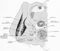
Fig. 657. Part of a transverse section through a human embryo of 60 mm. head-foot length. (Embryo R. Meyer 264; slide 44, row 2, section 2.) By the union of the mesonephric fold and testis to the anterior abdominal wall, formed by the chorda gubernaculi, both have been carried quite away from the posterior abdominal wall. Between the urogenital fold and the posterior wall a layer of loose mesenchyme has formed. Thereby the portion of the abdominal cavity which contains the urogenital fold is separated off from the rest of the cavity as the saccus vaginalis. The separation between the two is completed by a peritoneal fold formed by the a. umbilicalis.


Meyer References
Meyer R. : Ueber die fetale Uterusschleimhaut, Zeit. f . Geburtsh. u. Gynak., vol. 38, 1898. Meyer, R. : Zur Entstehung des doppelten Uterus, Zeit. f . Geburtsh. u. Gynak., vol. 38, 1898b.
Meyer R. : Ueber epitheliale Gebilde im Myometrium des fetalen und kindlichen Uterus einsehl. des Gartner'schen Ganges, Berlin, 1899.
Meyer R. : Ueber sogenannte Vornierenreste und das nephrogene Zwischenblastem bei mensehlichen Embryonen und ihre eventuelle pathologische Persistenz, Charite-Annalen, vol. 33, p. 649-656, with 2 text-figs., 1909.
Meyer R. : Zur Kenntnis des Gartner'schen Ganges besonders in der Vagina und dem Hymen des Menschen, Arch. f. mikr. Anat., vol. 73, 1909.
Meyer R. : Zur Entwicklungsgeschichte und Anatomie des utrieulus prostaticus beim Menschen, Arch. f. mikr. Anat. u. Entw., vol. 74, 1909b.
Meyer R. : Ueber erosio portionis uteri, Deut. patholog. Gesellsch. Erlangen, 1910.
Meyer R. : Die Erosion und Pseudoerosion des Erwachsenen, Arch. f. Gynak. vol. 91, part. 3, p. 1-35 with 1 plate and 3 text-figs., 1910.
Meyer R. : Die Epithelentwicklung der cervix und portio vaginalis und die pseudoerosio congenita (Kongenital. histol. Ectropium), Arch. f. Gynak., vol. 90, part 3, p. 1-20 with 1 plate, 1910.
Meyer R. : Beitrag zur Frage nach der Genese der im Urogenitalgebiet vorkommenden Mischgeschwiilste und Teratome, Reprint from the Charite Annalen, vol. 34, p. 1-23 with 10 text-figs., 1910.
References
Dallenbach-Hellweg G & Schmidt D. (2001). History of gynecological pathology. X. Dr. Robert Meyer. Int. J. Gynecol. Pathol. , 20, 289-308. PMID: 11444206
MEYER R. (1945). [Not Available]. Z Immun exp ther , 105, 349-59. PMID: 21007107
Autobiography of Dr. Robert Meyer (1864-1947).
Cite this page: Hill, M.A. (2026, April 18) Embryology Embryology History - Robert Meyer. Retrieved from https://embryology.med.unsw.edu.au/embryology/index.php/Embryology_History_-_Robert_Meyer
- © Dr Mark Hill 2026, UNSW Embryology ISBN: 978 0 7334 2609 4 - UNSW CRICOS Provider Code No. 00098G