BGDA Practical 7 - Week 6
Introduction
Key Events of Human Development during the sixth week (week 6) following fertilization or clinical GA week 8.
There are 2 Carnegie stages that show external embryo development during this week.


| Week 6 Embryo - Stage 16 Movies | |||||||||||||
|---|---|---|---|---|---|---|---|---|---|---|---|---|---|
|
<html5media height="620" width="600">File:Stage 16 MRI 3D01.mp4</html5media> |
These Week 6 movies shows the appearance of human embryo at Carnegie stage 16 (GA week 8).
Embryo Surface - Carnegie Stage 16 This movie shows an unlabeled MRI 3D volume embryo scan of the Kyoto embryo (stage 16, week 6). Compare this with the earlier stage 13 week 4 embryo. Note:
| ||||||||||||
| <html5media height="620" width="600">File:Stage 16 MRI 3D03.mp4</html5media> | Neural Development
This labelled version shows the Stage 16 embryo rotating with the CNS spinal cord and the secondary brain vesicles and flexures labeled. Note:
| ||||||||||||
| <html5media height="700" width="600">File:Stage16 MRI S01.mp4</html5media> | Sagittal Sections
This movie shows an unlabeled MRI sagittal scan passing from right to left through the Kyoto embryo (stage 16, week 6). Compare this with the earlier week 4 stage 13 embryo CNS. Note:
| ||||||||||||
|
| ||||||||||||
Limb Development

- By week 6 the upper limb bud has grown to the point where the primitive hand appears as a "paddle-like" structure at the end of an externally visible undifferentiated limb bud.
- Within this primitive hand the beginning of the fingers can be seen as "digital rays". Cell between the digits will die by a process called apoptosis (programmed cell death).
- The lower limb bud appears less developed at each embryo stage. Features that appear on the upper limb appear on the lower limb about 2 days later.
- Upper limb bud nerves (median nerve, radial nerve and ulnar nerve) entered into hand plate.
- Myoblasts spindle shaped and oriented parallel to limb bud axis.
- See additional information Mouse limb development
Note - Overall limb development will be covered in the week 8 section.
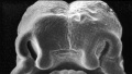
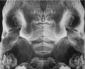
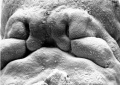
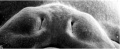
Face Development
- During week 6 there is fusion of the upper lip.
- Formed by the maxillary prominences of of the first pharyngeal arch and the frontonasal prominence.
- Failure of this embryonic process leads to cleft lip, this will be covered in more detail in BGDB.
Week 6 (Stage 16)
Week 6 (Stage 17)
Week 7 (Stage 18)
Week 7 (Stage 19)


Cardiovascular Development
Outflow Tract SeptationThe fused endocardial cushions are shown and the animation shows the growth of the spiral membranous septa that divide the single outflow tract into aortic and pulmonary outflows from the heart. Note the spiral nature of this septation process "swaps" the left/right ventricle to vessels anatomical relationship. Abnormalities in this process contribute to ventricular septal defects. |
|
|
Endocrine Development
These organs mainly develop in the embryonic period and function in the fetal period. (Covered in next practical)
- Thyroid - develops from thyroid median endodermal thickening in the floor of pharynx outpouch – thyroid diverticulum (day 24), functions from week 10
- Pituitary - develops from surface (anterior) and neural tube (posterior) ectoderm, connecting stalk between pouch and oral cavity degenerates
- Parathyroid - develop from endoderm of pharyngeal pouches, diverticulum elongate, hollow then solid, dorsal cell proliferation
- Thymus - develops from 2 origins for lymphoid thymocytes and thymic epithelial cells, diverticulum elongate, hollow then solid, ventral cell proliferation
- Adrenal - develops from 2 origins adrenal medulla (neural crest) and fetal cortex (surrounding mesenchyme), fetal cortex forms from mesothelium adjacent to dorsal mesentery, medulla neural crest cells from adjacent sympathetic ganglia
- Gonad - develops from 2 origins, germ cells migrate into the genital ridge (mesothelium and underlying mesenchyme)
- Links: Endocrine System Development
Sex Determination
At an earlier stage during week 4, the primordial germ cells began their migration into the region where the gonads will eventually form, the genital ridge, see this migration in the mouse.

Genital Ridge (Week 4)
- Humans (week 5-6), these germ cells do not determine sex, it is the support cells regulated by the presence of an X or Y chromosome.
- germ cells migrate into gonadal ridge
- Gonads (male - testis, female - ovary) identical at this stage, termed "indifferent"
Note - Sex determination and development will be covered in detail in BGDB.
Week 6 Interactive Component
| Attempt the Quiz - Week 6 | |
|---|---|

Here are a few simple Quiz questions that relate to Week 6 (GA week 8) from the lecture and practical. See your Quiz Result - Answer all the questions, then click "submit" to complete. The page will reload and you can then reopen this table to see your result and feedback.
|
Additional Information
| Additional Information - Content shown under this heading is not part of the material covered in this class. It is provided for those students who would like to know about some concepts or current research in topics related to the current class page. |
Timeline
| Week 6 - Human Embryo Stages and Events (GA week 8) | ||
|---|---|---|
| Embryo Week: Week 1 | Week 2 | Week 3 | Week 4 | Week 5 | Week 6 | Week 7 | Week 8 | Week 9 | ||
| Event | ||
| Pituitary - Week 6 connecting stalk between pouch and oral cavity degenerates
Parathyroid - Week 6 diverticulum elongate, hollow then solid, dorsal cell proliferation Thymus - Week 6 diverticulum elongate, hollow then solid, ventral cell proliferation Adrenal - Week 6 fetal cortex forms from mesothelium adjacent to dorsal mesentery, medulla neural crest cells from adjacent sympathetic ganglia Respire - Week 6 - descent of heart and lungs into thorax. Pleuroperitoneal foramen closes Tongue Week 6 - gustatory papilla, caudal midline near the foramen caecum (week 6 to 7 - nerve fibers approach the lingual epithelium) | ||
| Stage 16 | 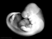 Limbs upper limb bud nerves median nerve, radial nerve and ulnar nerve entered into hand plate, myoblasts spindle shaped and oriented parallel to limb bud axis. Heart - outflow tract elliptical configuration with four cushions, the two larger fusing at this stage. Semilunar valve leaflets form at the downstream end of the cushions Head lip and palate components of the upper lip, medial nasal prominence and maxillary process present, median palatine process appears. | |
| Stage 17 | 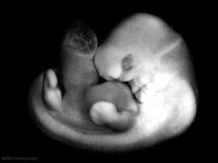 Neural - telencephalon areas of the future archicortex, paleocortex, and neocortex, visible. Beginning of future choroid plexus[2] Sense - Smell olfactory nerve fibres enter the brain[3] Neural - primordium of the epidural space appears first on the ventral part of the vertebral canal and develops rostro-caudally[4] Pancreas - rotation of the developing stomach moves ventral bud dorsally and fuses with the dorsal bud. | |
| Heart - separation of common cardiac outflow (aortic arch and pulmonary aorta) | ||
| Note - the day timing of stages is only approximate, system names link to first page of that specific system, and events are based upon the literature cited below. | ||
| References | ||
Neural Development

|
Summary of the cranial nerves. | ||||||||||||||||||||||||||||||||||||||||||||||||||||||||||||||||||||||
| Right lateral view of the central nervous system of embryo at Carnegie stage 16. Scale bar is 1 mm | |||||||||||||||||||||||||||||||||||||||||||||||||||||||||||||||||||||||
Mouse Limb Development

Mouse Limb Tissue Development[1]
| Carnegie | Stage | |||||||||||||||||||||||
| Human | Days | 1 | 2-3 | 4-5 | 5-6 | 7-12 | 13-15 | 15-17 | 17-19 | 20 | 22 | 24 | 28 | 30 | 33 | 36 | 40 | 42 | 44 | 48 | 52 | 54 | 55 | 58 |
| Mouse | Days | 1 | 2 | 3 | E4.5 | E5.0 | E6.0 | E7.0 | E8.0 | E9.0 | E9.5 | E10 | E10.5 | E11 | E11.5 | E12 | E12.5 | E13 | E13.5 | E14 | E14.5 | E15 | E15.5 | E16 |
| Rat | Days | 1 | 3.5 | 4-5 | 5 | 6 | 7.5 | 8.5 | 9 | 10.5 | 11 | 11.5 | 12 | 12.5 | 13 | 13.5 | 14 | 14.5 | 15 | 15.5 | 16 | 16.5 | 17 | 17.5 |
| Note these Carnegie stages are only approximate day timings for average of embryos. Links: Carnegie Stage Comparison | ||||||||||||||||||||||||
| ||||||||||||||||||||||||
Endocrine Development
BGDA: Lecture 1 | Lecture 2 | Practical 3 | Practical 6 | Practical 12 | Lecture Neural | Practical 14 | Histology Support - Female | Male | Tutorial
Glossary Links
- Glossary: A | B | C | D | E | F | G | H | I | J | K | L | M | N | O | P | Q | R | S | T | U | V | W | X | Y | Z | Numbers | Symbols | Term Link
Cite this page: Hill, M.A. (2026, July 6) Embryology BGDA Practical 7 - Week 6. Retrieved from https://embryology.med.unsw.edu.au/embryology/index.php/BGDA_Practical_7_-_Week_6
- © Dr Mark Hill 2026, UNSW Embryology ISBN: 978 0 7334 2609 4 - UNSW CRICOS Provider Code No. 00098G