Category:Mouse E12.5: Difference between revisions
m (→Events) |
m (→Events) |
||
| Line 12: | Line 12: | ||
* {{hearing}} {{inner ear}} - organ of Corti progenitor cells exit the cell cycle between {{ME12.5}} and {{ME14.5}} before the initiation of sensory receptor cell differentiation.{{#pmid:5861504|PMID5861504}} | * {{hearing}} {{inner ear}} - organ of Corti progenitor cells exit the cell cycle between {{ME12.5}} and {{ME14.5}} before the initiation of sensory receptor cell differentiation.{{#pmid:5861504|PMID5861504}} | ||
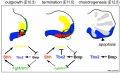
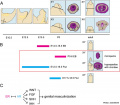
* {{tongue}} mesenchyme alters tongue shape and size, {{Hippo}} signalling and cell proliferation in a region- and stage-specific manner - Nf2cKO mutants had a decreased level of {{Hippo}} signalling transcription factor Yes-associated protein (Yap), Yap target genes and cell proliferation anteriorly, while having an increased Yap, Yap target genes and cell proliferation posteriorly, which lead to a tip-pointed and posteriorly widened tongue.{{#pmid:34697858|PMID34697858}} | * {{tongue}} mesenchyme alters tongue shape and size, {{Hippo}} signalling and cell proliferation in a region- and stage-specific manner - Nf2cKO mutants had a decreased level of {{Hippo}} signalling transcription factor Yes-associated protein (Yap), Yap target genes and cell proliferation anteriorly, while having an increased Yap, Yap target genes and cell proliferation posteriorly, which lead to a tip-pointed and posteriorly widened tongue.{{#pmid:34697858|PMID34697858}} | ||
Revision as of 13:09, 23 February 2022
The Embryology pages and media listed below relate to mouse embryonic day E12.5 of development. This staging by "days" relate to in the female presence of a vaginal plug indicating that the mating occurred, see timed pregnancy.
- Mouse Stages: E1 | E2.5 | E3.0 | E3.5 | E4.5 | E5.0 | E5.5 | E6.0 | E7.0 | E7.5 | E8.0 | E8.5 | E9.0 | E9.5 | E10 | E10.5 | E11 | E11.5 | E12 | E12.5 | E13 | E13.5 | E14 | E14.5 | E15 | E15.5 | E16 | E16.5 | E17 | E17.5 | E18 | E18.5 | E19 | E20 | Timeline | About timed pregnancy
| Carnegie | Stage | |||||||||||||||||||||||
| Human | Days | 1 | 2-3 | 4-5 | 5-6 | 7-12 | 13-15 | 15-17 | 17-19 | 20 | 22 | 24 | 28 | 30 | 33 | 36 | 40 | 42 | 44 | 48 | 52 | 54 | 55 | 58 |
| Mouse | Days | 1 | 2 | 3 | E4.5 | E5.0 | E6.0 | E7.0 | E8.0 | E9.0 | E9.5 | E10 | E10.5 | E11 | E11.5 | E12 | E12.5 | E13 | E13.5 | E14 | E14.5 | E15 | E15.5 | E16 |
| Rat | Days | 1 | 3.5 | 4-5 | 5 | 6 | 7.5 | 8.5 | 9 | 10.5 | 11 | 11.5 | 12 | 12.5 | 13 | 13.5 | 14 | 14.5 | 15 | 15.5 | 16 | 16.5 | 17 | 17.5 |
| Note these Carnegie stages are only approximate day timings for average of embryos. Links: Carnegie Stage Comparison | ||||||||||||||||||||||||
| ||||||||||||||||||||||||
| Timeline Links: human timeline | mouse timeline | mouse detailed timeline | chicken timeline | rat timeline | Medaka | Category:Timeline |
| Mouse Links: Introduction | Mouse Stages | Mouse Timeline | Mouse Timeline Detailed | Mouse Estrous Cycle | Mouse Heart | Mouse Knockout | Movie - Cephalic Plexus | Movie - Blastocyst Cdx2 | ANAT2341 Project 2009 | Category:Mouse | |||||||||||||||||||||||||||||||||||||||||||||||||||||||||||||||||||||||||||||||||||||||||||||||||||||||||||||||||||||||||||
|
| ||||||||||||||||||||||||||||||||||||||||||||||||||||||||||||||||||||||||||||||||||||||||||||||||||||||||||||||||||||||||||
Events
- Musculoskeletal System - Abdominal wall second body wall region containing myoblasts thicker (780 μm) than the primary body wall (180 μm). Now distinct inner and outer layers of tissue: inner layer shows unidirectional cell orientation; outer layer has no orientation.[1]
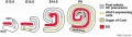
- palate - secondary palatal development begins. E12.5 to E14 grow vertically along the developing tongue.
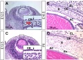
- Salivary Gland Development - indentations (clefts) start to form on the surface of the epithelial bud accompanied by alterations in the basement membrane. Clefts separate the primary bud into multiple buds and the epithelium proliferates. The base of the cleft becomes the primordial ductal structure, salivary branchi}}ng morphogenesis is repeated multiple times over the following days.[2]
- blood - Haematopoietic stem cells (HSCs) appear in the fetal liver.[3]
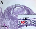
- hearing inner ear - organ of Corti progenitor cells exit the cell cycle between E12.5 and E14.5 before the initiation of sensory receptor cell differentiation.[4]
- tongue mesenchyme alters tongue shape and size, Hippo signalling and cell proliferation in a region- and stage-specific manner - Nf2cKO mutants had a decreased level of Hippo signalling transcription factor Yes-associated protein (Yap), Yap target genes and cell proliferation anteriorly, while having an increased Yap, Yap target genes and cell proliferation posteriorly, which lead to a tip-pointed and posteriorly widened tongue.[5]
References
- ↑ Nichol PF, Corliss RF, Yamada S, Shiota K & Saijoh Y. (2012). Muscle patterning in mouse and human abdominal wall development and omphalocele specimens of humans. Anat Rec (Hoboken) , 295, 2129-40. PMID: 22976993 DOI.
- ↑ Larsen M, Yamada KM & Musselmann K. (2010). Systems analysis of salivary gland development and disease. Wiley Interdiscip Rev Syst Biol Med , 2, 670-82. PMID: 20890964 DOI.
- ↑ Rybtsov S, Ivanovs A, Zhao S & Medvinsky A. (2016). Concealed expansion of immature precursors underpins acute burst of adult HSC activity in foetal liver. Development , 143, 1284-9. PMID: 27095492 DOI.
- ↑ Kikuchi K & Hilding D. (1965). The development of the organ of Corti in the mouse. Acta Otolaryngol. , 60, 207-22. PMID: 5861504 DOI.
- ↑ Ishan M, Chen G, Yu W, Wang Z, Giovannini M, Cao X & Liu HX. (2021). Deletion of Nf2 in neural crest-derived tongue mesenchyme alters tongue shape and size, Hippo signalling and cell proliferation in a region- and stage-specific manner. Cell Prolif , 54, e13144. PMID: 34697858 DOI.
Search Pubmed: Mouse E12.5
Pages in category 'Mouse E12.5'
The following 4 pages are in this category, out of 4 total.
Media in category 'Mouse E12.5'
The following 62 files are in this category, out of 62 total.
 Anderson2016-fig24b.jpg 800 × 800; 109 KB
 Anderson2016-fig25b.jpg 800 × 800; 83 KB
 Anderson2016-fig31.jpg 800 × 800; 99 KB
 Anderson2016-fig32a.jpg 800 × 800; 108 KB
 Anderson2016-fig32b.jpg 800 × 800; 89 KB
 Anderson2016-fig46a.jpg 800 × 800; 100 KB
 Anderson2016-fig50a.jpg 668 × 783; 139 KB
 Anderson2016-fig50b.jpg 798 × 786; 170 KB
 Hindlimb Tbx2 model.jpg 1,000 × 607; 84 KB
 Hoxa gene expression in limb bud 01.jpg 1,376 × 533; 85 KB
 Hoxa gene expression in limb bud 02.jpg 546 × 600; 40 KB
 Limb patterning factors 05.jpg 800 × 794; 37 KB
 Limb patterning factors 06.jpg 800 × 794; 41 KB
 Limb patterning factors 07.jpg 800 × 794; 44 KB
 Lung Fgf10 expression cartoon.jpg 1,280 × 821; 96 KB
 Mouse - forebrain Robo3 expression.jpg 675 × 1,280; 289 KB
 Mouse - palate MMP-25 expression.jpg 1,000 × 818; 243 KB
 Mouse bladder development E12.5-E16.5.jpg 1,105 × 1,000; 202 KB
 Mouse Bmp4 expression face 01.jpg 1,200 × 322; 58 KB
 Mouse Bmp4 expression limb and face 01.jpg 1,200 × 513; 91 KB
 Mouse cochlea development cartoon.jpg 1,000 × 280; 53 KB
 Mouse cornea development 01.jpg 1,200 × 880; 325 KB
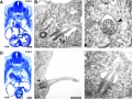
 Mouse cornea E12.5.jpg 700 × 558; 123 KB
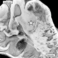
 Mouse CT E12.5 scalebar.jpg 221 × 344; 4 KB
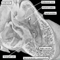
 Mouse CT E12.5.jpg 602 × 800; 62 KB
 Mouse E12.5 gene expression.jpg 1,823 × 1,100; 272 KB
 Mouse E12.5 Myog Expression.jpg 397 × 397; 17 KB
 Mouse E12.5 Sox9 Expression.jpg 397 × 397; 26 KB
 Mouse external genital development.jpg 800 × 701; 75 KB
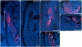
 Mouse eye neural crest.jpg 1,086 × 1,509; 483 KB
 Mouse face Bmp4.mp4 ; 480 KB
 Mouse gonad Gcnf expression 01.jpg 1,947 × 843; 304 KB
 Mouse gonad Gcnf expression E12.5.jpg 331 × 785; 68 KB
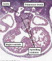
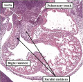
 Mouse heart primary cilia 01.jpg 1,200 × 908; 415 KB
 Mouse HOXA5 expression E12.5.jpg 1,000 × 577; 192 KB
 Mouse limb bone development timeline.jpg 1,256 × 469; 107 KB
 Mouse limb skeleton cartoon.jpg 1,000 × 487; 64 KB
 Mouse limb tissue development.jpg 1,280 × 767; 161 KB
 Mouse Lmx1b gene expression.jpg 1,624 × 550; 104 KB
 Mouse mammary development 01.jpg 1,200 × 773; 147 KB
 Mouse melanoblast distribution 01.jpg 697 × 1,000; 192 KB
 Mouse melanoblast distribution 02.jpg 777 × 1,121; 129 KB
 Mouse model of ovarian cord formation 01.jpg 800 × 491; 85 KB
 Mouse model of ovarian cord formation.jpg 800 × 491; 85 KB
 Mouse pancreas development.jpg 600 × 939; 261 KB
 Mouse REN expression 01.jpg 904 × 1,000; 293 KB
- Mouse tooth movie 02.mp4 ; 17.91 MB
 Mouse ventral body wall development 01.jpg 1,200 × 635; 130 KB
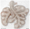 Mouse whole lung E12.5.jpg 600 × 594; 17 KB
 Mouse- facial branchiomotor neuron migration.jpg 600 × 841; 104 KB
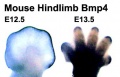 Mouse- hindlimb Bmp4 expression.jpg 374 × 241; 19 KB
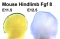 Mouse- hindlimb Fgf8 expression.jpg 374 × 243; 17 KB
 Mouse- intervertebral disc development 02.jpg 726 × 728; 36 KB
 Mouse- intervertebral disc development.jpg 1,454 × 728; 95 KB
 Mouse- placenta Hox13 expression.jpg 1,000 × 1,070; 150 KB
 Mouse- X-linked gene expression in primordial germ cells.jpg 800 × 632; 90 KB
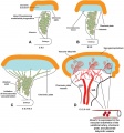
 Mouse-coronary vessel formation.jpg 800 × 282; 44 KB
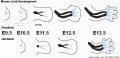
 Mouse-E12.5.png 496 × 600; 278 KB
 Mouse-E9.5 E10.5 E11.5 E12.5.png 599 × 407; 170 KB
 Mouse-kidney in vitro.jpg 955 × 461; 57 KB
 Mouse-pituitary Sox4 expression.jpg 596 × 448; 55 KB
 Telencephalon development signals.jpg 1,000 × 711; 62 KB


