Category:Human Embryo
From Embryology
This Embryology category shows pages and media related to human embryonic development occurring in the first 8 weeks of human development.
Subcategories
This category has the following 72 subcategories, out of 72 total.
C
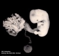
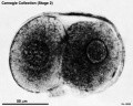
- Carnegie Collection
- Carnegie Embryo 1267
- Carnegie Embryo 1324
- Carnegie Embryo 1332
- Carnegie Embryo 1534
- Carnegie Embryo 175
- Carnegie Embryo 190
- Carnegie Embryo 199
- Carnegie Embryo 2114
- Carnegie Embryo 219
- Carnegie Embryo 2256
- Carnegie Embryo 293
- Carnegie Embryo 368
- Carnegie Embryo 390
- Carnegie Embryo 409
- Carnegie Embryo 423
- Carnegie Embryo 424
- Carnegie Embryo 426
- Carnegie Embryo 431
- Carnegie Embryo 4361
- Carnegie Embryo 437
- Carnegie Embryo 452
- Carnegie Embryo 484
- Carnegie Embryo 490
- Carnegie Embryo 491
- Carnegie Embryo 508
- Carnegie Embryo 509
- Carnegie Embryo 511
- Carnegie Embryo 576
- Carnegie Embryo 6150
- Carnegie Embryo 626
- Carnegie Embryo 657
- Carnegie Embryo 709
- Carnegie Embryo 7900
- Carnegie Embryo 84
- Carnegie Embryo 8913
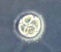
- Carnegie Stage 1
- Carnegie Stage 10
- Carnegie Stage 11
- Carnegie Stage 12
- Carnegie Stage 13
- Carnegie Stage 15
- Carnegie Stage 16
- Carnegie Stage 17
- Carnegie Stage 18
- Carnegie Stage 19
- Carnegie Stage 20
- Carnegie Stage 21
- Carnegie Stage 22
- Carnegie Stage 23
- Carnegie Stage 3
- Carnegie Stage 6
- Carnegie Stage 7
- Carnegie Stage 8
- Carnegie Stage 9
E
Pages in category 'Human Embryo'
The following 200 pages are in this category, out of 261 total.
(previous page) (next page)A
- Template:Abdominal Wall Muscle Timeline table
- Abnormal Development - Biological Toxins
- Abnormal Development - Chemicals
- Abnormal Development - Drugs
- Abnormal Development - Herbal Drugs
- Abnormal Development - Illegal Drugs
- Abnormal Development - Thalidomide
- Alpha-Fetoprotein
- Amniocentesis
- Assisted Reproductive Technology
- Assisted Reproductive Technology - Glossary
- Australian Drug Categories
- Australian In Vitro Fertilization and Donor Questions
B
C
- Carnegie stage 1
- Carnegie stage 10
- Carnegie stage 10 gallery
- Carnegie stage 11
- Carnegie stage 12
- Carnegie stage 13
- Carnegie stage 14
- Carnegie stage 15
- Carnegie stage 16
- Carnegie stage 17
- Carnegie Stage 17 Movie
- Carnegie stage 18
- Carnegie stage 19
- Carnegie stage 2
- Carnegie stage 20
- Carnegie stage 21
- Carnegie stage 22
- Carnegie stage 22 - selected serial sections
- Carnegie stage 22 - serial sections
- Carnegie stage 23
- Carnegie stage 3
- Carnegie stage 4
- Carnegie stage 5
- Carnegie stage 6
- Carnegie stage 7
- Carnegie stage 8
- Carnegie stage 9
- Carnegie stage 9 gallery
- Template:Carnegie stage table 1
- Template:Carnegie stages
- Carnegie Stages
- Template talk:Carnegie stages
- Chorionic villus sampling
- Comparative Genomic Hybridization
- Template:CRL
- Template:CS1
- Template:CS10
- Template:CS11
- Template:CS12
- Template:CS13
- Template:CS14
- Template:CS15
- Template:CS16
- Template:CS17
- Template:CS18
- Template:CS19
- Template:CS2
- Template:CS20
- Template:CS21
- Template:CS22
- Template:CS23
- Template:CS3
- Template:CS4
- Template:CS5
- Template:CS6
- Template:CS7
- Template:CS8
- Template:CS9
D
E
F
G
H
- Template:Harvard Collection table01
- Human Abnormal Development
- Human Chorionic Gonadotropin
- Template:Human embryo Hertwig G31 table
- Template:Human embryo Klb table
- Template:Human embryo Meyer 300 table
- Template:Human embryo Pfannenstiel III table
- Template:Human embryo Strahl 4mm table
- Template:Human Eyelid timeline table
M
O
P
- Paper - A fifteen-somite human embryo
- Paper - A human embryo of the pre-somite period from the uterine tube
- Paper - A Human Embryo of Thirteen Somites
- Paper - A Human Embryo with Seven Pairs of Somites Measuring about 2 mm in Length
- Paper - A Human Ovum at the Previllous Stage
- Paper - A previllous human embryo (1946)
- Paper - Congenital absence of ventrolateral abdominal musculature (1946)
- Paper - Description of a 4 mm human embryo (1906)
- Special:Badtitle/NS501:Paper - Description of a Human Embryo of 13-14 Mesodermic Somites
- Paper - Development of the human placenta in the first three months of gestation (1960)
- Paper - Development of the trigone of the bladder and the termination of the mesonephric ducts (1946)
- Paper - Growth allometry of the myocardium in human embryos from stages 15 to 23
- Paper - Hydatiform degeneration in an early human embryo (1946)
- Paper - Observations on the neural crest of a ten-somite human embryo (1939)
- Paper - On measuring human embryos
- Paper - On Ossification Centers in Human Embryos
- Paper - On The Age Of Human Embryos
- Paper - Principles of growth and repair in membrane bones (1946)
- Paper - Sequential innervation of the intestinal loop in the human embryo
- Paper - Simple formulae for estimating the age and size of human embryos
- Paper - Structural organization of the human cerebral cortex prior to the appearance of the cortical plate (1983)
- Paper - The Anatomy of a 17.8 mm Human Embryo
- Paper - The Anatomy of the Head End of a 20 mm Human Embryo
- Paper - The chondrocranium of a 20 mm human embryo
- Paper - The development of the human brain stage 12
- Paper - The development of the islands of Langerhans in the human embryo (1903)
- Paper - The Development of the Nose and of the Pharynx and its Derivatives in Man
- Paper - The development of the subcutaneous vascular plexus in the head of the human embryo (1923)
- Paper - The early development of the eye in staged human embryos
- Paper - The early development of the otic vesicle in staged human embryos
- Paper - The first appearance of the neural tube and optic primordium in the human embryo at stage 10
- Paper - The human brain at stages 18-20 including the choroid plexuses and the amygdaloid and septal nuclei (1990)
- Paper - The Nuclei of Origin of the Cranial Nerves in the 10 mm Human Embryo
- Paper - The Origin of the Otic and Optic Primordia in Man
- Paper - The timing and sequence of events in the development of the human endocrine system during the embryonic period proper
- Paper The development of the subcutaneous vascular plexus in the head of the human embryo (1923)
- Template:Pearce1903 table1
- Preimplantation Genetic Diagnosis
- Preimplantation Genetic Screening
- Prenatal Diagnosis
- Prenatal Genetic Diagnosis
R
- Template:Ref-Arey1925
- Template:Ref-Atwell1926b
- Template:Ref-Bartelmez1924
- Template:Ref-Bossy1981
- Template:Ref-Bremer1906
- Template:Ref-Chidester1908
- Template:Ref-FlorianHill1935
- Template:Ref-Hamilton1946b
- Template:Ref-Hamilton1946c
- Template:Ref-HamiltonBoyd1960
- Template:Ref-Ingalls1920
- Template:Ref-Ingalls1929
- Template:Ref-Johnson1914
- Template:Ref-Jordan1909a
Media in category 'Human Embryo'
The following 133 files are in this category, out of 738 total.
(previous page) (next page) Stage10 K12202-01.jpg 1,307 × 1,307; 240 KB
 Stage10 K12202-02.jpg 1,307 × 1,307; 242 KB
 Stage10 K12202-03.jpg 1,434 × 1,105; 191 KB
 Stage10 K12202-04.jpg 1,434 × 1,105; 227 KB
 Stage10 K12202-05.jpg 1,307 × 1,307; 278 KB
 Stage10 K12202-06.jpg 1,307 × 1,307; 299 KB
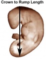 Stage13 crown rump length.jpg 288 × 371; 10 KB
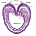
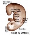 Stage13 surface bulges.jpg 337 × 389; 14 KB
 Stage14 human scale.jpg 646 × 530; 40 KB
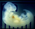
 Stage14 human.jpg 646 × 530; 32 KB
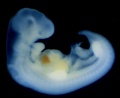
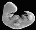 Stage14 SEM.jpg 646 × 530; 36 KB
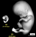 Stage14compare23.jpg 593 × 601; 32 KB
 Stage16 bf10.jpg 1,778 × 2,000; 319 KB
 Stage16 bf9.jpg 1,677 × 2,000; 319 KB
 Stage18 bf14.jpg 1,518 × 2,000; 311 KB
 Stage19 bf16.jpg 1,041 × 1,000; 99 KB
 Stage19 bf17.jpg 1,041 × 1,000; 68 KB
 Stage2 bf01.jpg 650 × 521; 49 KB
 Stage2.jpg 216 × 185; 4 KB
 Stage22 HPA1L.jpg 640 × 433; 65 KB
 Stage22 HPA2L.jpg 603 × 399; 82 KB
 Stage3 bf01.jpg 507 × 625; 39 KB
 Stage3 bf02.jpg 507 × 625; 31 KB
 Stage7 axes.jpg 500 × 375; 9 KB
 Stage7 axial process.jpg 500 × 375; 11 KB
 Stage7 bf5.jpg 1,712 × 1,206; 875 KB
 Stage7 bf51.jpg 600 × 450; 96 KB
 Stage7 bf52.jpg 600 × 450; 114 KB
 Stage7 bf53.jpg 500 × 375; 74 KB
 Stage7 bf5a.jpg 1,024 × 721; 690 KB
 Stage7 bf5b.jpg 500 × 352; 216 KB
 Stage7 bf6.jpg 347 × 599; 69 KB
 Stage7 bf7.jpg 400 × 341; 22 KB
 Stage7 bf8.jpg 400 × 341; 18 KB
 Stage7 bf9.jpg 1,200 × 1,039; 501 KB
 Stage7 features.jpg 500 × 375; 9 KB
 Stage7 folding.jpg 500 × 375; 10 KB
 Stage7 primitive streak labelled.jpg 500 × 375; 13 KB
 Stage7 SEM1.jpg 450 × 333; 40 KB
 Stage7 SEM2.jpg 450 × 300; 27 KB
 Stage7 SEM4.jpg 450 × 332; 60 KB
 Stage7-bf1.jpg 600 × 676; 34 KB
 Stage7-bf2.jpg 800 × 579; 53 KB
 Stage7-bf3.jpg 523 × 600; 45 KB
 Stage7-bf4.jpg 800 × 695; 53 KB
 Stage7-sem1.jpg 814 × 600; 100 KB
 Stage7-sem2.jpg 590 × 800; 98 KB
 Stage7-sem3.jpg 1,000 × 664; 89 KB
 Stage7-sem4.jpg 937 × 595; 79 KB
 Stage7-sem5.jpg 1,000 × 735; 202 KB
 Stage7.jpg 312 × 427; 7 KB
 Stage8 bf4.jpg 600 × 449; 17 KB
 Stage8 human.jpg 276 × 450; 14 KB
 Stage8 nodal cilia.jpg 450 × 321; 50 KB
 Stage9 dorsal.jpg 287 × 378; 5 KB
 Stage9 ventral.jpg 291 × 384; 5 KB
 Stage9sm.jpg 287 × 378; 6 KB
 Statistics-Utah heart defects.jpg 580 × 350; 35 KB
 Streeter1906 fig01.jpg 1,193 × 1,318; 167 KB
 Streeter1906 fig02.jpg 1,527 × 2,059; 244 KB
 Sudler1902-fig05.jpg 1,000 × 769; 182 KB
 Sudler1902-fig06.jpg 600 × 865; 146 KB
 Sudler1902-fig07.jpg 1,000 × 1,089; 148 KB
 Thyng1914 fig01.jpg 786 × 1,000; 140 KB
 Thyng1914 fig02.jpg 1,189 × 800; 112 KB
 Thyng1914 plate01.jpg 1,147 × 1,500; 370 KB
 Thyng1914 plate1a.jpg 1,148 × 1,500; 402 KB
 Thyng1914 plate1b.jpg 1,147 × 1,500; 0 bytes
 Thyng1914 plate2a.jpg 1,157 × 1,500; 338 KB
 Thyng1914 plate2b.jpg 1,157 × 1,500; 275 KB
 Thyng1914 plate3a.jpg 1,911 × 1,500; 389 KB
 Thyng1914 plate3b.jpg 1,911 × 1,500; 489 KB
 Thyng1914 plate4a.jpg 1,142 × 1,500; 315 KB
 Thyng1914 plate4b.jpg 1,142 × 1,500; 336 KB
 Thyng1914 plate5a.jpg 1,613 × 1,500; 257 KB
 Thyng1914 plate5b.jpg 1,613 × 1,500; 429 KB
 Thyng1914 plate6.jpg 2,000 × 1,098; 422 KB
 Trilaminar embryo cartoon.jpg 1,029 × 1,000; 107 KB
 Trisomy 21 newborn.jpg 320 × 240; 16 KB
 Trisomy18male.jpg 480 × 284; 10 KB
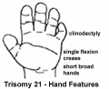 Trisomy21 hand.jpg 360 × 295; 12 KB
 Trisomy21arrow.gif 200 × 175; 5 KB
 Trisomy21female.jpg 480 × 284; 12 KB
 Trisomy21male.jpg 480 × 284; 10 KB
 Turner1920 fig01-4.jpg 1,000 × 651; 53 KB
 Turner1920 fig05-6.jpg 1,000 × 651; 35 KB
 Turner1920 fig07-8.jpg 1,000 × 651; 43 KB
 Turner1920 fig09-10.jpg 1,000 × 651; 37 KB
 Turner1920 fig11-12.jpg 1,000 × 651; 47 KB
 Turner1920 fig13-14.jpg 1,000 × 651; 41 KB
 Turner1920 fig15-16.jpg 1,000 × 651; 47 KB
 Turner1920 fig17-18.jpg 1,000 × 651; 37 KB
 Turner1920 fig19-20.jpg 1,000 × 651; 41 KB
 Turner1920 fig21-22.jpg 1,000 × 651; 36 KB
 Turner1920 fig23-24.jpg 1,000 × 651; 38 KB
 Turner1920 fig25-26.jpg 1,000 × 651; 35 KB
 Turner1920 fig27-28.jpg 1,000 × 651; 39 KB
 Turner1920 fig29-30.jpg 1,000 × 651; 33 KB
 Turner1920 fig31-32.jpg 1,000 × 651; 33 KB
 Turner1920 fig33-34.jpg 1,000 × 651; 26 KB
 Turner1920 fig35-36.jpg 1,000 × 651; 29 KB
 Turner1920 fig37-38.jpg 1,000 × 651; 26 KB
 Turner1920 fig39-40.jpg 1,000 × 651; 27 KB
 Turner1920 fig41-42.jpg 1,000 × 651; 25 KB
 Turner1920 fig43-44.jpg 1,000 × 651; 28 KB
 Turner1920 fig45-46.jpg 1,000 × 651; 26 KB
 Turner1920 fig47-48.jpg 1,000 × 651; 30 KB
 Turner1920 fig49-50.jpg 1,000 × 651; 26 KB
 Turner1920 fig51-53.jpg 1,000 × 651; 32 KB
 Turner1920 fig54-56.jpg 1,000 × 651; 30 KB
 Turner1920 fig57-59.jpg 1,000 × 651; 31 KB
 Turner1920 fig60-62.jpg 1,000 × 651; 28 KB
 Turner1920 fig63-65.jpg 1,000 × 651; 30 KB
 Turner1920 fig66-68.jpg 1,000 × 651; 30 KB
 Turner1920 fig69-72.jpg 1,000 × 651; 36 KB
 Turner1920 fig73-81.jpg 1,000 × 651; 37 KB
 Turner1920 plate01.jpg 1,000 × 651; 33 KB
 Ultrasound12wk 3D image.jpg 301 × 248; 8 KB
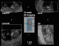 Ultrasound12wk 3D.jpg 512 × 398; 18 KB
 Wallin1913-fig01.jpg 1,339 × 1,736; 174 KB
 Wallin1913-fig02.jpg 1,191 × 1,743; 175 KB
 Wallin1913-fig03.jpg 1,189 × 1,665; 205 KB
 Wallin1913-fig04-07.jpg 1,413 × 2,231; 305 KB
 Wallin1913-fig04.jpg 1,347 × 736; 122 KB
 Wallin1913-fig05.jpg 596 × 561; 42 KB
 Wallin1913-fig06.jpg 607 × 522; 38 KB
 Wallin1913-fig07.jpg 659 × 558; 40 KB
 Waterston1915 fig01.jpg 628 × 805; 107 KB
 Waterston1915 fig02.jpg 923 × 669; 117 KB
 Waterston1915 fig03.jpg 767 × 937; 153 KB
 Waterston1915 fig04.jpg 633 × 919; 123 KB
 Waterston1915 fig05.jpg 898 × 559; 108 KB
 Williams1908-fig20.jpg 920 × 1,500; 96 KB


