Carnegie stage 14: Difference between revisions
| Line 111: | Line 111: | ||
==Hinrichsen Collection== | ==Hinrichsen Collection== | ||
{| | {| | ||
| width= | | width=510px|[[File:ME52 001.jpg|500px]] | ||
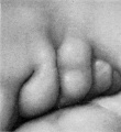
| valign=top|Hinrichsen collection Human Embryo ME52 ([[Carnegie stage 14|stage14]]) CRL 6.6 mm. | | valign=top|Hinrichsen collection Human Embryo ME52 ([[Carnegie stage 14|stage14]]) CRL 6.6 mm. | ||
Revision as of 10:37, 19 October 2016
| Embryology - 15 Jun 2024 |
|---|
| Google Translate - select your language from the list shown below (this will open a new external page) |
|
العربية | català | 中文 | 中國傳統的 | français | Deutsche | עִברִית | हिंदी | bahasa Indonesia | italiano | 日本語 | 한국어 | မြန်မာ | Pilipino | Polskie | português | ਪੰਜਾਬੀ ਦੇ | Română | русский | Español | Swahili | Svensk | ไทย | Türkçe | اردو | ייִדיש | Tiếng Việt These external translations are automated and may not be accurate. (More? About Translations) |
Introduction

Facts
Week 5, 31 - 35 days, 5 - 7 mm
Gestational Age GA - week 7
View: Lateral view. Amniotic membrane removed.
Summary
- Ectoderm: sensory placodes, lens pit, otocyst, nasal placode, primary/secondary vesicles, fourth ventricle of brain,
- Mesoderm: continued segmentation of paraxial mesoderm (more than 30 somite pairs), heart prominence
- Head: 1st, 2nd and 3rd pharyngeal arch, forebrain, site of lens placode, site of otic placode, stomodeum
- Body: heart, liver, umbilical cord, mesonephric ridge
- Limb: upper and lower limb buds
See also Carnegie stage 14 Events
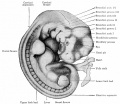
Features
- midbrain, nasal placode, lens pit, 1,2,3 pharyngeal arches, fourth ventricle of brain, 1st pharyngeal groove, heart prominence, cervical sinus, upper limb bud, mesonephric ridge, lower limb bud, umbilical cord.
- Identify: midbrain region, nasal placode, lens pit, 1st, 2nd and 3rd pharyngeal arches, 1st pharyngeal groove, maxillary and mandibular components of 1st pharyngeal arch, fourth ventricle of brain, heart prominence, cervical sinus, upper limb bud, mesonephric ridge, lower limb bud, umbilical cord.
- Links: Week 5 | Head | Lecture - Limb | Lecture - Gastrointestinal | Lecture - Head Development | Science Practical - Gastrointestinal | Science Practical - Head | Movie - Embryo stage 14 | Category:Carnegie Stage 14 | Stage 15
| Week: | 1 | 2 | 3 | 4 | 5 | 6 | 7 | 8 |
| Carnegie stage: | 1 2 3 4 | 5 6 | 7 8 9 | 10 11 12 13 | 14 15 | 16 17 | 18 19 | 20 21 22 23 |
- Carnegie Stages: 1 | 2 | 3 | 4 | 5 | 6 | 7 | 8 | 9 | 10 | 11 | 12 | 13 | 14 | 15 | 16 | 17 | 18 | 19 | 20 | 21 | 22 | 23 | About Stages | Timeline
Bright Field
| Lateral view | |
|---|---|

|

|
| Ventral view | |

|

|
Embryo Virtual Slide
|
|


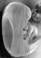
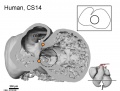
Scanning EM


Dorsolateral view
Cranial end
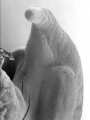
Cloacal region
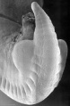
Caudal end


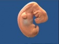
Kyoto Collection

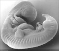
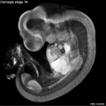
Lateral view of embryo surface at Carnegie stage 14.


|

|
| ||||||||||||||||||||||||||||||||||||||||||||||||||||||||||||||||||||||
| Lateral view of the central nervous system of embryo at Carnegie stage 14 (Scale bar is 1 mm). | ||||||||||||||||||||||||||||||||||||||||||||||||||||||||||||||||||||||||
Carnegie Collection


- Carnegie stage 14: 6830 left | 1380 left | 7333 left | 6502 left | 5654 left | 4154 left | 4629 right | 7394 right | 6848 left | 7400 right | 7394 left | 1620 left | 7400 left
| Carnegie Collection Embryos - Stage 14 | ||||||||||
|---|---|---|---|---|---|---|---|---|---|---|
| Serial No. | Size (mm) | Grade | Fixative | Embedding Medium | Plane | Thinness (µm) | Stain | Year | Notes | |
| 4 | E.,7 | Poor | p | P | Transverse | 10 | Al. coch. | 1892 | ||
| 18 | E.,7 Ch., 18x18 | Poor | p | P | Transverse | 20 | Al. coch. | 1895 | ||
| 80 | E., 5.0 Ch., 24x18x8 | Good | Alc. | P | Transverse | 20 | Al. coch. | 1897 | ||
| 187 | E.,7 Ch., 35x30x25 | Poor | ? | P | Sagittal | 20 | Al. coch. | 1902 | ||
| 1208 | E.,7 Ch., 22x11Xx1 | Poor | ? | P | Sagittal | 20 | Al. coch. | 1902 | ||
| 245 | E.,6 Ch., 13x12x10 | Poor | Formol, Zenker | ? | Transverse | 5 | (Stain - Haematoxylin Eosin) | 1904 | ||
| 372 | E.,7 | Fair | p | P | Transverse | 10 | H.-Congo red | 1902 | ||
| 380 | E,6 Ch., 20x20x14 | Poor | p | P | Sagittal | 20 | (Stain - Haematoxylin Eosin) | 1906 | ||
| 387 | E.,7 Ch., 45x40x50 | Good | Formalin | P | Transverse | 20 | (Stain - Haematoxylin Eosin) | 1907 | ||
| 442 | E.,6 Ch., 25x20 | Poor | Formalin | P | Coronal? | 50 | Al. coch. | 1908 | ||
| 552 | E.,6 Ch, 40x28Xx8 | Poor | p | P | Sagittal | 40 | Al. coch. | 1911 | Possible anencephaly | |
| 560 | E., 7.0 Ch, 24x24 | Poor | Formalin | P | Coronal | 40 | Al. coch. | 1912 | ||
| 676 | E., 6.0 Ch, 35x20x17 | Good | Carnoy | P | Tr.-Coronal | 20 | (Stain - Haematoxylin Eosin) | 1913 | Possible spina bifida | |
| 873 | E,6.0 Ch., 35x28x16 | Poor | Formalin | P | Sagittal | 20 | Al. coch. | 1914 | ||
| 988 | E,6.0 Ch., 38x30x23 | Good | Formol-corros. acetic | P | Transverse | 20 | Al. coch. | 1914 | ||
| 1380 | E , 5.7 Ch, 36x24x24 | Exc. | Formalin | P | Coronal | 20 | Al. coch. | 1916 | ||
| 1620 | E, 6.6 Ch, 35x30x8 | Good | Formalin | P | Sagittal | 20 | Al. coch. | 1916 | ||
| ?? | E, 6.68 | Fair | ? | ? | Transverse | 6 | Al. coch., or. G | 1919 | ||
| 2841 | E , sy Ch. 35x21 | Good | Alc. | P | Transverse | 20 | (Stain - Haematoxylin Eosin) or.G | 1920 | ||
| 3360 | E.6.0 | Good | Formalin | C | Transverse | 20 | (Stain - Haematoxylin Eosin) or.G | 1920 | In myomatous uterus. Advanced. | |
| 3805 | E., 5.9 | Exc. | Bouin | P | Transverse | 15 | (Stain - Haematoxylin Eosin) | 1921 | Evans embryo No. 168. Serial bromides only | |
| 3960 | E., 5.5 | Good | Formalin | C-P | Coronal | 20 | Al. coch. | 1922 | Blood vessels naturally injected | |
| 4154 | E, 6.8 Ch., 33x31x20 | Poor | Alc. | C-P | Transverse | 8 | (Stain - Haematoxylin Eosin) | 1923 | ||
| 4245-6 | E., 7.0 | Good | Formalin | P | Transverse | 15 | Al. coch. | 1923 | Univ. Pennsylvania No. 40. Ag added | |
| 4692 | E., 6.5 Ch., 32x23 | Good | Formalin | C-P | Sagittal | 10 | (Stain - Haematoxylin Eosin) | 1924 | ||
| 4672 | E,8.2 Ch., 40X34X25 | Good | Formalin | P | Transverse | 20 | Al. coch. | 1924 | Advanced | |
| 4805 | E., 7.3 Ch., 15X8X9 | Good | Formalin | C-P | Transverse | 10 | (Stain - Haematoxylin Eosin) | 1924 | Tubal | |
| 5437 | E., 7.0 | Good | Formalin | C-P | Transverse | 8 | (Stain - Haematoxylin Eosin) | 1927 | Advanced | |
| 5654 | E., 5.0 Ch., 30x23x17 | Good | Formalin | P | Transverse | 10 | Al. coch., eosin | 1928 | Less advanced | |
| 5787 | E., 6.8 Ch., 32x30x23 | Good | Formalin | P | Sagittal | 10 | Al. coch., eosin | 1928 | ||
| 6428 | E., 7.0 Ch., 30X28X25 | Good | Formalin | C-P | Coronal | 6, 10 | Al. coch. | 1931 | Advanced | |
| 6500 | E, 4.9* | Good | Souza? | C-P | Sagittal | 10 | Al. coch. | 1931 | E. Leitz, Berlin | |
| 6502 | E., 6.7* | Exc. | Souza? | C-P | Transverse | 5, 10 | (Stain - Haematoxylin Eosin) | 1931 | E. Leitz, Berlin. Ag added to slides 1-25 | |
| 6503 | E., 6.3* | Exc. | Souza? | C-P | Coronal | 10 | Al. coch. | 1931 | E. Leitz, Berlin | |
| 6739 | E.,8 | Poor | Formalin | C-P | Sagittal | 20 | (Stain - Haematoxylin Eosin) | 1933 | ||
| 6830 | E,5.5 Ch., 47x23x15 | Exc. | Formalin | C-P | Coronal | 8 | (Stain - Haematoxylin Eosin) | 1933 | ||
| 6848 | E., 7.8 | Good | Formalin | C-P | Coronal | 10 | (Stain - Haematoxylin Eosin) | 1934 | Tubal | |
| 7324 | E, 6.6 Ch., 17x13x10 | Good | Formalin | C-P | Transverse | 8 | (Stain - Haematoxylin Eosin) | 1936 | Low implantation | |
| 7333 | E, 6.3 | Good | Formalin | C-P | Transverse | 8 | (Stain - Haematoxylin Eosin) | 1936 | ||
| 7394 | E, 7.2 Ch., 45x20x20 | Exc. | Formalin | C-P | Transverse | 8 | (Stain - Haematoxylin Eosin) | 1937 | ||
| 7400 | E, 6.3 Ch., 35x25x20 | Good | Formalin | C-P | Coronal | 10 | (Stain - Haematoxylin Eosin) | 1937 | ||
| 7522 | E., 7.7 Ch., 33x16x16 | Good | Formalin | C-P | Transverse | 8 | (Stain - Haematoxylin Eosin) | 1938 | Natural blood injection | |
| 7598 | E., 7.0 Ch., 30x30x25 | Poor | Alc. | C-P | Transverse | 10 | (Stain - Haematoxylin Eosin) | 1938 | Macerated | |
| 7667 | E., 5 Ch., 16x14x12 | Fair | Formalin | P | Transverse | 8 | (Stain - Haematoxylin Eosin), phlox. | 1939 | ||
| 7829 | E., 7.0 | Exc. | Bouin | C-P | Transverse | 8 | (Stain - Haematoxylin Eosin) | 1940 | Advanced | |
| 7870 | E,7.2 Ch., 25x20x13 | Exc. | Bouin | C-P | Transverse | 8 | (Stain - Haematoxylin Eosin) | 1941 | On borderline of next stage. Ag added | |
| 8141 | E,7.3 Ch.,33x28 | Exc. | C-P | Coronal | 8 | (Stain - Haematoxylin Eosin) | 1943 | Shrinkage cracks in brain | ||
| 8306 | E.5.3 Ch., 27 | Exc. | Bouin | C-P | Transverse | 10, 20 | (Stain - Haematoxylin Eosin), phlox. | 1945 | ||
| 8308 | E, 5.85 Ch., 27x18x18 | Exc. | Formol & Bouin | C-P | Sagittal | 8 | Azan | 1945 | ||
| 8314 | E,8 Ch.,23x22 | Exc. | Formol | C-P | Transverse | 8 | Azan | 1945 | ||
| 8357 | E., 6.5 | Good | Formol | C-P | Sagittal | 8 | Azan | 1946 | ||
| 8552 | E,6.5 | Exc. | Alc. & Bouin | C-P | Transverse | 8 | Azan | 1947 | ||
| 8999 | E,6 Ch.,16x15 | Exc. | Alc. & Bouin | C-P | Sagittal | 8 | Azan | 1952 | ||
| 9695 | E,8.5 | ? | 1955 | Not cut | ||||||
Abbreviations
| ||||||||||
| iBook - Carnegie Embryos | |
|---|---|

|
|
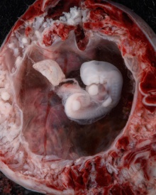
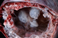
Hinrichsen Collection

|
Hinrichsen collection Human Embryo ME52 (stage14) CRL 6.6 mm.
Embryo right lateral view.
|

Image source: The Hinrichsen Collection images are reproduced with the permission of Prof. Beate Brand-Saberi, Head, Department of Anatomy and Molecular Embryology, Ruhr-Universität Bochum. Images are for educational purposes only and cannot be reproduced electronically or in writing without permission.
Stage 14 Embryo Movie
|
| Stage 14 Model |
| Page | Play |
Events
- Hearing - otic vesicle ventral portion elongates to form the cochlear duct and endolymphatic appendage becomes tapered.[1]
- Vision - (about 32 days) the lens placode is indented by the lens pit, cup-shaped and still communicates with the surface by a narrowing pore.[2]
- Cardiovascular
- Coronary circulation plexus of blind epicardial capillaries appears on the heart.[3]
- Cerebral artery - basilar artery forms from the consolidation of the neural arteries.[4]
References
- ↑ Streeter GL. Developmental horizons in human embryos. Description of age group XIII, embryos about 4 or 5 millimeters long, and age group XIV, period of indentation of the lens vesicle. (1945) Carnegie Instn. Wash. Publ. 557, Contrib. Embryol., Carnegie Inst. Wash., 31: 27-63.
- ↑ <pubmed>7364662</pubmed>
- ↑ <pubmed>3286038</pubmed>
- ↑ <pubmed>26060802</pubmed>| J Stroke.
Additional Images
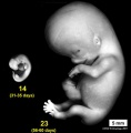
Stage 14 compare size to Stage 23
Stage 14 Optical Projection Tomography
Stage 14 heart MRI

External ear Stages 14-23 and adult
7mm embryo described by Mall
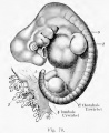
Kollmann Fig. 70
Streeter Fig. 13


- Carnegie Stages: 1 | 2 | 3 | 4 | 5 | 6 | 7 | 8 | 9 | 10 | 11 | 12 | 13 | 14 | 15 | 16 | 17 | 18 | 19 | 20 | 21 | 22 | 23 | About Stages | Timeline
Image Source: Scanning electron micrographs of the Carnegie stages of the early human embryos are reproduced with the permission of Prof Kathy Sulik, from embryos collected by Dr. Vekemans and Tania Attié-Bitach. Images are for educational purposes only and cannot be reproduced electronically or in writing without permission.
Image source: The Kyoto Collection images are reproduced with the permission of Prof. Kohei Shiota and Prof. Shigehito Yamada, Anatomy and Developmental Biology, Kyoto University Graduate School of Medicine, Kyoto, Japan for educational purposes only and cannot be reproduced electronically or in writing without permission.
Cite this page: Hill, M.A. (2024, June 15) Embryology Carnegie stage 14. Retrieved from https://embryology.med.unsw.edu.au/embryology/index.php/Carnegie_stage_14
- © Dr Mark Hill 2024, UNSW Embryology ISBN: 978 0 7334 2609 4 - UNSW CRICOS Provider Code No. 00098G