Lecture - Sensory Development
Introduction
This lecture will introduce development of the special sensory structures associated with hearing, vision, smell and taste. Due to time limitations the lecture will focus on hearing development and if time is available vision and other senses will be introduced in general.
|
|
|
|
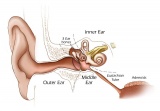
We use the sense of balance and hearing to position ourselves in space, sense our surrounding environment, and to communicate. Portions of the ear appear very early in development as specialized region (otic placode) on the embryo surface that sinks into the mesenchyme to form a vesicle (otic vesicle = otocyst) that form the inner ear.
This region connects centrally to the nervous system and peripherally through specialized bones to the external ear (auricle). This organisation develops different sources forming the 3 ear parts: inner ear (otic placode, otocyst), middle ear (1st pharyngeal pouch and 1st and 2nd arch mesenchyme), and outer ear (1st pharyngeal cleft and 6 surface hillocks).
This complex origin, organisation, and timecourse means that abnormal development of any one system can impact upon the development of hearing.
Objectives
- Understanding of sensory placode development
- Understanding of inner, middle and external ear origins
- Understanding of timecourse of auditory development
- Understanding of abnormalities of auditory development
- Brief understanding of other (vision) sensory development
Also review your Head development lecture.
Lecture Resources
| Movies and Virtual Slides | |||||||||||||||||||
|---|---|---|---|---|---|---|---|---|---|---|---|---|---|---|---|---|---|---|---|
|
|
|
|
| |||||||||||||||


| 2016 Lecture Video Recording |
|---|
| This 2016 lecture video recording is similar in content to the current 2017 online lecture.
<html5media height="600" width="800">File:2016Lecture-Hearing.mp4</html5media> Click to play new window - 2016 Lecture Video (51 MB) |
Development Timing

- Week 3 - otic placode, otic vesicle
- Week 5 - cochlear part of otic vesicle elongates (humans 2.5 turns)
- Week 9 - Mesenchyme surrounding membranous labryinth (otic capsule) chondrifies
- Week 12-16 - Capsule adjacent to membranous labryinth undegoes vacuolization to form a cavity (perilymphatic space) around membranous labrynth and fills with perilymph
- Week 16-24 - Centres of ossification appear in remaining cartilage of otic capsule form petrous portion of temporal bone. Continues to ossify to form mastoid process of temporal bone.
- 3rd Trimester - Vibration acoustically of maternal abdominal wall induces startle response in fetus.
Embryonic Origin Overview

| Outer Ear | Middle Ear | Inner Ear |
|---|---|---|
|
|
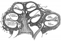
Sensory Placodes
| Week 4 | |
|---|---|

|

|
| Otic placodes (Stage 11 dorsal view) | Sensory placodes ((Stage 14 ventral view) |
- week 4 a series of thickened surface ectodermal patches form in pairs in the head region.
- Recent research suggests that all sensory placodes may arise from common panplacodal primordium origin around the neural plate, and then differentiate to eventually have different developmental fates. PMID 20801420
- sensory placodes will later contribute key components of each of our special senses (vision, hearing and smell).
- Other species have a number of additional placodes which form other sensory structures (fish, lateral line receptor).
- Note that their initial postion on the developing head is significantly different to their final position in the future sensory system.
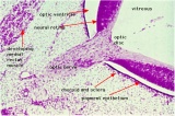
Otic Placode
- stage 13/14 embryo (shown below) the otic placode has sunk from the surface ectoderm to form a hollow epithelial ball, the otocyst, which now lies beneath the surface surrounded by mesenchyme (mesoderm).
- The epithelia of this ball varies in thickness and has begun to distort, it will eventually form the inner ear membranous labyrinth.

Lens Placode
- lies on the surface, adjacent to the outpocketing of the nervous system (which will for the retina) and will form the lens.

Nasal Placode
- 2 large components (medial and lateral), will form the nasal olefactory epithelium.
- Note the extension of the (neural tube) diencephalon forming the optic stalk underlying the optic placode.

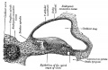
Inner Ear
Described in detail later in lecture.

Stage 13 otocyst

Stage 22 ear
- The inner ear is derived from a pair of surface sensory placodes (otic placodes) in the head region.
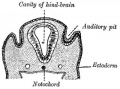
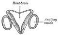
- These placodes fold inwards forming a depression, then pinch off entirely from the surface forming a fluid-filled sac or vesicle (otic vesicle, otocyst).
- The vesicle sinks into the head mesenchyme some of which closely surrounds the otocyst forming the otic capsule.
- The otocyst finally lies close to the early developing hindbrain (rhombencephalon) and the developing vestibulo-cochlear-facial ganglion complex.
- Links: Inner Ear | Neuroscience - The Inner Ear
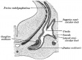
Middle Ear
- derived from first pharyngeal pouch and 1st and 2nd arch mesenchyme
- extends as tubotympanic recess - during week 5 recess contacts outer ear canal
- mesoderm between 2 canals forms tympanic membrane
- expands to form tympanic recess
- stalk of recess forms auditory tube(eustachian tube, pharyngotympanic tube)
Ossicles
|

Pharyngeal arch cartilages |


Tympanic Cavity
|

|

Cavities formed from the First Cleft |
Middle Ear Genes - gooscoid, RARs, Prx1, Otx2, Hoxa1, Hoxb1, endothelian related molecules
- Links: Middle Ear | Neuroscience - The Middle Ear
Outer Ear
- External ear is derived from 6 surface hillocks, 3 on each of pharyngeal arch 1 and 2.
- structure completed during embryonic period.
- External auditory meatus is derived from the 1st pharyngeal cleft.
- transiently blocked during development.
- The newborn external ear structure and position is an easily accessible diagnostic tool for potential abnormalities or further clinical screening.
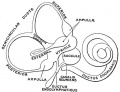
Pinna- Auricle
Develops from six aural hillocks
|

arch 1 and 2 hillocks |

| Pharyngeal Arch | Hillock | Auricle Component |
| Arch 1 | 1 | tragus |
| 2 | helix | |
| 3 | cymba concha | |
| Arch 2 | 4 | concha |
| 5 | antihelix | |
| 6 | antitragus |

External ear stages 14-23 and adult (not to scale)
External auditory meatus
- derived from first pharyngeal cleft
- ectodermal diverticulum
- week 5 - extends inwards to pharynx
- until week 18 has ectodermal plug - plug forms stratified squamous epithelia of canal and outer eardrum
Timeline
(EAM data - Nishimura, 1992 PMID 1441991) |

outer ear and external auditory meatus |
Links: Outer Ear | Neuroscience - The External Ear
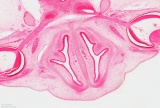
Inner


Otocyst


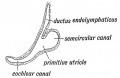
beginning of cochlea and semicircular canals
Week 8 cochlea


- week 3 otic placode forms on surface ectoderm
- otic placode sinks into mesoderm
- forms otocyst (otic vesicle)
- branches form and generate endolymphatic duct and sac
- forms vestibular (dorsal) and cochlear (ventral) regions
- differentiation of otic vesicle to membranous labyrinth
Vestibular Sac
- generates 3 expansions - form semicircular ducts
- remainder forms utricle
- epithelia lining generates - hair cells, ampullary cristae, utricular macula
- Vestibular - Otoconia, otoconin- inner ear biominerals
Cochlear sac
|

|

Bony Labyrinth
- formed from chrondified mesoderm
- Periotic Capsule
- mesenchyme within capsule degenerates to form space filled with perilymph
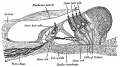
Vestibulocochlear Nerve
- forms beside otocyst
- from wall of otocyst and neural crest cells
- bipolar neurons
- vestibular neurons
- outer end of internal acoustic meatus
- innervate hair cells in membranous labyrinth
- axons project to brain stem and synapse in vestibular nucleus
- cochlear neurons
- cell bodies lie in modiolus
- central pillar of cochlear
- innervate hair cells of spiral organ
- axons project to cochlear nucleus
Inner Ear Genes
- hindbrain segmentation occurs at same time placode arises
- otocyst adjacent to rhombomere 5
- may influence development
- Hoxa1, kreisler, Fgf3
- genes regulating neural crest cells (neural genes)
- Pax2 Ko affects cochlear and spiral ganglion, but not vestibular apparatus
- nerogenin 1 affects both ganglia
Semicircular canal
- Otx1- cochlear and vestibular normal
- Hmx3, Prx1, Prx2
Sensory Organs
- thyroid hormone receptor beta
- Zebrafish-mindbomb mutant has excess hair cells but not supporting cells, Notch-Delta signaling
- Gene Expression-inner ear
- Brn-3c and Hair cell development
- Supporting Cells- p27kip
- Thyroid Hormone
- Ganglion neurons require growth factors
- vestibular neurons- BDNF, NT3
- survival not development
Postnatal Changes

Newborn to adult Eustachian (auditory, otopharyngeal or pharyngotympanic) tube.
- Connects middle ear cavity to nasopharynx portion of pharynx
Functions
- Ventilation - pressure equalization in the middle ear
- Clearance - allow fluid drainage from the middle ear Tube is normally closed and opened by muscles
At birth
- shorter (17-18 mm), narrower and runs almost horizontal Tube is opened by a single muscle, tensor palati muscle
Adult
- longer (twice as long), wider and runs at approximately 45 degrees to the horizontal. Tube is opened by two separate muscles, tensor palati and levator palati
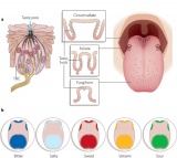
Vision
Timeline
|
Stage 13 (week 5)

|
|

|

|
|

|

|
| B1L | B2L | B3L | B4L | B5L | B6L | B7L |
Lens
Surface ectoderm -> lens placode (optic placode) -> lens pit -> lens vesicle -> lens fibres -> lens capsule and embryonic/fetal nucleus.
Retina
Neural plate ectoderm -> prosencephalon (forebrain) eye fields -> neural plate growth carries eye field region forward -> eye field invaginates forming optic grooves (sulci) -> diencephalon optic groove interacts with surface ectoderm (induces optic placode) -> optic stalk -> optic vesicle -> folds inward (optic cup) forming double layer -> inner neural retina, outer pigmented retina

Links: Embryo Images - Eye Development
Neural Crest
Eye connective tissue
Abnormalities

- Inner - common cavity, severe cochlear hypoplasia
- Large vestibular aqueduct syndrome (LVAS) can be one of the common causes of hearing loss
- Middle - rare and can be part of first arch syndrome, Malleus, Incus and Stapes Fixation
- Cholesteatoma- Epithelium trapped within skull base in development, erosion of bones: temporal bone, middle ear, mastoid
- Outer - Several genetic effects and syndromes, Environmental Effects
Outer Ear Abnormalities


- Microtia - abnormally small external ear
- Preauricular sinus - occurs in 0.25% births, bilateral (hereditary) 25-50%, unilateral (mainly the left), duct runs inward can extend into the parotid gland, Postnatally sites for infection
Fetal Alcohol Syndrome

- Postion- Lower or uneven height, "railroad track” appearance, curve at top part of outer ear is under-developed, folded over parallel to curve beneath
Congenital Deafness
Sensorineural - cochlear or central auditory pathway
- Hereditary
- recessive- severe
- dominant- mild
- can be associated with abnormal pigmentation (hair and irises)
- Acquired
- rubella (German measles), maternal infection during 2nd month of pregnancy, vaccination of young girls
- streptomycin
- antibiotic
- thalidomide
Conductive - disease of outer and middle ear
- produced by otitis media with effusion, is widespread in young children.
- temporary blockage of outer or middle ear
Bionic Ear
Cochlear Implant - Professor Graeme Clark (1960s, Australia) Array of electrodes implanted within cochlea, direct electrical stimulation to auditory nerve fibres
Conductive Hearing Loss
- Conductive Hearing Loss Produces a Reversible Binaural Hearing Impairment David R. Moore, Jemma E. Hine, Ze Dong Jiang, Hiroaki Matsuda, Carl H. Parsons, and Andrew J. King J. Neurosci. 1999;19 8704-8711 http://www.jneurosci.org/cgi/content/abstract/19/19/8704
- tested ferrets by lon-term plugging of ear canal
- Repeated testing during the 22 months after unplugging revealed a gradual return to normal levels of unmasking.
- Results show that a unilateral conductive hearing loss, in either infancy or adulthood, impairs binaural hearing both during and after the hearing loss.
- Show scant evidence for adaptation to the plug and demonstrate a recovery from the impairment that occurs over a period of several months after restoration of normal peripheral function.
References
Textbooks
- Before We Are Born (5th ed.) Moore and Persaud Chapter 20: p460-479
- Essentials of Human Embryology, Larson Chapter 12: p252-272
Online Textbooks
- Developmental Biology (6th ed.) Gilbert, Scott F. Sunderland (MA): Sinauer Associates, Inc.; c2000. Evolution of the mammalian middle ear bones from the reptilian jaw | Chick embryo rhombomere neural crest cells | Some derivatives of the pharyngeal arches | Formation of the Neural Tube | Differentiation of the Neural Tube | Tissue Architecture of the Central Nervous System | Neuronal Types | Snapshot Summary: Central Nervous System and Epidermis
- Neuroscience Purves, Dale; Augustine, George J.; Fitzpatrick, David; Katz, Lawrence C.; LaMantia, Anthony-Samuel; McNamara, James O.; Williams, S. Mark. Sunderland (MA): Sinauer Associates, Inc. ; c2001 The Auditory System | The Inner Ear | The Middle Ear | The External Ear | Early Brain Development | Construction of Neural Circuits | Modification of Brain Circuits as a Result of Experience
- Molecular Biology of the Cell (4th Edn) Alberts, Bruce; Johnson, Alexander; Lewis, Julian; Raff, Martin; Roberts, Keith; Walter, Peter. New York: Garland Publishing; 2002. Neural Development | The three phases of neural development
- Clinical Methods 63. Cranial Nerves IX and X: The Glossopharyngeal and Vagus Nerves | The Tongue | 126. The Ear and Auditory System | An Overview of the Head and Neck - Ears and Hearing | Audiometry
- Health Services/Technology Assessment Text (HSTAT) Bethesda (MD): National Library of Medicine (US), 2003 Oct. Developmental Disorders Associated with Failure to Thrive
- Eurekah Bioscience CollectionCranial Neural Crest and Development of the Head Skeleton
Search
- Bookshelf hearing development
- Pubmed hearing development
External Links
External Links Notice - The dynamic nature of the internet may mean that some of these listed links may no longer function. If the link no longer works search the web with the link text or name. Links to any external commercial sites are provided for information purposes only and should never be considered an endorsement. UNSW Embryology is provided as an educational resource with no clinical information or commercial affiliation.
- NIDCD - Balance Disorders
- Embryo Images Online
- Eye Development - Eye Development Unit | Eye Fields-Optic Vesicle (Weeks 3-4) | Optic Cup, Lens Vesicle, Choroid Fissure, Hyaloid Artery (Weeks 5-6) | Cornea, Anterior Chamber, Pupillary Membrane, Lens, Retina (Weeks 7-8) | Iris, Cilliary Body (Weeks 9-15) | Eyelids (Weeks 8-10)
- Ear Development - Ear Development Unit | Inner Ear | [http://www.med.unc.edu/embryo_images/unit-ear/ear_htms/ear012.htm Middle Ear | External Ear
Terms
| Hearing Terms | ||
|---|---|---|
Hearing and Balance Development
|
| Vision Terms | ||
|---|---|---|
|
| 2018 ANAT2341 - Timetable | Course Outline | Moodle | Tutorial 1 | Tutorial 2 | Tutorial 3 |
Labs: 1 Preimplantation and Implantation | 2 Reproductive Technology Revolution | 3 Group Projects | 4 GM manipulation mouse embryos | 5 Early chicken eggs | 6 Female reproductive tract | 7 Skin regeneration | 8 Vertebral development | 9 Organogenesis Lab | 10 Cardiac development | 11 Group projects | 12 Stem Cell Journal Club |
|
Lectures: 1 Introduction | 2 Fertilization | 3 Week 1/2 | 4 Week 3 | 5 Ectoderm | 6 Placenta | 7 Mesoderm | 8 Endoderm | 9 Research Technology | 10 Cardiovascular | 11 Respiratory | 12 Neural crest | 13 Head | 14 Musculoskeletal | 15 Limb | 16 Renal | 17 Genital | 18 Endocrine | 19 Sensory | 20 Fetal | 21 Integumentary | 22 Birth | 23 Stem cells | 24 Revision |
| Student Projects: Group Projects Information Project 1 | Project 3 | Project 4 | Project 5 | 2018 Test Student | Copyright |