| All human and animal embryos go through very similar stages of early development. See also Humans and Animal Embryology.
What are the key things in development that we share?
This page introduces a few of the concepts of comparative development shared with all animals.
- Ernst Haeckel (1834 – 1919) "ontogeny recapitulates phylogeny" claimed that an individual organism's biological development (ontogeny), parallels and summarises its species evolutionary development (phylogeny). First a single-celled organism, then evolve into a fish, then an amphibian, then a reptile, then a bird, and finally reach a mammal.
- Current developmental biology shows that animals follow similar developmental programs, but do not go through Haeckel's "species change" during development.
|
Meiosis | Mitosis | Gastrulation | Body Plan | Limbs | Tissue Development | Organ Development | Animal Models | Human and Other Animal Development
| Teacher Note
|
 This is currently only a draft designed to help K12 students understand comparative embryology. This is currently only a draft designed to help K12 students understand comparative embryology.
I am currently looking to simplify concepts and include images on this page. I am happy to receive feedback as too what you may like to be included here. I have also begun to add some simple exercises that can be used in class to help understand concepts in embryonic development and comparison. Note some of the links on this page leave the K12 notes section and may be beyond the level of your students, bookmark this page to easily return here.
This page can be printed using the menu from the list at the bottom of the page "Printable version" or Printable version.
Also look at the K12 Human and Other Animal Development that simply compares development times and embryo structures.
See also online textbook - Gilbert SF. Developmental Biology. 6th edition. Sunderland (MA): Sinauer Associates; 2000. Comparative Embryology.
K12 Professional Development 2016 | K12 Professional Development 2014
|
Meiosis
| In the male and female in all animals (and plants) that reproduce sexually to form an embryo, these very first cells form by meiosis.
Meiosis a reductive form of cell division that only occurs in the egg (oocyte) and sperm (spermatozoa) and allows new genetic combinations of offspring to be generated.
Meiosis has 2 key components:
- Genetic reorganisation - the genetic material (chromosomes) that you have from your mother and father are recombined.
- Genetic reductive - the chromosome number is halved and only fertilisation will allow the paired chromosomes that we all contain in all our cells.

|
Meiosis in all mammals:
- Female embryo XX - X chromosome from egg and X chromosome from sperm
- Male embryo XY - X chromosome from egg and Y chromosome from sperm
- Both male and female embryos - get the recombined 22 autosomes and the mother's mitochondrial DNA
|
|
<html5media height="200" width="300">File:Oocyte_Meiosis_01.mp4</html5media>
This movie shows the egg (oocyte) completing the first part of meiosis (meiosis I). The chromosomes are coloured blue, the cell membrane is green. The small structure on the left are chromosomes that will not be used in the embryo.
|
Mitosis
| Mitosis is the type of normal cell division that allows growth and development of all animal embryos.
After the first cell has been formed by the egg (oocyte) and sperm (spermatozoa) fusing, every cell division in the embryo forms 2 genetically identical daughter cells. Every mitosis in all animal cells has the same features.
Mitosis has 2 key components:
- Chromosome duplication - has to occur before cell division can occur.
- Mitosis - a set of 5 standard phases dividing the nucleus (and the chromosomes it contains) before the cytoplasm divides.
| Mitosis is a biological process that is the same (evolutionarily conserved) in all plants and animals.
|
|
<html5media height="300" width="300">File:Mitosis 11.mp4</html5media>
This movie shows a cell that has already begun mitosis separating the chromosomes in the nucleus then the cell cytoplasm. The chromosomes are white.
|
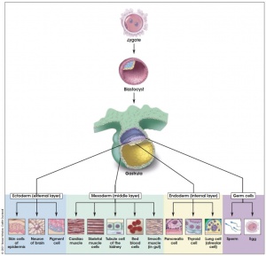
Gastrulation
| Development of any animal requires the differentiation of different cell types and tissues from essentially the same initial cells. One of the first shared steps is the process of "gastrulation" (means gut formation). This forms the 3 cell layers that will form all the embryo, they are often described as the "germ layers" and this stage of development as the "gastrula". The same layers form the same structures in all these animals.
3 Germ Layers:
- Ectoderm - outer layer of skin (epidermis) and nervous system (central nervous system and peripheral nervous system).
- Mesoderm - bone, muscle, cartilage, blood (connective tissues)
- Endoderm - lining of the gut, lungs, endocrine organs.
| Gastrulation is a process that occurs in nearly all animals and each germ layer contributes the same body components to all animal embryos.
|
|
|
| Teacher Note
|
| Triploblastic (three layer) organisms describe vertebrate embryos with these 3 germ layers. Diploblastic (two layers) lacy a mesoderm layer and occurs in some marine invertebrate animals: porifera (sponges), cnidarians (sea anemones, hydra, jellyfish) and ctenophores (comb jellies).
|
Body Plan
| The next step in development of any animal requires the embryo body plan (axes) by a patterning process.
Body plan (axes)
- head and body
- left and right side
- front and back
Many signals used to establish this patterning have been identified and are shared (similar or the same) between different animals. Note that some of the signals used to establish the overall body plan can be reused later at other stages in embryo development and within the body organs and tissues.
| Body plan genes and signaling pathways are similar for all animals.
|
|
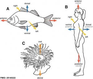
Different animal body plan axes.
|
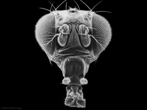
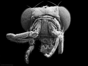
Below is an example of what happens in a fly if these patterning signals get disrupted, putting legs from the body on the head where antenna should be located.
|
|
| Normal fly head with antenna
|
Abnormal fly head with legs for antenna
|
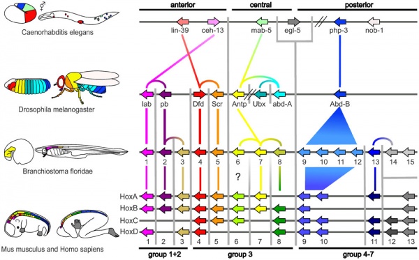
This fly mutation identified a common patterning gene family called Hox that establishes the head to tail axes in the embryo.
This complicated picture shows how different Hox genes are expressed at different embryo levels in different species (worms, flies, mouse and human).
| Teacher Note
|
| The fly head picture is easy to explain, and you can also talk about the 1995 Nobel Prize for this discovery.
The Hox gene picture will be difficult for students to understand. Just understand that in all animal embryos, different Hox genes are expressed along the head to tail axis of the embryo and that this establishes an initial pattern in all embryos.
"Hox" is the acronym for "Homeobox", a large family of similar genes that control the formation of many body structures during early embryonic development.
Here is a page with University level student information on Homeobox
|
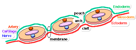
Pharyngeal Arches
The vertebrate early embryo head region develops a series of transient structures called pharyngeal arches (branchial arches and gill arches). Each arch has a similar structure and is formed from all 3 germ layers. In fish, these arches develop into the gill apparatus. In mammals, these arches contribute many different head structures including the jaws, hearing components and endocrine organs.
|
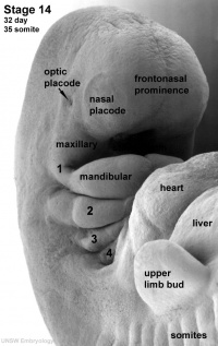
This is a human embryo (week 5) showing the pharyngeal arches numbered 1, 2, 3, 4.
- The arches developmentally form in sequence, top to bottom (1 to 6).
- The arches are then remodelled over the next 3 developmental weeks to form many different components of the head.
- Other mammals form similar structures from each arch, but over a different time course.
The cartoon below shows the repeated structure within each arch, germ layer contribution, and the special names given to each region.
|
| Pharyngeal Arches are formed by all vertebrates, are temporary structures, contain all 3 germ layer, and contribute similar head components to all animal embryos.
|
| Teacher Note
|
| Note that humans do not have a 5th arch (numbered on the basis of structures formed) and arch 4 and 6 are present as a single structure on the above surface embryo view.
The table below gives a detailed overview of adult structures formed from each arch in the human embryo.
| Pharyngeal Arch
|
Nerve
|
Artery
|
Neural Crest
(Skeletal Structures)
|
Muscles
|
Ligaments
|
1
(maxillary/mandibular)
|
trigeminal (CN V)
|
maxillary artery (terminal branches)
|
mandible, maxilla, malleus, incus
|
muscles of mastication, mylohyoid, tensor tympanic, ant. belly digastric
|
ant lig of malleus, sphenomandibular ligament
|
2
(hyoid)
|
facial (CN VII)
|
stapedial (embryonic)
corticotympanic (adult)
|
stapes, styloid process, lesser cornu of hyoid, upper part of body of hyoid bone
|
muscles of facial expression, stapedius, stylohyoid, post. belly digastric
|
stylohyoid ligament
|
| 3
|
glossopharyngeal (CN IX)
|
common carotid, internal carotid arteries
|
greater cornu of hyoid, lower part of body of hyoid bone
|
stylopharyngeus
|
|
| 4
|
vagus (CN X) superior laryngeal branch
|
part of aortic arch (left), part right subclavian artery (right)
|
thyroid, cricoid, arytenoid, corniculate and cuneform cartilages
|
crycothyroid, soft palate levator veli palatini (not tensor veli palatini)
|
|
| 6
|
vagus (CN X) recurrent laryngeal branch
|
part of left pulmonary artery (left), part of right pulmonary artery (right)
|
thyroid, cricoid, arytenoid, corniculate and cuneform cartilages
|
larynx intrinsic muscles (not cricothyroid muscle)
|
|
Pharyngeal arches this page contains more detailed information about these structures.
|
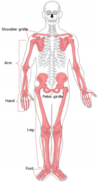
Limbs
| Many different animals form limbs (arms and legs). During embryo development there are common signalling molecules and regions that form the initial limb structure. A similar process occurs for both the upper (arm) and low (leg) limb development.
The final limb structures formed can appear different, but the bones shows that they share a pattern of development.
|
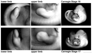
Early human embryo upper and lower limbs.
|
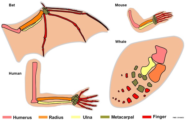
Upper limb bones of 4 different species. Each limb is significantly different in size and function, but all contain the same basic skeletal structures.
|
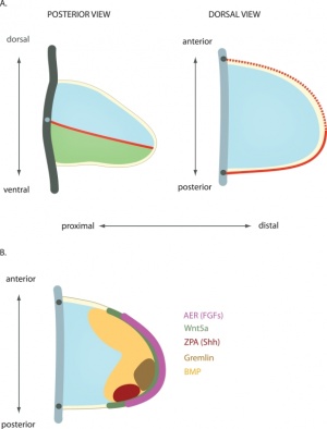
Cartoon of the early limb showing regions that establish the developmental patterning.
|
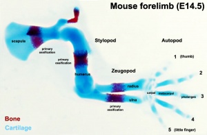
Mouse Embryo Limb
|
Limbs even though animal arms, legs, flippers and wings appear externally different their skeletons show common features and have a common function (motility).
|
Tissue Development
| All animal embryos have similar tissues (connective, muscle, nervous, and epithelial) that that develop in different regions of the embryo. The development of these tissues is different from embryo patterning and is separate for each tissue.
The developmental signals that control say a connective tissue, like bone, are the same for all bones throughout the body. These mechanisms are the same in other animals. Therefore bone development will go through similar stages in all animals bodies even though different bones are eventually formed.
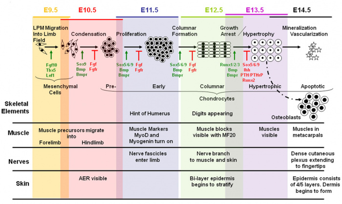
Mouse limb tissue development
| Tissue Development - the genes and signals that control embryonic development of these tissues are closely related in all animals and differ from the "patterning genes".
|
|
The limb skeleton.
|
| Teacher Note
|
| If the students have already done some biology they should understand these 4 main tissue types: connective, muscle, nervous, and epithelial. It is beyond the scope of this current page to go through these developmental molecular mechanisms.
|
Organ Development
Many animals have common organs used for digestion (stomach, liver, pancreas), breathing (lungs), waste (kidneys) and cardiovascular (heart).
The signalling mechanisms that are used to control their initial development and later internal patterning are the same in many different species embryos.
| Teacher Note
|
| Organ development is difficult to explain easily to students. The diagram below shows how the mouse liver development is controlled by different genes. Most easily explained as, similar to what is seen in the human liver development.
|
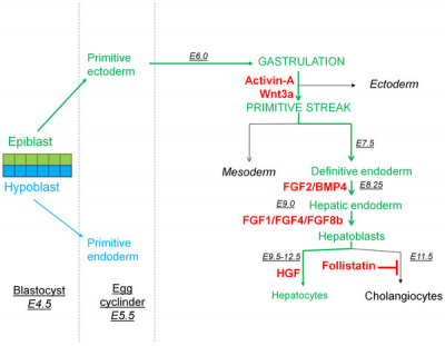
Mouse liver development
|
- E - means the embryonic day of development.
- Red names - are the genes controlling specific development steps.
- The other colours refer to developmental stages and features.
|
Animal Models
These shared signalling mechanisms allow us to use animal development to study both normal and abnormal human development.
Common animal "models" used include mouse (mammal), chicken (bird) and zebrafish (fish). Though very different species, current research shows that these embryos share common signalling mechanisms that form similar structures in different animals.
- Mouse - (mammal) good model for easy genetic manipulation.
- Chicken - (bird) develops in an egg so the earlier stages of development can be observed in the living embryo.
- Zebrafish - (fish) good model of vertebrate (backbone) animal development as the embryos are "see through" and can be observed while growing.
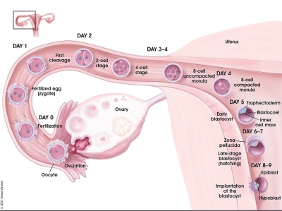
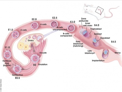
| Fertilisation to Implantation
|
| Human (9 days)
|
Mouse (5 days)
|
|
|
Human embryo summary of the events from fertilisation not implantation.
Humans take about 37 weeks to develop before birth.
|
Mouse embryo summary of the events from fertilisation not implantation.
Mice take about 3 weeks to develop before birth.
|
| Human and Mouse - In mammals, there is a difference in overall timing but the processes, organs and tissues are shared.
|
| Zebrafish embryo development from the 1-cell stage to 85 hours post fertilisation.
|
| <html5media height="300" width="948">File:zebrafish movie01.mp4</html5media>
Zebrafish take about 3 days to develop before hatching.
|
| Animal Models - because of these shared (common) mechanisms we can use animals to model human development.
|
Comparing Embryos
| Whats difference in humans?
The long fetal growth period allows extensive neural growth and development, though humans are not the longest prenatal period, so its not just about time.
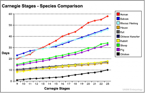
| Developmental Times - the graph compares different animals time to reach the same stage in embryo development.
|
|
|
| Animal Development Time
|
| Animal
|
Average Days
|
| Bear (Black)
|
210
|
| Bison
|
270
|
| Budgerigar
|
18
|
| Camel
|
410
|
| cat
|
65
|
| cow
|
281
|
| chicken
|
21
|
| Chimpanzee
|
236
|
| Chinchilla
|
111
|
| Coyote
|
63
|
| deer (Mule)
|
200
|
| dog
|
63
|
| Donkey
|
365
|
| Duck
|
28
|
| Duck (Muscovy)
|
35
|
| elephant
|
660
|
| Elk, Wapiti
|
255
|
| Ferret
|
42
|
| Finch
|
14
|
| Fox
|
52
|
| Giraffe
|
425
|
| goat
|
150
|
| Goose
|
28
|
| Gorilla
|
270
|
| Guinea fowl
|
28
|
| guinea pig
|
68
|
| Hare
|
36
|
| Hippopotamus
|
240
|
| horse
|
338
|
| Human
|
274
|
| Leopard
|
95
|
| Lion
|
108
|
| Llama
|
350
|
| Marmoset
|
150
|
| Mink (European)
|
41
|
| monkey (Macaque)
|
180
|
| Moose
|
240
|
| mouse
|
20
|
| Muskox
|
255
|
| Muskrat
|
29
|
| Nutria, Coypu
|
130
|
| opossum
|
12
|
| Otter
|
285
|
| Panther
|
90
|
| Parrot
|
26
|
| Pheasant
|
24
|
| Pig
|
114
|
| Pigeon
|
18
|
| Porcupine
|
210
|
| Pronghorn
|
230
|
| Quail
|
16
|
| rabbit
|
31
|
| Raccoon
|
63
|
| rat
|
21
|
| Reindeer
|
225
|
| Rhinoceros (African)
|
480
|
| Seal
|
330
|
| sheep
|
150
|
| Shrew
|
20
|
| Skunk
|
63
|
| Squirrel (Gray)
|
40
|
| Swan
|
35
|
| Tapir
|
390
|
| Tarsier
|
182
|
| Tiger
|
103
|
| Turkey
|
28
|
| Walrus
|
450
|
| whale (Sperm)
|
450
|
| Wolf
|
63
|
| Woodchuck
|
31
|
|
Animal Notes and Table Data Sources
- Each animal species has different variations +/- the average values shown in the table.
- Gestation is the carrying of an animal embryo or fetus inside a female viviparous animal. Except in the case of human gestational age GA.
- Incubation is the laying of an egg (birds, reptiles, monotremes) with development occurring outside the female animal.
See also - Timeline Comparisons
Additional Data Sources
- Theiler K. The House Mouse: Atlas of Mouse Development (1972, 1989) Springer-Verlag, NY. Online
- Witschi E. Rat Development. In: Growth Including Reproduction and Morphological Development. (1962) Altman PL. and Dittmer DS. ed. Fed. Am. Soc. Exp. Biol., Washington DC, pp. 304-314.
- The Genetics of the Dog. E Ostrander, E. and Ruvinsky, A. ISBN: 9781845939403 (2012)
- Merck Veterinary Manual. Aiello, S.E. and Moses, M.A. (ed) ISBN: 0911910506 (2013) Online
- Witschi, E. (1962) Development: Rat. In: Growth Including Reproduction and Morphological Development. Altman, P. L. , and D. S. Dittmer, ed. Fed. Am. Soc. Exp. Biol., Washington DC, pp. 304-314.
|
Species Embryonic Comparison Timeline
| Carnegie
|
Stage
|
1
|
2
|
3
|
4
|
5
|
6
|
7
|
8
|
9
|
10
|
11
|
12
|
13
|
14
|
15
|
16
|
17
|
18
|
19
|
20
|
21
|
22
|
23
|
| Human
|
Days
|
1
|
2-3
|
4-5
|
5-6
|
7-12
|
13-15
|
15-17
|
17-19
|
20
|
22
|
24
|
28
|
30
|
33
|
36
|
40
|
42
|
44
|
48
|
52
|
54
|
55
|
58
|
| Mouse
|
Days
|
1
|
2
|
3
|
E4.5
|
E5.0
|
E6.0
|
E7.0
|
E8.0
|
E9.0
|
E9.5
|
E10
|
E10.5
|
E11
|
E11.5
|
E12
|
E12.5
|
E13
|
E13.5
|
E14
|
E14.5
|
E15
|
E15.5
|
E16
|
| Rat
|
Days
|
1
|
3.5
|
4-5
|
5
|
6
|
7.5
|
8.5
|
9
|
10.5
|
11
|
11.5
|
12
|
12.5
|
13
|
13.5
|
14
|
14.5
|
15
|
15.5
|
16
|
16.5
|
17
|
17.5
|
| Note these Carnegie stages are only approximate day timings for average of embryos. Links: Carnegie Stage Comparison
|
| Table References
|
| Human
O'Rahilly R. (1979). Early human development and the chief sources of information on staged human embryos. Eur. J. Obstet. Gynecol. Reprod. Biol. , 9, 273-80. PMID: 400868
Otis EM and Brent R. Equivalent ages in mouse and human embryos. (1954) Anat Rec. 120(1):33-63. PMID 13207763
Mouse
Theiler K. The House Mouse: Atlas of Mouse Development (1972, 1989) Springer-Verlag, NY. Online
OTIS EM & BRENT R. (1954). Equivalent ages in mouse and human embryos. Anat. Rec. , 120, 33-63. PMID: 13207763
Rat
Witschi E. Rat Development. In: Growth Including Reproduction and Morphological Development. (1962) Altman PL. and Dittmer DS. ed. Fed. Am. Soc. Exp. Biol., Washington DC, pp. 304-314.
Pérez-Cano FJ, Franch À, Castellote C & Castell M. (2012). The suckling rat as a model for immunonutrition studies in early life. Clin. Dev. Immunol. , 2012, 537310. PMID: 22899949 DOI.
|
|
Now looks at Human and Other Animal Development
| Teacher Note
|
| This next page allows students to directly compare the appearance of embryos from different species.
|
Cite this page: Hill, M.A. (2026, July 25) Embryology K12 Comparative Embryology. Retrieved from https://embryology.med.unsw.edu.au/embryology/index.php/K12_Comparative_Embryology
- What Links Here?
- © Dr Mark Hill 2026, UNSW Embryology ISBN: 978 0 7334 2609 4 - UNSW CRICOS Provider Code No. 00098G
|


