Category:Heart: Difference between revisions
No edit summary |
|||
| Line 1: | Line 1: | ||
{{Template:Heart Links}} | |||
The links in | |||
The links in this next section are to the original 2008 online notes pages for [http://embryology.med.unsw.edu.au/Notes/heart.htm Cardiovascular System Development]. | |||
====Cardiovascular Notes==== | ====Cardiovascular Notes==== | ||
Revision as of 02:46, 27 March 2010
The links in this next section are to the original 2008 online notes pages for Cardiovascular System Development.
Cardiovascular Notes
Introduction | Abnormalities | Stage 13/14 | Stage 22 | Stage 22 Selected Highpower | Heart | Heart Rate | BloodBlood Vessels | Molecular | Lymphatic | Text only page | WWW Links | Postnatal | History - Harvey
Cardiovascular Movies
Heart Movies | Heart Looping | Atrial Septation | Realignment | Ventricular Septation | Heart Septation Models | Historic Heart Movie |
Other Cardiac and Vascular Movies Fetal Circulation (Before Birth) | Circulation (After Birth) | Aortic Branches to Glands (Kidneys only) | Aortic Branches to Glands (Gonads only)
Notes
- Pages section on this current page include lectures, laboratories, notes, quizzes and educational module sections that relate to cardiovascular development.
Subcategories
This category has the following 4 subcategories, out of 4 total.
Pages in category 'Heart'
The following 57 pages are in this category, out of 257 total.
(previous page) (next page)R
- Template:Ref-Mandarim-de-Lacerda1991
- Template:Ref-McBride1981
- Template:Ref-Morrill1916
- Template:Ref-Murray1919
- Template:Ref-NobaokRehman1941
- Template:Ref-Odgers1938
- Template:Ref-Patten1922
- Template:Ref-Patten1929
- Template:Ref-Patten1930
- Template:Ref-Patten1931
- Template:Ref-Patten1938
- Template:Ref-Patten1949
- Template:Ref-PMID1018009
- Template:Ref-PMID5165416
- Template:Ref-Pohlman1907
- Template:Ref-Pohlman1909
- Template:Ref-Retzer1920
- Template:Ref-Robinson1902
- Template:Ref-ScammonNorris1918
- Template:Ref-Schulte1916
- Template:Ref-Shaner1929
- Template:Ref-Shaner1930
- Template:Ref-Takahashi1923
- Template:Ref-Tandler1912
- Template:Ref-Vernall1962
- Template:Ref-Walmsley1931
- Template:Ref-Wang1917
- Template:Ref-Wang1918
- Template:Ref-Waterston1917
- Template:Ref-Watson1924
- Template:Ref-West1915
- Template:Ref-Witte1919
- Template:Ref-Yater1929
- Template:Ref-Yoshinaga1921
- Template:Ref-Zimmerman1927
- RPAH Cardiac Embryology 2014
T
U
V
Media in category 'Heart'
The following 200 files are in this category, out of 434 total.
(previous page) (next page) Gray0462.gif 350 × 403; 49 KB
 Gray0464.gif 450 × 455; 51 KB
 Gray0467.jpg 600 × 464; 47 KB
 Gray0468.jpg 500 × 436; 43 KB
 Gray0470.jpg 800 × 371; 40 KB
 Gray0472.jpg 550 × 653; 57 KB
 Gray0474.jpg 459 × 600; 33 KB
 Gray0475.jpg 2,042 × 1,363; 350 KB
 Gray0476.jpg 600 × 609; 99 KB
 Gray0492.jpg 600 × 508; 117 KB
 Gray0498.jpg 475 × 416; 39 KB
 Gray0502.jpg 1,000 × 1,329; 215 KB
 Gray0506.jpg 375 × 464; 25 KB
 Gray0556.jpg 600 × 533; 86 KB
 Gray0971.jpg 800 × 583; 166 KB
 Head and heart muscle cartoon.jpg 874 × 800; 129 KB
 Heart chicken embryo stage 12.jpg 1,000 × 1,182; 221 KB
 Heart chicken embryo stage 16.jpg 1,000 × 1,307; 234 KB
 Heart chicken embryo stage 21.jpg 1,000 × 1,197; 207 KB
 Heart chicken embryo stage 25.jpg 1,000 × 1,154; 219 KB
 Heart conduction system-bird-monotreme-placental.jpg 1,040 × 625; 76 KB
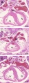
 Heart histology 003.jpg 400 × 500; 136 KB
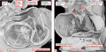
 Heart human embryo CRL10mm 01.jpg 1,000 × 1,389; 537 KB
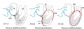
 Heart innervation 01.jpg 1,280 × 599; 92 KB
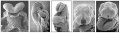
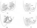
 Heart Looping Sequence (SEMs).jpg 1,928 × 776; 212 KB
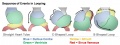 Heart Looping Sequence.jpg 1,548 × 577; 85 KB
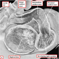
 Heart outflow tract stage 14 01.jpg 2,039 × 996; 274 KB
 Heart outflow tract stage 14 02.jpg 996 × 996; 139 KB
 Heart outflow tract stage 14 03.jpg 989 × 996; 134 KB
 Heart Tube Fusion.jpg 1,551 × 1,139; 125 KB
 Heart Tube Segments.jpg 1,082 × 771; 63 KB
 Heart valve histology 01.jpg 1,008 × 1,280; 318 KB
 Heart valve histology 02.jpg 800 × 456; 103 KB
 Heart valve histology 03.jpg 800 × 489; 111 KB
 Heart-cartoon-001.jpg 600 × 697; 40 KB
 Heart-ventricular-septum-03.jpg 320 × 240; 14 KB
 Heart1 atrium.gif 350 × 373; 243 KB
 Heart1 ventricle.mov ; 209 KB
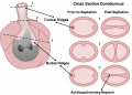 HeartILP draft otcrosssection.jpg 1,433 × 1,026; 119 KB
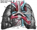 Historic-lungs.jpg 600 × 493; 79 KB
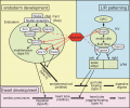
 Human heart developmental functional networks.jpg 833 × 614; 424 KB
 Human heart SEM1.jpg 1,200 × 330; 47 KB
 Human stage14 heart MRI - ventricular septation.jpg 864 × 652; 39 KB
 Human stage18 heart MRI - ventricular septation.jpg 1,014 × 652; 48 KB
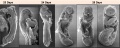
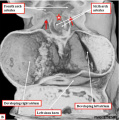
 Human week 10 fetus 05.jpg 1,600 × 1,200; 612 KB
 Human- fetal week 10 heart ABCD.jpg 600 × 450; 133 KB
 Human- fetal week 10 upper body A.jpg 600 × 450; 104 KB
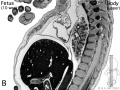 Human- fetal week 10 upper body B.jpg 600 × 450; 105 KB
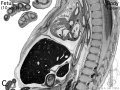 Human- fetal week 10 upper body C.jpg 600 × 450; 109 KB
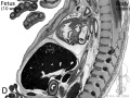 Human- fetal week 10 upper body D.jpg 600 × 450; 106 KB
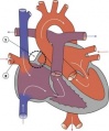
 Human-heart-E3L.jpg 639 × 393; 79 KB
 Hypoplastic Left Heart.jpg 297 × 350; 17 KB
 Ingalls1920plate04.jpg 762 × 1,044; 52 KB
 Intermediate Heart Development Timeline.jpg 1,772 × 687; 133 KB
 Interrupted Aortic Arch.jpg 290 × 350; 17 KB
 Johnson1917 plate01fig02.jpg 495 × 800; 52 KB
 Johnson1917 plate03fig01.jpg 800 × 613; 57 KB
 Johnson1917 plate03fig02.jpg 800 × 620; 42 KB
 Keith1902 fig194.jpg 829 × 750; 50 KB
 Keith1902 fig212.jpg 1,123 × 750; 139 KB
 Kollmann528.jpg 744 × 587; 91 KB
 Kollmann529.jpg 737 × 593; 94 KB
 Kollmann530.jpg 733 × 524; 83 KB
 Kollmann531.jpg 688 × 570; 88 KB
 Kollmann532.jpg 702 × 409; 59 KB
 Kollmann533.jpg 717 × 594; 76 KB
 Kollmann534.jpg 684 × 599; 74 KB
 Kollmann539.jpg 736 × 696; 132 KB
 Kollmann549.jpg 689 × 573; 75 KB
 Kollmann555.jpg 605 × 585; 61 KB
 Kollmann561.jpg 739 × 591; 82 KB
 Kollmann562.jpg 670 × 581; 68 KB
 Kollmann563.jpg 754 × 848; 143 KB
 Kramer1942 fig01.jpg 1,280 × 1,108; 280 KB
 Kramer1942 fig02.jpg 1,000 × 1,526; 398 KB
 Kramer1942 fig03.jpg 1,000 × 854; 259 KB
 Kramer1942 fig04.jpg 1,000 × 654; 107 KB
 Kramer1942 fig05.jpg 1,000 × 417; 100 KB
 Kramer1942 fig06.jpg 1,000 × 482; 86 KB
 Kramer1942 fig07.jpg 1,000 × 708; 121 KB
 Kramer1942 fig08.jpg 1,000 × 457; 83 KB
 Kramer1942 fig09.jpg 1,000 × 832; 120 KB
 Le Gros Clark01.jpg 807 × 820; 159 KB
 Licata1954 fig01.jpg 1,000 × 725; 90 KB
 Licata1954 fig02.jpg 1,000 × 610; 71 KB
 Licata1954 fig03.jpg 1,000 × 708; 92 KB
 Licata1954 fig04.jpg 1,000 × 765; 84 KB
 Licata1954 fig05.jpg 1,000 × 760; 100 KB
 Licata1954 fig06.jpg 1,000 × 834; 99 KB
 Licata1954 fig07.jpg 1,000 × 931; 111 KB
 Licata1954 fig08.jpg 1,000 × 670; 82 KB
 Licata1954 fig09.jpg 1,000 × 813; 93 KB
 Licata1954 fig10.jpg 1,000 × 1,484; 367 KB
 Licata1954 fig11.jpg 1,000 × 1,502; 285 KB
 Licata1954 fig12.jpg 1,000 × 755; 82 KB
 Licata1954 fig13.jpg 1,000 × 915; 114 KB
 Licata1954 fig14.jpg 1,000 × 1,347; 177 KB
 Licata1954 fig15.jpg 1,000 × 1,324; 179 KB
 Licata1954 fig16.jpg 1,000 × 1,212; 297 KB
 Licata1954 fig16a.jpg 699 × 877; 107 KB
 Licata1954 fig16b.jpg 784 × 874; 117 KB
 Licata1954 fig16c.jpg 697 × 922; 140 KB
 Licata1954 fig16d.jpg 779 × 928; 146 KB
- Looping animation 002.mov ; 212 KB
 Mall1912-fig01.jpg 969 × 1,100; 365 KB
 Mall1912-fig02.jpg 797 × 730; 180 KB
 Mall1912-fig03.jpg 627 × 600; 95 KB
 Mall1912-fig04.jpg 832 × 650; 151 KB
 Mall1912-fig05.jpg 900 × 836; 177 KB
 Mall1912-fig06.jpg 1,000 × 991; 184 KB
 Mall1912-fig07.jpg 652 × 600; 93 KB
 Mall1912-fig08.jpg 800 × 471; 82 KB
 Mall1912-fig09.jpg 700 × 800; 158 KB
 Mall1912-fig10.jpg 800 × 550; 136 KB
 Mall1912-fig11.jpg 1,000 × 725; 124 KB
 Mall1912-fig12.jpg 816 × 800; 212 KB
 Mall1912-fig13.jpg 800 × 631; 173 KB
 Mall1912-fig14.jpg 538 × 600; 66 KB
 Mall1912-fig15.jpg 600 × 385; 33 KB
 Mall1912-fig16.jpg 735 × 650; 59 KB
 Mall1912-fig17.jpg 574 × 500; 39 KB
 Mall1912-fig18.jpg 600 × 425; 38 KB
 Mall1912-fig19.jpg 900 × 629; 77 KB
 Mall1912-fig20.jpg 400 × 494; 56 KB
 Mall1912-fig21.jpg 760 × 582; 141 KB
 Mall1912-fig22.jpg 800 × 600; 116 KB
 Mall1912-fig23.jpg 800 × 594; 159 KB
 Mall1912-fig24-25.jpg 850 × 1,000; 241 KB
 Mall1912-fig26.jpg 800 × 769; 150 KB
 Mall1912-fig27.jpg 542 × 600; 41 KB
 Mall1912-fig28.jpg 731 × 1,000; 396 KB
 Mall1912-fig29.jpg 472 × 580; 47 KB
 Mall1912-fig30.jpg 438 × 500; 58 KB
 Mall1912-fig31-33.jpg 963 × 1,000; 94 KB
 Mall1912-fig34-37.jpg 1,000 × 774; 96 KB
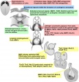 Molecular & Genetic Cardiac Development Factors.jpg 1,475 × 1,541; 269 KB
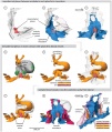
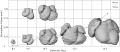 Mouse 3D Heart external E8.5-14.5.jpeg 1,280 × 559; 90 KB
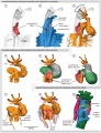
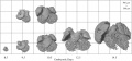 Mouse 3D Heart internal E8.5-14.5.jpeg 1,280 × 599; 106 KB
 Mouse E9 cervical intersomitic vessels A.jpg 1,714 × 1,142; 209 KB
 Mouse E9 cervical intersomitic vessels B.jpg 1,714 × 1,142; 175 KB
 Mouse E9 cervical intersomitic vessels C.jpg 1,714 × 1,142; 245 KB
 Mouse E9 cervical intersomitic vessels D.jpg 1,714 × 1,142; 147 KB
 Mouse E9 cervical intersomitic vessels.jpg 2,989 × 2,300; 777 KB
 Mouse embryo vascular.png 600 × 576; 518 KB
 Mouse heart E9.5.jpg 600 × 558; 31 KB
 Mouse heart primary cilia 01.jpg 1,200 × 908; 415 KB
 Mouse-coronary vessel formation.jpg 800 × 282; 44 KB
 Mouse-heart E17.5.jpg 353 × 1,000; 145 KB
 Nipbl heart and organ patterning.png 600 × 502; 164 KB
- Outflow tract 001.mov ; 1,020 KB
 Partial Anomalous Pulmonary Venous Drainage.jpg 290 × 350; 16 KB
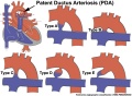 Patent ductus arteriosus classification.jpg 1,200 × 880; 138 KB
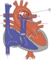 Patent Ductus Arteriosus.jpg 294 × 350; 16 KB
 Patten024.jpg 1,047 × 451; 75 KB
 Patten027.jpg 722 × 872; 123 KB
 Patten049.jpg 807 × 864; 119 KB
 Patten050.jpg 800 × 890; 109 KB
 Persistent fifth aortic arch and patent ductus arteriosus CT01.jpg 589 × 800; 69 KB
 Platypus right auricle 01.jpg 800 × 640; 124 KB
 Platypus ventricular septum 01.jpg 683 × 800; 94 KB
 Pulmonary Atresia.jpg 301 × 350; 17 KB
 Pulmonary Stenosis.jpg 289 × 350; 16 KB
 Quain595.jpg 1,200 × 803; 123 KB
 Quain596.jpg 869 × 819; 97 KB
 Robert Anderson.jpg 307 × 400; 16 KB
 Rugh 107.jpg 1,073 × 800; 188 KB
 Rugh 156.jpg 700 × 655; 86 KB
 Rugh 157.jpg 993 × 1,000; 183 KB
 Rugh 158.jpg 1,000 × 623; 158 KB
 Rugh 159.jpg 768 × 800; 87 KB
 Rugh 160.jpg 1,000 × 568; 97 KB
 Rugh 161.jpg 1,000 × 568; 104 KB
 Rugh 163.jpg 651 × 800; 75 KB
 Rugh 164.jpg 941 × 800; 108 KB
 Rugh 165.jpg 598 × 800; 132 KB
 Rugh 166.jpg 638 × 1,000; 175 KB
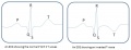 Schematic ECG normal and inverted T-wave.jpg 1,001 × 384; 32 KB
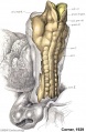
 Stage 10 historic-Corner1929-1.jpg 654 × 1,000; 145 KB
 Stage 10 historic-Corner1929-1a.jpg 523 × 800; 87 KB
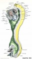 Stage 11 historic-Atwell1930-1.jpg 538 × 1,000; 75 KB
 Stage 11 historic-Atwell1930-1a.jpg 430 × 800; 46 KB
 Stage 11 historic-Atwell1930-1b.jpg 323 × 600; 26 KB
 Stage 11 historic-Atwell1930-1c.jpg 215 × 400; 14 KB
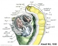
 Stage 11 historic-Atwell1930-2.jpg 800 × 639; 87 KB
 Stage 11 historic-Atwell1930-2a.jpg 800 × 639; 87 KB
 Stage 11 historic-Atwell1930-2b.jpg 600 × 479; 50 KB
 Stage 11 historic-Atwell1930-2c.jpg 400 × 319; 23 KB
 Stage 11 historic-Atwell1930-4.jpg 1,000 × 1,121; 114 KB
 Stage 11 historic-Davis1923-3.jpg 1,000 × 615; 84 KB
 Stage 11 historic-Davis1923-3a.jpg 800 × 492; 59 KB
 Stage 11 historic-Davis1923-3b.jpg 600 × 369; 41 KB
 Stage 11 historic-Davis1923-3c.jpg 400 × 246; 22 KB
 Stage 13 image 022.jpg 1,000 × 473; 101 KB
 Stage 13 image 023.jpg 1,000 × 544; 110 KB
 Stage 13 image 061.jpg 1,000 × 600; 101 KB
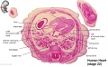
 Stage 22 image 175.jpg 1,000 × 666; 158 KB
 Stage 22 image 178.jpg 1,000 × 658; 148 KB
 Stage 22 image 179.jpg 1,000 × 658; 131 KB
 Stage10 sem11.jpg 1,000 × 636; 76 KB
 Stage11 historic-Atwell1930-2.jpg 1,000 × 799; 131 KB


.jpg)