Category:Carnegie Embryo 43: Difference between revisions
mNo edit summary |
m (→Larynx) |
||
| Line 29: | Line 29: | ||
{{Carnegie Collection stage 19 table}} | {{Carnegie Collection stage 19 table}} | ||
==Limb== | |||
{{Ref-Lewis1902}} | |||
Embryo [[:Category:Carnegie Embryo 43|'''XLIII''']] measures 16 mm. V. B. and 11 mm. jST. B. It is about six weeks old. Many changes have taken place during the sixth Aveck. The entire arm has migrated posteriorly, dragging muscles and nerves with it. The brachial plexus has a decided posterior inclination. The skeletal system is much farther advanced and consists for the most part of cartilage; its individual elements are assuming more the adult form. The clavicle now unites the arm and thoracic skeletons. | |||
The muscular tissues have become more clearly differentiated and except in the hand are easily distinguished. Muscles, such as the trapezius, serratus, and pectoral, have spread out into sheets and acquired more their permanent attachments, in the case of the trapezius, latissimus and pectorals by migTation or extension of their fibers. | |||
In the hand, however, we find the interossei still in an undifferentiated condition like that of the deep flexor layer in embryo CIX or the serratus and infrahyoids in embryo CLXIIL' | |||
===The Skeletal System=== | |||
The vertebral column. The intervertebral discs are of still more compact tissue than in embryo CIX, but they occupy only about one-fourth height of the segment, while in CIX they occupied nearly one-half the anterior-posterior length of the segment. The body of each vertebra contains a large mass of cartilage, which is continuous with the cartilage in the transverse and neural processes. Indications of the hypochordal brace of Froriep are present in connection with the ventral side of the first three discs. The anterior one is the largest, the others decreasing rapidly in size. | |||
The ribs are composed of long, slender cartilages, surrounded by a thick perichondrium. This is continuous with the condensed tissue of the tips of the ribs. The tips of the first seven ribs are connected by a narrow strip of condensed tissue which appears to be formed by the turning anteriorly of their tips until they touch the rib above and fuse with it. Thus is formed the' anlage of one-half the sternum on either side some little distance from the median line. There is at present no sign of union of the two halves of the sternal anlagen. The first rib is fused with the median end of the clavicle. The ribs show a marked increase in their lateral convexity, as in embryo CIX there was scarcely any. There are no joint cavities between the ribs and vertebrte. | |||
The scapula is composed largely of cartilage. It has migrated posteriorly so that less than one-half of it lies above the level of the first rib. The whole scapula is larger than in embryo CIX. There is a thick layer of perichondriuui around the cartilage and a considerable mass of condensed tissue along the vertebral border, and at the posterior angle, the cartilage reaches to the level of the third rib and the condensed tissue nearly to the fifth. The anterior border is somewhat irregular and thickened and gives origin in part to the supraspinatus muscle. The lateral lip of this border probably represents the spine and the median lip the anterior border. Projecting from the lateral side of the head and continuous with the lateral lip of the anterior border is the acromion process. It is large, curved and mostly of condensed tissue and contains a slender core of cartilage continuous with the cartilage of the body. The coracoid process arises from the median side of the head, is larger than the acromion, and contains a much larger cartilao-inous core, which is continuous with the cartilage of the body. The acromio-clavicular ligament is strongly developed. | |||
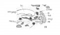
'''Fig. 12.''' Cartilaginous slceleton of tlie arm of embryo [[:Category:Carnegie Embryo 43|'''XLIII''']], lateral view. X 20 diameters. | |||
The clavicle consists of a thick mass of condensed tissue, extending from the acromion to the tip of the first rib, where it continues with the half sternal anlage. There is no line of separation at either end. There is a small core of a peculiar precartilaginous tissue. | |||
The humerus is larger, longer and more slender in proportion to its length than in the preceding stage. The two ends are enlarged. The main portion is of cartilage snrronnded by a thick perichondrihm which is continuous with that of the head of the scapnla, forming the Ijeginning of the capsular ligament. There is also a strip of perichondrium between scapula and humerus in which there are no signs of a joint cavity. At the proximal end the perichondrium shows thickenings for the tuberosities, while at the distal end the condyles are for the most part of cartilage continuous with that of the main portion. Considerable masses of condensed tissue, however, help to increase the size of the condyles. A portion of the head of the humerus rests against the base of the eoracoid process, indicating that a portion of this is to be incorporated with the head of the scapula. | |||
The ulna and radius are of cartilage surrounded by a thick perichondrium. This is continuous with that of the distal end of the humerus, forming the beginning of the capsule. The perichondrium of the proximal end of the radius is continuous with that of tlie adjoining surface of the ulna. The cartilages of the humerus, radius and ulna are separated from each other by condensed tissue in which no signs of cavities are present. The olecranon is quite well developed and consists mostly of cartilage. The coronoid process is mostly of condensed tissue. The great sigmoid fossa is rather shallow. The bicipital tuberosity is of condensed tissue. The distal ends of these bones are enlarged and separated from each other by condensed tissue continuous with the perichondrium of each. | |||
The carpus consists of a condensed tissue matrix in which lie imbedded the various cartilages. The distal row is complete, the trapezium, trapezoid, os magnum and unciform. The latter has spread in between the fifth metacarpal and the cuneiform (pyramidal). In the proximal row the cuneiform and scaphoid are of cartilage and the hmar and pisiform of condensed tissue. | |||
The metacarpus shows five slender cartilages surrounded by very thick condensed tissue layer or perichondrium. The first metacarpal cartilage is only about one-half the length of the others. | |||
The ulnar four plialanges of the first row are present as short slender cartilages deeply imbedded in condensed tissue. In the first digit condensed tissue takes the place of the cartilage. At the tip of each digit is a mass of condensed tissue. | |||
There are no joint cavities between the cartilages of the hand, each one is separated from its neighbor by an area of condensed tissue. | |||
Hagen has reconstnicted the cartilaginous skeletal system of a hnman embryo of abont this age. A comparison of the drawings from the reconstructions shows that there is considerable variation in the carpal region. In none of my stages does the metacarpal come irt contact with the radius, either before or after the cartilages of thecarpus and metacarpus appear, and there is a considerable area of dense mesenchyma between metacarpus and radius. I am inclined to believe what he calls metacarpal I, may be trapezium and his so-called firstphalanx the metacarpal. | |||
===The Muscular System=== | |||
Plates I and II, Figs. A and B. The trapezius muscle has both clavicular and acromial attachments. The muscle has extended posteriorly so that the muscle fibers run from theocciput to the level of the fifth rib. They are connected by a considerable interval of fascia with the dorsal ends of the cervical and all the thoracic neural processes. | |||
The levator scapulce and serratus anterior muscles are greatly altered iit shape. The latter forms a broad, thin sheet between the dorsal border of the scapula and the first nine ribs, being attached by a digitation tO' each rib. The scapular attachment is into the condensed tissue along its dorsal border. | |||
The pectoral mass is now spread out into a large, thin sheet, which has split into the major and minor muscles. The clavicular and sternocostal portions of the pedoralis major are separated by a considerable interval. The clavicular fibers arise from the median one-third of the clavicle and pass to the humerus. They overlap the humeral ends of the sterno-costal fibers which arise from the first six ribs and the sternal anlage. | |||
The pedoralis minor is a distinct muscle arising from the second, third and fourth ribs and passing to the coracoid process. | |||
The subelavius muscle is quite Avell developed and runs from the first ril) to the clavicle, having a course nearly at right angles to the latter. | |||
The latissimus dorsi has spread out into a broad, thin sheet of muscle fibers, which are connected by fascia with the lower thoracic and lumbar neirral processes. Its humeral end is closely miited with the teres major. | |||
The teres major muscle has about the relations found in the adult. It and the latissimus dorsi are inserted together into the humerus. | |||
Tlie deltoid muscle is very much like the adult in its attachments and shape. | |||
Hagen, Die Bildung des Knorpelskeletes beim mensclalicben Embryo, Arch, fiir Anat. u. Pbys., 1900. | |||
The infraspinatus nniscle arises from the anterior portion of the lateral sin-face of the scapula and can bo easily traced to its insertion into the great tuberosity of the humerus. The teres minor cannot be separated from it. | |||
The supraspinal us muscle arises from the anterior thickened border of the scapula and passes to the great tuberosity of the humerus. | |||
The suhscapularis muscle occupies the central portion of the median surface of the scapula. It is separated from the teres major. It passes beneath coracoid process to the lesser tuberosity of the humerus. | |||
The triceps muscle is easily traced from its origin by the three heads to its insertion into the olecranon process. The three heads are quite easily distinguished. The long head is smaller in proportion than in the adult. | |||
The biceps muscle is more elongated and shows more of a separation of its two heads than in embryo CIX. The long head still arises from the base of the coracoid process. The two heads join about the middle of the humerus and pass to a thickening of condensed tissue on the radius. The short head arises in common with the coracobrachialis muscle from the tip of the coracoid process. This latter muscle is inserted into the middle of the median surface of the humerus. It is closely connected with the biceps for most of its length. | |||
The hracJiiaUs muscle is spread out more over the distal portion of the humerus and its muscle fibers extend farther toward the insertion into the coronoid process of the ulna than in the preceding stage. | |||
The flexor mass of the forearm and hand show a most marked advance over the preceding stage. The various muscles of the superficial layer which arise from the internal condyle are easily recognized. They are more or less fused at their origin and for some little distance from it. | |||
The pahnaris longus muscle, the most superficial one, is thin and wide, ends in the condensed tissue of the palmar fascia. | |||
The pronator teres muscle passes to the middle of the shaft of the radius. | |||
The flexor carpi radialis muscle lies mostly on the radial side of the forearm, towards the distal end of which it bends under the deep flexor and ends in a condensed tissue tendon which fuses with the condensed tissue near the proximal end of the second metacarpal. This portion of the muscle is not yet clearly differentiated from the condensed tissue on the palmar surface of the caqms. | |||
The flexor digitonim suhlimis muscle arises beneath the palmaris longus in connection with it from the internal condyle, and also from the shaft of tlie ulna, for a little distance distal of the coronoid ]~)rocess. | |||
It is very broad and spreads out over the middle of the forearm and carpus, where it divides into fonr broad, thin tendons which fnse with the condensed tissue surrounding the distal end of the four ulnar metacarpals and first row of phalanges. The muscle fibers continue distal as far as the middle of the carpus, where the muscle becomes wider and thicker. The tendons do not show the split which is later to appear and enclose the deep flexor tendon. The strongest part of the tendons lie on the ulnar side of digits. | |||
The deep layer of the preceding stage has undergone marked changes. | |||
The flexor carpi ulnaris muscle is quite distinct. It arises partly from the internal condyle superficial to the sublimis and closely connected with it and the palmaris longus and partly from the ulna. The muscle at its origin is broad and thin but narrows into a condensed tissue tendon which is inserted into the os pisiform. | |||
The flexor digitorum profundus and the flexor polUcis longus muscles arise from the surfaces of the radius and ulna and the internal condyle. They are closely imited and pass to the carpal region where division takes place into five well-formed oval tendons, which pass beneath the tendons of the sublimis, and fuse with the condensed tissue about the ends of the digits. | |||
The pronator quadraius muscle is a small, oval mass connecting the distal ends of the ulna and radius. | |||
The lumhride muscles are fonned. They arise from the profundus near the angles formed by the iive tendons. They are short and contain distinct muscle fibers which end in tendons that fuse with the condensed tissue on the radial side of the ulnar four digits. | |||
The intrinsic muscles of the hand, the interossei, and muscles of the thumb and little finger, are represented by a late premuscle tissue in which a few muscle fibers are beginning to appear. These masses are more or less continuous with each other and lie on the palmar surface of the carpus and metacarpus and partially in between the latter. The distal ends of these masses fuse with the less differentiated condensed tissue about the digits. | |||
The extensor muscles of the forearm show considerable advance over the preceding stage, but the development does not seem to have been as rapid as in the case of the flexor muscles. | |||
Of the first group, the extensor communis digitorum and the extensor minimi digiti are united into a broad, thin sheet which divides in the metacarpal region into four broad, thin tendons that end in the condensed tissue of the four ulnar digits. The extensor carpi ulnaris closely associated with this muscle at its origin from the external condyle arises also partly from the ulna and is inserted into the condensed tissue at the proximal end of the fifth metacarpal. It is quite separate from the common extensor for the greater part of its length. | |||
Of the second group, the hracMoradinlis is quite distinct from the extensor- carpi radialis longior et hrevior for most of its length, but at their origin, however, the two are closely connected. Both muscles are broader and larger than in the preceding stage. The extensor passes beneath the third gTOup and ends in the condensed tissue near the proximal ends of the second and third metacarpals. | |||
The third group, which arises beneath the first from both radius and ulna, has split more or less into four parts. The proximal one, which is the most completely separated, is the supinator and passes from the ulna and external condyle to the radius. It is united with rest of this group along their ulnar origins, forming thus a continuous sheet for a short distance. The next two pass over the extensor carpi radii tendon, and fuse with the condensed tissue of the first digit. They are the abductor pollicis longits, extensor poUicis brevis and the extensor poUicis longus muscles. The fourth division is broad and thin and soon joins the deep surface of the tendon of the extensor communis and goes with it to be inserted into the condensed tissue of the second digit. | |||
===The Nerves=== | |||
By the migration of the arm posteriorly the brachial plexus has been pulled caudally and given a decided posterior inclination. It has also divided into the various cords more than in the preceding stage. | |||
The distribution of the muscle and cutaneous nerves is much as in. the adult and as in the next stage. | |||
==Larynx== | ==Larynx== | ||
| Line 111: | Line 229: | ||
to the glossopharyngeal and to the hypoglossus (also figs. 15, 16, | to the glossopharyngeal and to the hypoglossus (also figs. 15, 16, | ||
19, 20). | 19, 20). | ||
==Caudal end of the spinal cord== | ==Caudal end of the spinal cord== | ||
Revision as of 09:42, 20 October 2020
This Embryology category shows pages and images that relate to the Carnegie Collection Embryo No. 43. This embryo was classified as Carnegie stage 19 occurring during Week 7.
| Carnegie Collection - Stage 19 | |||||||||||
|---|---|---|---|---|---|---|---|---|---|---|---|
| Serial No. | Size (mm) | Grade | Fixative | Embedding Medium | Plane | Thinness (µm) | Stain | Score | Sex | Year | Notes |
| 43 | E, 16 | Good | Alc. | P | 50 | Al. coch. | 10 | M 1 | 1894 | ||
Abbreviations
| |||||||||||
References
Bardeen CR. and Lewis WH. The development of the limbs, body-wall and back. (1901) Amer. J Anat. 1: 1-36.
Lisser H. Studies on the development of the human larynx. (1911) Amer. J Anat. 12: 27-66.
Kunitomo K. The development and reduction of the tail and of the caudal end of the spinal cord (1920) Contrib. Embryol., Carnegie Inst. Wash. Publ. 272, 9: 163-198.
| Carnegie Collection - Stage 19 | |||||||||||
|---|---|---|---|---|---|---|---|---|---|---|---|
| Serial No. | Size (mm) | Grade | Fixative | Embedding Medium | Plane | Thinness (µm) | Stain | Score | Sex | Year | Notes |
| 17 | E, 18 Ch, 40x30x20 | Poor | Alc. | P | 50, 100 | Al. carm. | 16.5 | Male | 1894 | ||
| 43 | E, 16 | Good | Alc. | P | 50 | Al. coch. | 10 | Male | 1894 | ||
| 293 | E, 19 | Poor | Ale. | P | Sagittal | 50 | Coch. | 16.5 | S | 1905 | |
| 390 | E, 19 | Good | Formol? | P | Sagittal | 20, | (Stain - Haematoxylin Eosin) | 11.5 | Male | 1906 | Tubal Injected
50 |
| 409 | E.18 Ch, 50x40x40 | Good | Formalin | P | Transverse | 20 | Copper, iron H. & erythrosin | 14.5 | Male | 1907 | |
| 432 | E..18.5 Ch , 45x35x20 | Good | Formalin | P | Sagittal | 20 | H. & Congo red | 13.5 | Male | 1910 | Tubal |
| 576 | E. 17 Ch, 60x40 | Good | Formalin | P | Sagittal | 15, 20 | (Stain - Haematoxylin Eosin) | 14.5 | d | 1912 | Tubal |
| 626 | E., 21.5 Ch., 40x30x21 | Good | Formalin | P | Transverse | 100 | Al. coch. | 14_5 | 6 | 1913 | |
| 6??8 | E, 20 Ch, ca. 30 | Poor | Formalin | P | Sagittal | 50 | Al. coch. | 12 | 9 | 1913 | Head damaged |
| 709 | E, 19 Ch. 40x35x25 | Poor | Alc. | P | Coronal | 40 | Al. coch, Lyons blue | 15 | 49 | 1913 | |
| 837 | E. 21 Ch. 65x45x | Good | Formalin | P | Sagittal | 40 | Al. coch. | 14.5 | P | 1914 | |
| 1324 | E., 18 50x30x18 | Good | Formalin | C | Coronal | 40 | (Stain - Haematoxylin Eosin), aur, or. G | 125 | 79 | 1915 | |
| 1332 | E., 19 Ch., 40x43x22 | Poor | Formalin | C | Coronal | 40 | (Stain - Haematoxylin Eosin) aur, or. G. | 15 | Male | 1915 | |
| 1390 | E., 18 Ch, 40x38x15 | Good | Formalin | P | Sagittal | 20 | Al. coch. | 10_5 | Male | 1915 | Tubal |
| 1534 | E., 13 Ch.,35x31x25 | Poor | Formalin | P | 53% | 50 | Al. coch. | 13.5 | F | 1916 | Protruding midbrain |
| 2114 | E., 19.3 Ch., 49x42x33 | Good | Formol | P | Transverse | 40 | A1. coch. | 12 | M | 1918 | |
| 4405 | E., 15.5 | Good | Formalin | P | Transverse | 10 | Coch, Mallory | 13.5 | <3 | 1923 | Midbrain injured |
| 4501 | E, 18 | Exc. | Bouin | P | Transverse | 15 | Coch, or. G. | 14.6 | 1924 | Cystic left kidney | |
| 5609 | E., 18 | Exc. | Formalin | P | Coronal | 25 | A1. coch. | 13.5 | Male | ||
| 6150 | E., 17 Ch., 40x39x30 | Good | Bouin | C-P | Transverse | 15 | (Stain - Haematoxylin Eosin) | 16.5 | Male | 1930 | Tubal |
| 6824 | E., 18.5 Ch., 45x40x25 | Good | Formalin | C-P | Sagittal | 12 | (Stain - Haematoxylin Eosin) | 14.5 | Female | 1933 | |
| 7900 | E., 16.5 | Good | Bouin | C-P | Sagittal | 20 | (Stain - Haematoxylin Eosin), phlox. | 11.5 | . . | 1941 | Tubal |
| 8092 | E., 16.3 Ch., 52 x 47 | Exc. | Bouin | C-P | Transverse | 20 | (Stain - Haematoxylin Eosin), phlox. | 13 | Male | 1942 | |
| 8913 | E.,? Ch, 34 | Poor | Formalin | p | Transverse | 10 | Alan . | 7 | 1951 | rubella. Medical abortion. Isolated head damaged | |
| 8965 | E, 19.1 Ch, 42x32x19 | Good | Formol—Zenker | C-P | Transverse | 10 | Borax, carm, or. G. | 1952 | Univ. Chicago No. H 173 | ||
| 9097 | E, 21 | Exc. | Formol—glucose | C-P | Coronal | 10 | Azan ? . | ? | 1930 | Univ. Chicago No H 1380 | |
| 9113 | E, 185 Ch, 24 | Exc. | Formalin | C-P | Transverse | 10 | Alan > 6 | 1953 | Rubella. Medical abortion | ||
| 9325 | E, 17.0 Ch, 32x28x20 | Good | Formalin | —acetic p | Transverse | 15& 8-10 | Azan & Ag | ? | - | 1955 | Tubal |
Abbreviations
| |||||||||||
Limb
Lewis WH. The development of the arm in man. (1902) Amer. J Anat. 1(2): 145-184.
Embryo XLIII measures 16 mm. V. B. and 11 mm. jST. B. It is about six weeks old. Many changes have taken place during the sixth Aveck. The entire arm has migrated posteriorly, dragging muscles and nerves with it. The brachial plexus has a decided posterior inclination. The skeletal system is much farther advanced and consists for the most part of cartilage; its individual elements are assuming more the adult form. The clavicle now unites the arm and thoracic skeletons.
The muscular tissues have become more clearly differentiated and except in the hand are easily distinguished. Muscles, such as the trapezius, serratus, and pectoral, have spread out into sheets and acquired more their permanent attachments, in the case of the trapezius, latissimus and pectorals by migTation or extension of their fibers.
In the hand, however, we find the interossei still in an undifferentiated condition like that of the deep flexor layer in embryo CIX or the serratus and infrahyoids in embryo CLXIIL'
The Skeletal System
The vertebral column. The intervertebral discs are of still more compact tissue than in embryo CIX, but they occupy only about one-fourth height of the segment, while in CIX they occupied nearly one-half the anterior-posterior length of the segment. The body of each vertebra contains a large mass of cartilage, which is continuous with the cartilage in the transverse and neural processes. Indications of the hypochordal brace of Froriep are present in connection with the ventral side of the first three discs. The anterior one is the largest, the others decreasing rapidly in size.
The ribs are composed of long, slender cartilages, surrounded by a thick perichondrium. This is continuous with the condensed tissue of the tips of the ribs. The tips of the first seven ribs are connected by a narrow strip of condensed tissue which appears to be formed by the turning anteriorly of their tips until they touch the rib above and fuse with it. Thus is formed the' anlage of one-half the sternum on either side some little distance from the median line. There is at present no sign of union of the two halves of the sternal anlagen. The first rib is fused with the median end of the clavicle. The ribs show a marked increase in their lateral convexity, as in embryo CIX there was scarcely any. There are no joint cavities between the ribs and vertebrte.
The scapula is composed largely of cartilage. It has migrated posteriorly so that less than one-half of it lies above the level of the first rib. The whole scapula is larger than in embryo CIX. There is a thick layer of perichondriuui around the cartilage and a considerable mass of condensed tissue along the vertebral border, and at the posterior angle, the cartilage reaches to the level of the third rib and the condensed tissue nearly to the fifth. The anterior border is somewhat irregular and thickened and gives origin in part to the supraspinatus muscle. The lateral lip of this border probably represents the spine and the median lip the anterior border. Projecting from the lateral side of the head and continuous with the lateral lip of the anterior border is the acromion process. It is large, curved and mostly of condensed tissue and contains a slender core of cartilage continuous with the cartilage of the body. The coracoid process arises from the median side of the head, is larger than the acromion, and contains a much larger cartilao-inous core, which is continuous with the cartilage of the body. The acromio-clavicular ligament is strongly developed.
Fig. 12. Cartilaginous slceleton of tlie arm of embryo XLIII, lateral view. X 20 diameters.
The clavicle consists of a thick mass of condensed tissue, extending from the acromion to the tip of the first rib, where it continues with the half sternal anlage. There is no line of separation at either end. There is a small core of a peculiar precartilaginous tissue.
The humerus is larger, longer and more slender in proportion to its length than in the preceding stage. The two ends are enlarged. The main portion is of cartilage snrronnded by a thick perichondrihm which is continuous with that of the head of the scapnla, forming the Ijeginning of the capsular ligament. There is also a strip of perichondrium between scapula and humerus in which there are no signs of a joint cavity. At the proximal end the perichondrium shows thickenings for the tuberosities, while at the distal end the condyles are for the most part of cartilage continuous with that of the main portion. Considerable masses of condensed tissue, however, help to increase the size of the condyles. A portion of the head of the humerus rests against the base of the eoracoid process, indicating that a portion of this is to be incorporated with the head of the scapula.
The ulna and radius are of cartilage surrounded by a thick perichondrium. This is continuous with that of the distal end of the humerus, forming the beginning of the capsule. The perichondrium of the proximal end of the radius is continuous with that of tlie adjoining surface of the ulna. The cartilages of the humerus, radius and ulna are separated from each other by condensed tissue in which no signs of cavities are present. The olecranon is quite well developed and consists mostly of cartilage. The coronoid process is mostly of condensed tissue. The great sigmoid fossa is rather shallow. The bicipital tuberosity is of condensed tissue. The distal ends of these bones are enlarged and separated from each other by condensed tissue continuous with the perichondrium of each.
The carpus consists of a condensed tissue matrix in which lie imbedded the various cartilages. The distal row is complete, the trapezium, trapezoid, os magnum and unciform. The latter has spread in between the fifth metacarpal and the cuneiform (pyramidal). In the proximal row the cuneiform and scaphoid are of cartilage and the hmar and pisiform of condensed tissue.
The metacarpus shows five slender cartilages surrounded by very thick condensed tissue layer or perichondrium. The first metacarpal cartilage is only about one-half the length of the others.
The ulnar four plialanges of the first row are present as short slender cartilages deeply imbedded in condensed tissue. In the first digit condensed tissue takes the place of the cartilage. At the tip of each digit is a mass of condensed tissue.
There are no joint cavities between the cartilages of the hand, each one is separated from its neighbor by an area of condensed tissue.
Hagen has reconstnicted the cartilaginous skeletal system of a hnman embryo of abont this age. A comparison of the drawings from the reconstructions shows that there is considerable variation in the carpal region. In none of my stages does the metacarpal come irt contact with the radius, either before or after the cartilages of thecarpus and metacarpus appear, and there is a considerable area of dense mesenchyma between metacarpus and radius. I am inclined to believe what he calls metacarpal I, may be trapezium and his so-called firstphalanx the metacarpal.
The Muscular System
Plates I and II, Figs. A and B. The trapezius muscle has both clavicular and acromial attachments. The muscle has extended posteriorly so that the muscle fibers run from theocciput to the level of the fifth rib. They are connected by a considerable interval of fascia with the dorsal ends of the cervical and all the thoracic neural processes.
The levator scapulce and serratus anterior muscles are greatly altered iit shape. The latter forms a broad, thin sheet between the dorsal border of the scapula and the first nine ribs, being attached by a digitation tO' each rib. The scapular attachment is into the condensed tissue along its dorsal border.
The pectoral mass is now spread out into a large, thin sheet, which has split into the major and minor muscles. The clavicular and sternocostal portions of the pedoralis major are separated by a considerable interval. The clavicular fibers arise from the median one-third of the clavicle and pass to the humerus. They overlap the humeral ends of the sterno-costal fibers which arise from the first six ribs and the sternal anlage.
The pedoralis minor is a distinct muscle arising from the second, third and fourth ribs and passing to the coracoid process.
The subelavius muscle is quite Avell developed and runs from the first ril) to the clavicle, having a course nearly at right angles to the latter.
The latissimus dorsi has spread out into a broad, thin sheet of muscle fibers, which are connected by fascia with the lower thoracic and lumbar neirral processes. Its humeral end is closely miited with the teres major.
The teres major muscle has about the relations found in the adult. It and the latissimus dorsi are inserted together into the humerus.
Tlie deltoid muscle is very much like the adult in its attachments and shape.
Hagen, Die Bildung des Knorpelskeletes beim mensclalicben Embryo, Arch, fiir Anat. u. Pbys., 1900.
The infraspinatus nniscle arises from the anterior portion of the lateral sin-face of the scapula and can bo easily traced to its insertion into the great tuberosity of the humerus. The teres minor cannot be separated from it.
The supraspinal us muscle arises from the anterior thickened border of the scapula and passes to the great tuberosity of the humerus.
The suhscapularis muscle occupies the central portion of the median surface of the scapula. It is separated from the teres major. It passes beneath coracoid process to the lesser tuberosity of the humerus.
The triceps muscle is easily traced from its origin by the three heads to its insertion into the olecranon process. The three heads are quite easily distinguished. The long head is smaller in proportion than in the adult.
The biceps muscle is more elongated and shows more of a separation of its two heads than in embryo CIX. The long head still arises from the base of the coracoid process. The two heads join about the middle of the humerus and pass to a thickening of condensed tissue on the radius. The short head arises in common with the coracobrachialis muscle from the tip of the coracoid process. This latter muscle is inserted into the middle of the median surface of the humerus. It is closely connected with the biceps for most of its length.
The hracJiiaUs muscle is spread out more over the distal portion of the humerus and its muscle fibers extend farther toward the insertion into the coronoid process of the ulna than in the preceding stage.
The flexor mass of the forearm and hand show a most marked advance over the preceding stage. The various muscles of the superficial layer which arise from the internal condyle are easily recognized. They are more or less fused at their origin and for some little distance from it.
The pahnaris longus muscle, the most superficial one, is thin and wide, ends in the condensed tissue of the palmar fascia.
The pronator teres muscle passes to the middle of the shaft of the radius.
The flexor carpi radialis muscle lies mostly on the radial side of the forearm, towards the distal end of which it bends under the deep flexor and ends in a condensed tissue tendon which fuses with the condensed tissue near the proximal end of the second metacarpal. This portion of the muscle is not yet clearly differentiated from the condensed tissue on the palmar surface of the caqms.
The flexor digitonim suhlimis muscle arises beneath the palmaris longus in connection with it from the internal condyle, and also from the shaft of tlie ulna, for a little distance distal of the coronoid ]~)rocess.
It is very broad and spreads out over the middle of the forearm and carpus, where it divides into fonr broad, thin tendons which fnse with the condensed tissue surrounding the distal end of the four ulnar metacarpals and first row of phalanges. The muscle fibers continue distal as far as the middle of the carpus, where the muscle becomes wider and thicker. The tendons do not show the split which is later to appear and enclose the deep flexor tendon. The strongest part of the tendons lie on the ulnar side of digits.
The deep layer of the preceding stage has undergone marked changes.
The flexor carpi ulnaris muscle is quite distinct. It arises partly from the internal condyle superficial to the sublimis and closely connected with it and the palmaris longus and partly from the ulna. The muscle at its origin is broad and thin but narrows into a condensed tissue tendon which is inserted into the os pisiform.
The flexor digitorum profundus and the flexor polUcis longus muscles arise from the surfaces of the radius and ulna and the internal condyle. They are closely imited and pass to the carpal region where division takes place into five well-formed oval tendons, which pass beneath the tendons of the sublimis, and fuse with the condensed tissue about the ends of the digits.
The pronator quadraius muscle is a small, oval mass connecting the distal ends of the ulna and radius.
The lumhride muscles are fonned. They arise from the profundus near the angles formed by the iive tendons. They are short and contain distinct muscle fibers which end in tendons that fuse with the condensed tissue on the radial side of the ulnar four digits.
The intrinsic muscles of the hand, the interossei, and muscles of the thumb and little finger, are represented by a late premuscle tissue in which a few muscle fibers are beginning to appear. These masses are more or less continuous with each other and lie on the palmar surface of the carpus and metacarpus and partially in between the latter. The distal ends of these masses fuse with the less differentiated condensed tissue about the digits.
The extensor muscles of the forearm show considerable advance over the preceding stage, but the development does not seem to have been as rapid as in the case of the flexor muscles.
Of the first group, the extensor communis digitorum and the extensor minimi digiti are united into a broad, thin sheet which divides in the metacarpal region into four broad, thin tendons that end in the condensed tissue of the four ulnar digits. The extensor carpi ulnaris closely associated with this muscle at its origin from the external condyle arises also partly from the ulna and is inserted into the condensed tissue at the proximal end of the fifth metacarpal. It is quite separate from the common extensor for the greater part of its length.
Of the second group, the hracMoradinlis is quite distinct from the extensor- carpi radialis longior et hrevior for most of its length, but at their origin, however, the two are closely connected. Both muscles are broader and larger than in the preceding stage. The extensor passes beneath the third gTOup and ends in the condensed tissue near the proximal ends of the second and third metacarpals.
The third group, which arises beneath the first from both radius and ulna, has split more or less into four parts. The proximal one, which is the most completely separated, is the supinator and passes from the ulna and external condyle to the radius. It is united with rest of this group along their ulnar origins, forming thus a continuous sheet for a short distance. The next two pass over the extensor carpi radii tendon, and fuse with the condensed tissue of the first digit. They are the abductor pollicis longits, extensor poUicis brevis and the extensor poUicis longus muscles. The fourth division is broad and thin and soon joins the deep surface of the tendon of the extensor communis and goes with it to be inserted into the condensed tissue of the second digit.
The Nerves
By the migration of the arm posteriorly the brachial plexus has been pulled caudally and given a decided posterior inclination. It has also divided into the various cords more than in the preceding stage.
The distribution of the muscle and cutaneous nerves is much as in. the adult and as in the next stage.
Larynx
Lisser H. Studies on the development of the human larynx. (1911) Amer. J Anat. 12: 27-66.
Embryo no. 43- 16 mm Sagittal sections Embryo no. 43 measures 16 mm V.B. and 4 mm N.B. about six weeks old.
The Cartilages
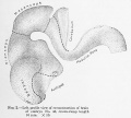
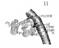
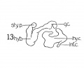
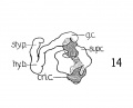
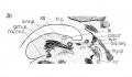
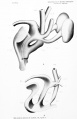
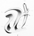
The thyreoid cartilage is a peculiar structure at this stage, still consisting purely of condensed mesenchyma. The lateral alae are united ventrally, but it is to the odd shape of the lateral masses, that I would call attention. Fig. 13 shows a graphic reconstruction of this cartilage from the side view. The superior cornu, fig. 13, is in evidence, and is in correct relation to the greater cornu of the hyoid bone, between which develops the thyreohyoid ligament. Another point in favor of this being the superior cornu of the thyreoid, is the attachment to it of the inferior constrictor pharyngis, as seen in fig. 11. Posterior, and below them, protrudes a curious cylindrical mass of condensed mesenchyma, which undoubtedly forms the rudiment of the inferior cornu of the thyreoid. It overlaps the cricoid and is in close apposition to it as shown in fig. 14. Probably there is no actual articular facet at this stage. Anteriorly, there is a strange projection, without apparent attachment to anything ; it seems to be evidence of greater activity of growth in the ventral part of this lateral mass, just as the superior and inferior cornua are the results of active growth in the posterior portion of this lateral mass. Apparently then, the directly lateral part lags behind temporarily, and the peculiar gap between the anterior cornu (as I call this odd projection) and the superior cornu, is filled in during the next week or so.
In the 20 mm. stage, there appears to be a slight tendency to condensation in this area, not marked enough to be included in the reconstruction of this stage.
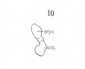
The cricoid cartilage (fig. 10) consists of pure condensed mesenchyma, with no evidence as yei of chondrification. Although rather crude in outline, yet it begins to suggest roughly the maturer form. Its relation to the thyreoid cartilage resembles the adult rather closely, and the continued ring, ventral and dorsal, is now complete. Also, the posterior portion is enlarging and begins to show advances over the relatively slower growth of the anterior arcus, conforming with the adult type. Certainly, it is further advanced than the thyreoid cartilage.
The aryiaenoids are still represented by a roughly oval mass of condensed mesenchyma, with no accuracy of form or outline.
The aryiaenoids are still represented by a roughly oval mass of condensed mesenchyma, with no accuracy of form or outline. Fig. 10 is a reconstruction of one of them from a lateral view. The mass is continuous with the cricoid mass, as indicated by the cross lined area, (fig. 14) but its main arytaenoid portion is stained deeply enough to differentiate it absolutely from being a part of the cricoid mass.
The epiglottis shows but little advance over its condition in the earlier embryos, except for some gain in length over breadth, but the mass out of which it assumes its adult shape, is easily recognized.
The hyoid hone and styloid process begin to show small areas of chondrification, and their appearance is seen in a reconstruction fig. 13 and 14. The attachment of the middle constrictor to the greater cornu is shown in fig. 11.
The Muscles
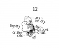
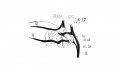
In a 14 mm. embryo Strazza says that a muscle mass, of spindle shaped formation can be made out laterally in cross sections, but that no distinct muscles can be isolated at this stage, and that the mass simply represents laryngeal musculature. And at 16 mm. he distinguishes a muscle band, bending around with the posterior convexity, which in its upper portions he calls the arytaenoideus transversus (interarytaenoideus), but considers this band a continuous muscle mass, only differentiated later by the development of the cartilages. Further down he thinks it to be the cricoary-taenoideus lateralis and thyreoarytaenoideus, but says it is con- tinuous with the above, only spread over a greater area. So he calls the larynx musculature an arch, which however, is not entirely horizontal, but goes from above and behind, to below and in front. Now it is true that considerable interlacing of fibres exists at this stage, but not much more than occurs in careful dissections of the adult larynx. It is also true that further separation and development of the cartilages will bring about clearer differentiation of the muscles. But that a continuous muscle band, which cannot be differentiated into individual muscles, exists at this stage is not in accord with the results shown, especially in fig. 12, a reconstruction of the 16 mm. stage, and in figs. 2, 8, 9, faithful drawings of the sections of the 14 mm. embryo, and fig. 15 of the 16 mm. embryo.
The cricothyreoideus, the cricoarytaenoideus posterior, and the
interarytaenoideus are definitely isolated, while the cricoarytaenoideus lateralis and thyreoarytaenoideus are clearly separated from the
others aiid are about as much separated from each other as they
are in the adult. There is no need of describing them any further.
The figures show them all in sufficient detail.
The constrictor muscles of the pharynx and oesophagus have
been reconstructed and the middle and inferior constrictors stand
out rather clearly. Nicolas states that the pharynx musculature
only unites from two independent lateral halves at 3 cm. I have
found perfect continuity at 10.5 mm. in the lower pharyngeal
portion, more union at 12.5 mm. and complete union at 14 nun.
Several of the tongue and pharynx muscles have been included
in the illustrations, and it will be seen that they are very clearly
isolated and well developed even at the 14 mm. stage (figs. 7-9).
For the identity of these muscles, I am under obligations to Dr.
Lewis, who very kindly gave me his own sketches, from which
figs. 7, 8 and 9 were developed.
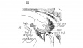
The Nerves
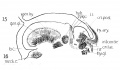
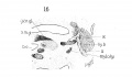
The nerves, are reconstructed in fig. 14 and in addition to showing the superior laryngeus and n. recurrens and n. laryngeus inferior, which can be followed to their respective innervations and to their anastomosis, there is included the relations of these, to the glossopharyngeal and to the hypoglossus (also figs. 15, 16, 19, 20).
Caudal end of the spinal cord
Kunitomo K. The development and reduction of the tail and of the caudal end of the spinal cord (1920) Contrib. Embryol., Carnegie Inst. Wash. Publ. 272, 9: 163-198.
Embryo No. 43, 16 mm Crown-Rump Length. This specimen has 37 cartilaginous vertebrae, the last being divided into three parts. There are 32 spinal ganglia. A graphic reconstruction was made of this embryo, but it is not illustrated in the figures.
| Carnegie Collection - Carnegie Embryos Sorted by Stage | |||||||||||||||||||||||||||||||||||||||||||||||||||||||||||||||||||||||||||||||||||||||||||||||||||||||||||||||||||||||||||||||||||||||||||||||||||||||||||||||||||||||||||||||||||||||||||||||||||||||||||||||||||||||||||||||||||||||||||||||||||||||||||||||||||||||||||||||||||||||||||||||||||||||||||||||||||||||||||||||||||||||||||||||||||||||||||||||||||||||||||||||||||||||||||||||||||||||||||||||||||||||||||||||||||||||||||||||||||||||||||||||||||||||||||||||||||||||||||||||||||||||||||||||||||||||||||||||||||||||||||||||||||||||||||||||||||||||||||||||||||||||||||||||||||||||||||||||||||||||||||||||||||||||||||||||||||||||||||||||||||||||||||||||||||||||||||||||||||||||||||||||||||||||||||||||||||||||||||||||||||||||||||||||||||||||||||||||||||||||||||||||||||||||||||||||||||||||||||||||||||||||||||||||||||||||||||||||||||||||||||||||||||||||||||||||||||||||||||||||||||||||||||||||||||||||||||||||||||||||||||||||||||||||||||||||||||||||||||||||||||||||||||||||||||||||||||||||||||||||
|---|---|---|---|---|---|---|---|---|---|---|---|---|---|---|---|---|---|---|---|---|---|---|---|---|---|---|---|---|---|---|---|---|---|---|---|---|---|---|---|---|---|---|---|---|---|---|---|---|---|---|---|---|---|---|---|---|---|---|---|---|---|---|---|---|---|---|---|---|---|---|---|---|---|---|---|---|---|---|---|---|---|---|---|---|---|---|---|---|---|---|---|---|---|---|---|---|---|---|---|---|---|---|---|---|---|---|---|---|---|---|---|---|---|---|---|---|---|---|---|---|---|---|---|---|---|---|---|---|---|---|---|---|---|---|---|---|---|---|---|---|---|---|---|---|---|---|---|---|---|---|---|---|---|---|---|---|---|---|---|---|---|---|---|---|---|---|---|---|---|---|---|---|---|---|---|---|---|---|---|---|---|---|---|---|---|---|---|---|---|---|---|---|---|---|---|---|---|---|---|---|---|---|---|---|---|---|---|---|---|---|---|---|---|---|---|---|---|---|---|---|---|---|---|---|---|---|---|---|---|---|---|---|---|---|---|---|---|---|---|---|---|---|---|---|---|---|---|---|---|---|---|---|---|---|---|---|---|---|---|---|---|---|---|---|---|---|---|---|---|---|---|---|---|---|---|---|---|---|---|---|---|---|---|---|---|---|---|---|---|---|---|---|---|---|---|---|---|---|---|---|---|---|---|---|---|---|---|---|---|---|---|---|---|---|---|---|---|---|---|---|---|---|---|---|---|---|---|---|---|---|---|---|---|---|---|---|---|---|---|---|---|---|---|---|---|---|---|---|---|---|---|---|---|---|---|---|---|---|---|---|---|---|---|---|---|---|---|---|---|---|---|---|---|---|---|---|---|---|---|---|---|---|---|---|---|---|---|---|---|---|---|---|---|---|---|---|---|---|---|---|---|---|---|---|---|---|---|---|---|---|---|---|---|---|---|---|---|---|---|---|---|---|---|---|---|---|---|---|---|---|---|---|---|---|---|---|---|---|---|---|---|---|---|---|---|---|---|---|---|---|---|---|---|---|---|---|---|---|---|---|---|---|---|---|---|---|---|---|---|---|---|---|---|---|---|---|---|---|---|---|---|---|---|---|---|---|---|---|---|---|---|---|---|---|---|---|---|---|---|---|---|---|---|---|---|---|---|---|---|---|---|---|---|---|---|---|---|---|---|---|---|---|---|---|---|---|---|---|---|---|---|---|---|---|---|---|---|---|---|---|---|---|---|---|---|---|---|---|---|---|---|---|---|---|---|---|---|---|---|---|---|---|---|---|---|---|---|---|---|---|---|---|---|---|---|---|---|---|---|---|---|---|---|---|---|---|---|---|---|---|---|---|---|---|---|---|---|---|---|---|---|---|---|---|---|---|---|---|---|---|---|---|---|---|---|---|---|---|---|---|---|---|---|---|---|---|---|---|---|---|---|---|---|---|---|---|---|---|---|---|---|---|---|---|---|---|---|---|---|---|---|---|---|---|---|---|---|---|---|---|---|---|---|---|---|---|---|---|---|---|---|---|---|---|---|---|---|---|---|---|---|---|---|---|---|---|---|---|---|---|---|---|---|---|---|---|---|---|---|---|---|---|---|---|---|---|---|---|---|---|---|---|---|---|---|---|---|---|---|---|---|---|---|---|---|---|---|---|---|---|---|---|---|---|---|---|---|---|---|---|---|---|---|---|---|---|---|---|---|---|---|---|---|---|---|---|---|---|---|---|---|---|---|---|---|---|---|---|---|---|---|---|---|---|---|---|---|---|---|---|---|---|---|---|---|---|---|---|---|---|---|---|---|---|---|---|---|---|---|---|---|---|---|---|---|---|---|---|---|---|---|---|---|---|---|---|---|---|---|---|---|---|---|---|---|---|---|---|---|---|---|---|---|---|---|---|---|---|---|---|---|---|---|---|---|---|---|---|---|---|---|---|---|---|---|---|---|---|---|---|---|---|---|---|---|---|---|---|---|---|---|---|---|---|---|---|---|---|---|---|---|---|---|---|---|---|---|---|---|---|---|---|---|---|---|---|---|---|---|---|---|---|---|---|---|---|---|---|---|---|---|---|---|---|---|---|---|---|---|---|---|---|---|---|---|---|---|---|---|---|---|---|---|---|---|---|---|---|---|---|---|---|---|---|---|---|---|---|---|---|---|---|---|---|---|---|---|---|---|---|---|---|---|---|---|---|---|---|---|---|---|---|---|---|---|---|---|---|---|---|---|---|---|---|---|---|---|---|---|---|---|---|---|---|---|---|---|---|---|
| |||||||||||||||||||||||||||||||||||||||||||||||||||||||||||||||||||||||||||||||||||||||||||||||||||||||||||||||||||||||||||||||||||||||||||||||||||||||||||||||||||||||||||||||||||||||||||||||||||||||||||||||||||||||||||||||||||||||||||||||||||||||||||||||||||||||||||||||||||||||||||||||||||||||||||||||||||||||||||||||||||||||||||||||||||||||||||||||||||||||||||||||||||||||||||||||||||||||||||||||||||||||||||||||||||||||||||||||||||||||||||||||||||||||||||||||||||||||||||||||||||||||||||||||||||||||||||||||||||||||||||||||||||||||||||||||||||||||||||||||||||||||||||||||||||||||||||||||||||||||||||||||||||||||||||||||||||||||||||||||||||||||||||||||||||||||||||||||||||||||||||||||||||||||||||||||||||||||||||||||||||||||||||||||||||||||||||||||||||||||||||||||||||||||||||||||||||||||||||||||||||||||||||||||||||||||||||||||||||||||||||||||||||||||||||||||||||||||||||||||||||||||||||||||||||||||||||||||||||||||||||||||||||||||||||||||||||||||||||||||||||||||||||||||||||||||||||||||||||||||
| Week: | 1 | 2 | 3 | 4 | 5 | 6 | 7 | 8 |
| Carnegie stage: | 1 2 3 4 | 5 6 | 7 8 9 | 10 11 12 13 | 14 15 | 16 17 | 18 19 | 20 21 22 23 |
Cite this page: Hill, M.A. (2024, April 27) Embryology Carnegie Embryo 43. Retrieved from https://embryology.med.unsw.edu.au/embryology/index.php/Category:Carnegie_Embryo_43
- © Dr Mark Hill 2024, UNSW Embryology ISBN: 978 0 7334 2609 4 - UNSW CRICOS Provider Code No. 00098G
Pages in category 'Carnegie Embryo 43'
The following 5 pages are in this category, out of 5 total.
Media in category 'Carnegie Embryo 43'
The following 14 files are in this category, out of 14 total.
 Jenkins002.jpg 1,230 × 1,102; 321 KB
 Lisser1911 fig10.jpg 791 × 637; 18 KB
 Lisser1911 fig11.jpg 791 × 637; 89 KB
 Lisser1911 fig12.jpg 791 × 637; 47 KB
 Lisser1911 fig13.jpg 791 × 637; 36 KB
 Lisser1911 fig14.jpg 791 × 637; 43 KB
 Lisser1911 fig15.jpg 1,581 × 916; 181 KB
 Lisser1911 fig16.jpg 1,581 × 916; 179 KB
 Lisser1911 fig17.jpg 1,581 × 916; 80 KB
 Lisser1911 fig18.jpg 1,581 × 916; 168 KB
 Lisser1911 fig19.jpg 1,581 × 916; 209 KB
 Lisser1911 fig20.jpg 1,581 × 916; 271 KB
 Pohlman1911 plate3.jpg 2,171 × 3,361; 454 KB
 Pohlman1911 plate3F.jpg 1,418 × 1,512; 98 KB


