Lecture - 2016 Course Introduction: Difference between revisions
mNo edit summary |
mNo edit summary |
||
| Line 43: | Line 43: | ||
|} | |} | ||
==History== | |||
[[History - Embryologists]] | [[Embryology History]] | [[Human Embryo Collections]] | |||
[[File:BrauneB1.jpg|400px|alt=The Position of the Uterus and Fetus at Term (1872)]] | |||
[[Embryology_History_-_17th_and_18th_Century_Anatomies|17-18C]] Braune - The Position of the Uterus and Fetus at Term (1872) | |||
{| class="wikitable mw-collapsible mw-collapsed" | |||
! colspan="3"| [[Human Embryo Collections]] | |||
|- | |||
| [[File:Wilhelm_His.jpg|link=Embryology History - Wilhelm His|200px]] | |||
[[Embryology History - Wilhelm His|Wilhelm His]] (1831-1904) | |||
His's Normentafel (Normal Table) | |||
[[Book - Anatomy Of Human Embryos|Anatomie menschlicher Embryonen]] (1882) | |||
| [[File:Keibel_Mall_034a.jpg|200px]] | |||
| [[File:Keibel_Mall_034b.jpg|300px]] | |||
|- | |||
| [[File:Franz Keibel.jpg|link=Embryology History - Franz Keibel|150px]] | |||
[[Embryology History - Franz Keibel|Franz Keibel]] (1861 - 1929) | |||
Franz Keibel and Curt Elze (1908) Normal Plates of the Development of the Human Embryo | |||
| [[File:Keibel1908_plate01.jpg|200px]] | |||
| [[File:Keibel1908_plate02.jpg|200px]] | |||
|- | |||
| [[File:Franklin Mall 03.jpg|150px]] | |||
[[Embryology History - Franklin Mall|Franklin Mall]] (1862-1917) | |||
[[Carnegie Collection]] | |||
| colspan=2|[[File:Human Carnegie stage 10-23.jpg|400px]] | |||
|- | |||
| Begun by [[:File:Hideo Nishimura.jpg|Dr. Hideo Nishimura]] (1912–1995) | |||
[[File:Hideo Nishimura.jpg|150px]] | |||
Developed by Kohei Shiota and currently curated by Shigehito Yamada. | |||
[[File:Shiota_Hill_Yamada.jpg|200px]] | |||
[[Kyoto Collection]] | |||
| colspan=2|[[File:Human_Carnegie_stage_1-23.jpg|400px]] | |||
|} | |||
{| class="wikitable mw-collapsible mw-collapsed" | |||
! col span=2|[[Animal_Development|Animal Models]] | |||
|- | |||
| [[File:Frog-icon.png|right|80px|link=Frog Development]] | |||
| {{Frog links}} | |||
* The frog was used by many of the early embryology investigators and currently there are many different molecular mechanisms concerning development of the frog. | |||
* The eggs develop independently, in relative synchrony and are relatively see-through making staging and observation fairly easy. | |||
* The frog was a key model for the study of the process of gastrulation. | |||
|- | |||
| [[File:Chick icon.jpg|80px|link=Chicken Development]] | |||
| | |||
{{Chicken}} | |||
* The chicken embryo develops and hatches in 20-21 days and historically these were one of the first embryos to be studied. Cutting a window in the egg shell allows direct observation of the embryo. The Hamburger & Hamilton chicken development staging allowed researchers to develop this model as a key embryological tool. | |||
* Key research involved the transplanting of quail cells into chick embryos, to later identify their contribution to different embryonic structures, particularly for somite, neural tube and neural crest development. | |||
|- | |||
| [[File:Mouse.jpg|right|80px|link=Mouse Development]] | |||
| {{Mouse}} | |||
* The mouse has always been a good embryological model, easy to generate (litters 8-20) and quick (21d). | |||
* Mouse embryology really expanded when molecular biologists used mice for gene knockouts. | |||
|- | |||
| [[File:Fly-icon.png|right|80px|link=Fly Development]] | |||
| [[Fly Development|Fly Development]] - The fruitfly (drosophila) was and is the traditional geneticist's tool. It has been transformed to an magnificent embryologist's tool, with developmental mechanisms being uncovered in this system combined with homolgy gene searches in other species. The fly genome was one of the first to be been completely sequenced. In early development nurse cells ''sacrifice'' their cytoplasmic contents to allow egg growth and early pattern formation is through the localization of maternal messenger RNAs (mRNAs). | |||
|- | |||
| | [[File:C elegans.jpg|right|80px|link=Worm Development]] | |||
| [[Worm Development|Worm Development]] - Early embryological studies of the worm ''Caenorhabditis elegans'' (C.Elegans, so called because of its "elegant" curving movement) characterized the fate of each and every cell in the worm through all stages of development. This worm has recently had its entire genome sequenced. | |||
|- | |||
| [[File:Zebrafish-icon.png|right|80px|link=Zebrafish Development]] | |||
| [[Zebrafish Development|Zebrafish Development]] - Zebrafish are seen as the latest and greatest "model' for embryological development studies. They can be easily genetically altered and develop as practically "see through" embryos, all internal development can be clearly observed from the outside in the living embryo. | |||
|} | |||
{| | |||
|- | |||
| [[Assisted_Reproductive_Technology|In Vitro Fertilization]] (1978) | |||
| [[Stem Cells]] (1981) | |||
| [[Embryology History - Shinya Yamanaka|Induced Stem Cells]] (2006) | |||
| [[Molecular Development]] | |||
|- | |||
| [[File:Intracytoplasmic_sperm_insemination.jpg|200px]] | |||
| [[File:Hematopoietic_and_stromal_cell_differentiation.jpg|200px]] | |||
| [[File:Mouse- embryonic stem cell signaling regulation.jpg|200px]] | |||
| [[File:Hedgehog signaling pathway.jpg|200px]] | |||
|} | |||
Content to be added here. | Content to be added here. | ||
Revision as of 13:20, 21 July 2016
| Embryology - 24 May 2026 |
|---|
| Google Translate - select your language from the list shown below (this will open a new external page) |
|
العربية | català | 中文 | 中國傳統的 | français | Deutsche | עִברִית | हिंदी | bahasa Indonesia | italiano | 日本語 | 한국어 | မြန်မာ | Pilipino | Polskie | português | ਪੰਜਾਬੀ ਦੇ | Română | русский | Español | Swahili | Svensk | ไทย | Türkçe | اردو | ייִדיש | Tiếng Việt These external translations are automated and may not be accurate. (More? About Translations) |
Course Introduction

Course coordinator |
This first lecture will be a general introduction to the course and the subject of Embryology.
|
Lecture Objectives

- Understand the course objectives and assessment.
- Brief understanding of the historic background of embryology.
- Brief understanding of Australian data.
- Broad overview of human development.
<html5media height="384" width="352">File:Human development 001.mp4</html5media>
Here is the whole course in One Minute.
| Introduction Movies | |||||||||||
|---|---|---|---|---|---|---|---|---|---|---|---|
|
|
| |||||||||
| Fertilization | Embryonic Development (week 1 - 8) | Birth (week 37) |
History
History - Embryologists | Embryology History | Human Embryo Collections

17-18C Braune - The Position of the Uterus and Fetus at Term (1872)
| Human Embryo Collections | ||
|---|---|---|
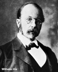
Wilhelm His (1831-1904) His's Normentafel (Normal Table) |

|

|
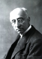
Franz Keibel (1861 - 1929) Franz Keibel and Curt Elze (1908) Normal Plates of the Development of the Human Embryo |

|

|

Franklin Mall (1862-1917) |

| |
| Begun by Dr. Hideo Nishimura (1912–1995)
Developed by Kohei Shiota and currently curated by Shigehito Yamada.
|

| |


| Animal Models | |
|---|---|
| |
| |
 |
mouse
|
| Fly Development - The fruitfly (drosophila) was and is the traditional geneticist's tool. It has been transformed to an magnificent embryologist's tool, with developmental mechanisms being uncovered in this system combined with homolgy gene searches in other species. The fly genome was one of the first to be been completely sequenced. In early development nurse cells sacrifice their cytoplasmic contents to allow egg growth and early pattern formation is through the localization of maternal messenger RNAs (mRNAs). | |
 |
Worm Development - Early embryological studies of the worm Caenorhabditis elegans (C.Elegans, so called because of its "elegant" curving movement) characterized the fate of each and every cell in the worm through all stages of development. This worm has recently had its entire genome sequenced. |
| Zebrafish Development - Zebrafish are seen as the latest and greatest "model' for embryological development studies. They can be easily genetically altered and develop as practically "see through" embryos, all internal development can be clearly observed from the outside in the living embryo. |
| In Vitro Fertilization (1978) | Stem Cells (1981) | Induced Stem Cells (2006) | Molecular Development |

|

|

|

|
Content to be added here.
Glossary Links
- Glossary: A | B | C | D | E | F | G | H | I | J | K | L | M | N | O | P | Q | R | S | T | U | V | W | X | Y | Z | Numbers | Symbols | Term Link
Cite this page: Hill, M.A. (2026, Mayıs 24) Embryology Lecture - 2016 Course Introduction. Retrieved from https://embryology.med.unsw.edu.au/embryology/index.php/Lecture_-_2016_Course_Introduction
- © Dr Mark Hill 2026, UNSW Embryology ISBN: 978 0 7334 2609 4 - UNSW CRICOS Provider Code No. 00098G