V
| Embryology - 27 Apr 2024 |
|---|
| Google Translate - select your language from the list shown below (this will open a new external page) |
|
العربية | català | 中文 | 中國傳統的 | français | Deutsche | עִברִית | हिंदी | bahasa Indonesia | italiano | 日本語 | 한국어 | မြန်မာ | Pilipino | Polskie | português | ਪੰਜਾਬੀ ਦੇ | Română | русский | Español | Swahili | Svensk | ไทย | Türkçe | اردو | ייִדיש | Tiếng Việt These external translations are automated and may not be accurate. (More? About Translations) |
Glossary Links
- Glossary: A | B | C | D | E | F | G | H | I | J | K | L | M | N | O | P | Q | R | S | T | U | V | W | X | Y | Z | Numbers | Symbols | Term Link
V
vaccination
- (Latin, vacca = cow) Term used to describe inoculation, because the first vaccine was obtained from cows, the cowpox virus, by Edward Jenner. Vaccination is the process of inoculation of a substance (vaccine) into the body for the purpose of producing active immunity (immune response) against a disease. Vaccination is therefore a misnomer, used initially by Louis Pasteur, for inoculation.
- (More? Viral Infection | Rubella | The Jenner Museum | Search Pubmed)
VACTERL
- An acronym for a group of developmental abnormalities: Vertebral anomalies, Anal atresia, Cardiac defect (often ventricular septal defect), TracheoEsophageal fistula, Renal (kidney) abnormalities and Limb abnormalities. Recently defined as a multiple polytopic developmental field defect.
- (More? VACTERL | Abnormal Development | PMID 21846383)
vacuum extractor
- Clinical birth aid, referring to a suction cap device used on fetal head to aid birth (parturition).
- (More? birth)
vagina
- The female genital tract muscular tube between the uterus and vestibule, part of female external genitalia.
- (More? vagina | Lecture - Genital Development)
vaginal seeding
- A term describing the practice of transfer of maternal vaginal fluid (by gauze swab) and associated vaginal microbiota to an infant born by caesarean section. This is thought to introduce microbiota associated with vaginal birth as apposed to caesarean section microbiota of maternal skin. It should be noted that there is little clinical evidence to support the practice and some associated risks of infection transfer.
- (More? Birth - Caesarean Delivery)
vaginosis
- See bacterial vaginosis (BV).
vaginitis
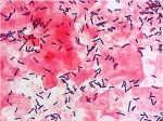
- (candidiasis, bacterial vaginosis, trichomoniasis) Clinical term for a variety of infections which lead to vaginal inflammation. Infections can be caused by several organisms including; yeast (candidiasis), abnormal bacteria (bacterial vaginosis including gonorrhea, chlamydia or mycoplasma), parasitic protozoa (trichomoniasis), virus (herpes).
vagus
- (Latin, vagus = wandering) cranial nerve X (CN X) A mixed nerve that leaves the head and neck to innervate gastrointestinal tract (pharynx, esophagus, stomach) respiratory tract (larynx, lungs), cardiac (heart) and abdominal viscera. This mixed nerve has sensory, motor and autonomic functions of viscera (glands, digestion, heart rate).
valproic acid
- An anticonvulsants or antiepileptic drug used to prevent and minimize seizures. This drug, like other anticonvulsants, is also teratogenic and increases the occurance of neural tube defects (NTDs). (Other anticonvulsants: Carbamazepine, Clonazepam, Ethosuximide, Phenobarbital, Phenytoin, Primidone)
- (More? Neural System - Abnormalities | drugs)
valsartan
- Commercial name for a drug used to block the renin-angiotensin system and reduce blood pressure. It is an angiotensin II receptor antagonist (angiotensin receptor blocker) leading to the dilation of blood vessels and subsequent reduction in blood pressure. A recent study of valsartan use for 5 years "by patients with impaired glucose tolerance and cardiovascular disease or risk factors, along with lifestyle modification, led to a relative reduction of 14% in the incidence of diabetes but did not reduce the rate of cardiovascular events."
- (More? PMID 20228403)
vanilloid receptor
- (VR) receptor was cloned in 1997, VR agonists (capsaicin and RTX) are currently utilized for a number of clinical syndromes (intractable neuropathic pain, spinal detrusor hyperreflexia, and bladder hypersensitivity). VR antagonist (capsazepine) are also currently being considered for therapeutic applications related to pain.
vanishing testis syndrome
- (anorchia, embryonic testicular regression) See anorchia.
- (More? testis)
varicella zoster virus
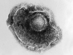
- (VZV) the chickenpox virus which can severely affect pregnant woman, fetus, and newborn babies. Fetal varicella syndrome (FVS) A viral infection by varicella zoster virus (chickenpox) virus following a maternal infection that is transmitted transplacentally to the fetus. Congenital defects can result from fetal infection including: skin lesions, limb defects (hypoplasia or paresis), and neurological (microcephal, ophthalmic lesions) .
- (More? Varicella Zoster Virus | Viral Infection)
varicoceles
- An abnormality of male gonad venous system, resulting from veins surrounding the testicular artery (in spermatic cord) becoming dilated (varicose), affecting testis temperature and fertility.
VARTA
- Acronym for the Victorian Assisted Reproductive Treatment Authority, located in the Australian state of Victoria. This group provides public education and resources for professionals and the community on fertility and issues related to assisted reproductive treatment, including IVF, surrogacy and donor-conception.
- (More? Assisted Reproductive Technology | VARTA)
VAS
- Clinical acronym for vibroacoustic stimulation.
vasa previa
- (vasa praevia) A placental abnormality where the fetal vessels either lie close too or cross the inner cervical os (opening) ahead of the fetal presenting part and unsupported by the placenta tissue or umbilical cord.
- (More? placenta | Placenta - Abnormalities | Lecture - Placenta Development | PMID 19772710)
vascular
- Term used in relation to blood vessels often combined with the heart, cardiovascular.
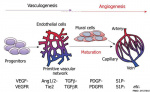
vascular endothelial growth factor
- (VEGF) A secreted protein growth factor family, which stimulates the proliferation of vasular endotheial cells and therefore blood vessel growth. VEGF's have several roles in embryonic development. The VEGF family has 7 members (VEGF-A, VEGF-B, VEGF-C, VEGF-D, VEGF-E, VEGF-F, and PlGF) that have a common VEGF homology domain. PIGF is the placental growth factor. The growth factors act through 3 VEGF tyrosine kinase membrane receptors (VEGFR-1 to 3) with seven immunoglobulin-like domains in the extracellular domain, a single transmembrane region, and an intracellular tyrosine kinase sequence.
- (More? Blood Vessel Development | Vascular Endothelial Growth Factor | PMID 15602010 | Search Pubmed)
vascular endothelial protein tyrosine phosphatase
- (VE-PTP) A phosphatase involved in vascular development through modulation of a receptor tyrosine kinase (Tie2) activity, the receptor of angiopoietin. Phosphatases remove and kinases add a phosphate group to proteins, phosphorylation is a common intracellular signaling pathway.
- (More? Blood Vessel Development)
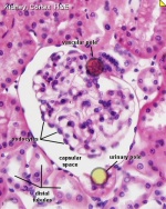
vascular pole
- Renal term for the side of the nephron Bowman's capsule where the afferent arteriole and efferent arteriole enter the glomerulus. The opposite "pole" is the urinary pole.
vasculogenesis
- Term describing the formation of new blood vessels from pluripotent precursor cells. In contrast to angiogenesis, which is the formation of new vessels from pre-existing vessels.
- (More? Blood Vessel Development | Placenta Development)
vas deferens
- (ductus deferens, Latin, deferens = carrying-away vessel)
- Spermatozoa Duct Pathway: seminiferous tubule - straight tubule - rete testis - ductuli efferentes - ductus epididymidis - ductus deferens
vasohibin
- Signaling protein family (Vasohibin-1, VASH1 and vasohibin-2, VASH2) involved in angiogenesis having a role in placentation. Acts as a negative-feedback regulator of angiogenesis induced in endothelial cells by angiogenesis stimulators such as VEGF and FGF-2. Proteins bind to small vasohibin binding protein (SVBP) that facilitates the secretion of vasohibins. Endothelial cell expression of VASH1 in the termination zone halts angiogenesis. Bone marrow derived mononuclear cells express VASH2 at the sprouting front to stimulate angiogenesis.
VATER syndrome
- A developmental abnormality, acronym for V (vertebral anomalies), A (anal atresia), TE (tracheo-esophageal fistula) and R (radial dysplasia). (V and R also include vascular and renal anomalies). Note that the acronym VACTERL syndrome now used, includes C (cardiac) and the L (limb anomalies).
- (More? Human Abnormal Development | Search Pubmed PMID 21846383)
VCX-A
- An acronym for Variably Charged, X chromosome A, a member of the family of proteins encoded by distinct genes located on the X chromosome (Xp22) and implicated in X-linked mental retardation. This RNA-binding protein post-transcriptionally regulates protein synthesis by binding the 5' end of capped mRNAs, preventing decapping and decay.
- (More? PMID 19812318 | Neural System Development)
vein
- Part of the circulatory system, returning blood to the heart. Nomenclature based upon direction of flow, towards the heart. In the embryo, the placental vein brings oxygenated blood to the heart. In the adult, pulmonary vein brings oxygenated blood from lungs to heart. Vein walls have histologically a different overall structure from ateries.
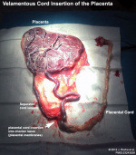
velamentous cord insertion
- (velamentous insertion) Clinical term for describing a placental abnormality where the placental cord inserts into the chorion laeve (placental membranes) away from the edge of the placenta. The placental vessels are therefore unprotected by Wharton's jelly where they traverse the membranes before they come together into the umbilical cord. This can cause hemorrhage if the vessels are damaged when the membranes are ruptured prior to birth. The condition is more common in monozygotic twins (15%) and triplets.
vellus hair
- The short hairs seen on newborn infant, these hairs are only a centimetre or two long and contain little or no pigment. Follicles that produce these hairs do not have sebaceous glands and never produce any other kind of hairs.
- (More? hair | integumentary | Lecture - Integumentary Development)
velocardiofacial syndrome
- See DiGeorge syndrome.
ventral mesogastrium
- The ventral mesentery attached at the level of the stomach lying between the stomach and the septum transversum then liver. This embryonic structure will later form both the lesser omentum, lying between the stomach and liver, and the falciform and coronary ligaments lying between the liver and the abdominal wall and diaphragm. Below the level of the stomach this ventral mesentery is absent and the midgut is attached only dorsally.
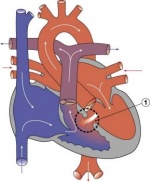
ventricle
- The two ventricles form the lower muscular-walled blood discharging chambers of the heart. The upper heart chambers (atria) receive blood.
ventricular septation
- Term describing the complex developmental formation of the interventricular septum, the wall between the right and left ventricles of the heart. In humans, this septation process begins in week 5 of development the ventricular wall initially has two separate components; a superior membranous septum and an inferior muscular septum. The muscular septum grows from the inferior cardiac wall and fuses with three membranous septum components; the right and left bulbar ridges and the dorsal endocardial cushion. The membranous septum is continuous with the septation of the cardiac outflow tract, the initial truncus arteriosus is separated into the aortic and pulmonary outflow. Abnormalities of this process are called Ventricular Septal Defects (VSD) and usually occurs in the membranous rather than muscular interventricular septum, and is more frequent in males that females. The process of separating the atria is called atrial septation.
- (More? cardiovascular | heart | Cardiac Tutorial)
ventricular septal defect
- (VSD) Term describing a cardiac abnormality due to due to partial or complete failure of interventricular septum formation, and separation of the right and left ventricles. Usually occurring in the membranous (perimembranous) rather than muscular interventricular septum and more frequent in males that females.
- (More? Ventricular Septal Defect | heart | cardiovascular)
vermiform
- (Latin, vermis = "a worm", and forma = "shape") having a worm-like shape, as in the appendix (vermiform appendix).
vermiform appendix
- See appendix.
vernix
- (vernix caseosa) See vernix caseosa.
vernix caseosa
- (vernix, Latin, vernix = varnish, caseous = cheese-like) term describing a coating formed on the fetal skin surface by: secreted sebum, cells sloughed off the fetus's skin, and shed lanugo hair. The coating also has a high water content (80%) largely compartmentalized within fetal corneocytes (cells forming the stratum corneum). This coating develops intially in a cranio-caudal direction and can be absent in preterm infants. Some functions include: protection of the fetal skin from extraembryonic fluids amnion, urine; providing a slippery surface helps with parturition; acting as a biofilm barrier against infection.
vernix caseosa peritonitis
- Clinical term for a rare post-caesarean section complication due to spilt vernix caseosa at the time of caesarean section mediating a maternal inflammatory reaction.
vertebra
- Individual bones (33 human) of the axial skeleton that enclose the spinal cord and form, with the intervertebral discs, the vertebral column. Vertebra bodies form first by endochondrial ossification of segmental sclerotomal mesenchyme which in turn came from pairs of somites. During vertebra formation there is a "segmental shift" out of register with the original somites. In development, the vertebral arch (enclosing the spinal cord) remains open dorsally and tied by a ligament to allow growth of the spinal cord.
- Differentiation: mesoderm -> paraxial mesoderm -> somites -> paired sclerotome -> vertebra
vertebral canal
- The bony tunnel formed by vertebral arch enclosing the spinal cord.
vertebral column
- (backbone) Term given to the complete structure formed from the alternating segments of vertebra and intervertebral discs which surround and support the spinal cord. The term used to define animals with a backbone (vertebra).
vertex presentation
- (cephalic presentation) Clinical (obstetric) birth term, referring to when the fetal head is closest to the cervix, hence "presentation". The most common and safest human birth position.
- (More? Birth)
venous thromboembolism
- Blood clotting within a vein, has been described as a rare risk for some women taking the combined oral contraceptive.
ventral telencephalon
- The neural tube telencephalon is the secondary brain vesicle formed from the prosencephalon and the ventral region generates several types of neurons (striatal, olfactory, cortical interneurons). Structurally, the ventral telencephalon has three proliferative zones forming ganglionic eminences (lateral, medial, caudal) that can migrate into the cortex or olfactory bulb.
- (More? telencephalon)
vesicle
- A general term describing an expanded fluid-filled region enclosed by a membrane or layer. Developmental examples include: acrosomal vesicle, brain (cephalic) vesicles, optic (lens) vesicle, otic vesicle (otocyst). Cell biology examples include: transport vesicles, secretory vesicles.
vestibular aqueduct
- (Latin, aquaeductus vestibuli) A component of the inner ear, describing the intraosseous part of the endolymphatic duct and endolymphatic sac system.
vestibular ganglion
- (Scarpa's ganglion) The primary afferent vestibular neuron ganglion of the vestibular nerve. Located within the internal auditory meatus.
vestibular membrane
- (Reissner's) Membranous structure within the cochlea of the inner ear, extends from the spiral lamina to the outer wall and divides the cochlea into an upper scala vestibuli, a lower scala tympani.
vestibulocochlear nerve
- Alternative name for cranial nerve eight (CN VIII) associated with balance (vestibulo) and hearing (cochlear).
vestibular neurons
- Neurons of the vestibular ganglia of cranial nerve 8 (CN VIII) involved with balance and motion detection. These neurons form as part of the anterior ventromedial otocyst epithelium and migrate into the periotic mesenchyme at the anterior part of the developing otocyst. At this location they differentiate and extend dendrites towards the developing sensory epithelia and axons to connect with central brainstem targets.
vibroacoustic stimulation
- (VAS) Clinical test in the third trimester where a sound is applied to maternal abdomen over the region of the fetal head. This generates a fetal "startle reflex", the fetal equivalent of the neonatal Moro reflex. It has been suggested that VAS can also raise fetal heart rate (FHR) acceleration or transient tachycardia.
- (More? Hearing Development | Fetal Development | Third Trimester | Moro reflex | PMID 3901769)
villi
- Plural of villus, which is a thin projection from a surface. A term used to describe the many functional units together of the fetal placenta.
villitis of unknown etiology
- (VUE) A placenta pathological condition where the origin is unknown.
villous cytotrophoblast
- (VCT) Trophoblast cells located on chorionic villi (as opposed to extravillous cytotrophoblasts) that can also fuse to form the syncytiotrophoblasts.
villus
- A single thin projection from a surface. A term used to describe the functional unit of the fetal placenta.
- (More? Placenta - Villi Development | Lecture - Placenta Development | placenta)
vinblastine
- A vinca alkaloid used as a chemotherapeutic antitumour drug that targets tubulin. The molecule forms a wedge at the interface of two tubulin molecules, interfering with tubulin and then microtubule assembly and therefore disrupts mitotic spindle formation.
Vinclozolin
- A dicarboximide fungicide shown to cause gonad tumours (Leydig cell) and atrophy in adult rat. Perinatal exposure in rats also inhibits morphological sex differentiation.
- (More? Endocrine System - Abnormalities | PMID 16417039)
virulence
- (Latin, virulenctia = virus poison) morbidity and mortality of a host caused by parasites and pathogens (relative infectiousness).
- (More? Viral Infection)
virus
- An infective agent that cannot reproduce by itself and therefore it infect cells to use the cell machinery to produce more virus. Different viruses have genetic material as single- or double-stranded RNA or DNA. The infectious virus particle is called a "virion" and is the genetic material packed in a protein shell. Viruses come in many genetic sizes, as little as 4 proteins up to 200 proteins. Many cancers can be caused by viruses (papilloma viruses, hepatitis B and C viruses, Epstein-Barr virus and human T-cell lymphotropic virus). Virus-induced cancers account for about 20% of worldwide cancer incidence.
- (More? Viral Infection | Rubella)
viscera
- The organs located in the ventral body cavity. The term visceral (respiratory, renal) relates to the layer closest to an organ.
visceral layer
- Renal term for the cells (podocytes) of the inner of Bowman's capsule that form extremely complex shapes. Cytoplasm form a fenestrated epithelium around the fenestrated capillaries of the glomerulus. The openings between the pedicles are called filtration slits. They are spanned by a thin membrane, the filtration slit membrane. The outer layer is the parietal layer.
- (More? Renal System Development)
visceral pleura
- The inner lining of pleural cavity derived from contact epithelia with lung bud of pericardioperitoneal canals from intraembryonic coelom.
visceral smooth muscle
- The muscle in walls of visceral organs. One of the 3 types of muscle in the body (skeletal, cardiac, smooth).
vitamins
- (Latin, vita = "life") The Template:Vitamins form 2 major classes the fat soluble (D, E, A, and K) and water soluble (B, and C). These organic compounds are required at very low concentrations and function as cellular metabolic regulators. The specific roles for vitamins, other than B (folic acid), during embryonic development (other than in maternal nutrition) is unknown.
- (More? Template:Vitamin | nutrition)
vitamin A
- (More? Nutrition)
vitamin B
- The Vitamin B group of eight separate water-soluble vitamins that play important roles in cell metabolism. Folic acid or vitamin B9 (folacin) and folate (naturally occurring form) are an important dietary requirement for normal neural development. Low levels have been show associated with neural tube defects including spina bifida.
- (More? Nutrition | Folic Acid and Neural Tube Defects | Neural System Development | Neural System - Abnormalities)
vitamin C
vitamin D
- Vitamin D is important Circulating 25-hydroxyvitamin D3 (25[OH]D), the most commonly used index of vitamin D status, is converted to the active hormone 1,25 dihydroxyvitamin D3 (1,25[OH]2D), which, operating through the vitamin D receptor (VDR). The vitamin D receptor belongs to the nuclear receptor superfamily. USA Institute of Medicine expert committee "calcium requirements varied with age, from 700 mg a day for children aged 1-3 years up to 1200 mg a day for women aged 51 to 70 and 1300 mg a day for teenagers and pregnant and lactating women."
- (More? Vitamin D| Nutrition| Dietary Reference Intakes for Calcium and Vitamin D | Paediatric Endocrine Group; Paediatric Bone Australasia. Prevention and treatment of infant and childhood vitamin D deficiency in Australia and New Zealand: a consensus statement. 2006 PMID16948623 | Vitamin D - a review 2008 PMID19142273 | AFP)
vitamin E
vitamin K
- Vitamin K is a generic term for derivatives of 2-methyl-1,4-naphthoquinone that have coagulation activity. Daily requirement for vitamin K is about 1 µg/kg. In newborns vitamin K nutrition is at risk.
vitrification
- A term used in Assisted Reproductive Technology techniques and cell culture for an ultra-rapid cryopreservation method that prevents ice formation within the suspension which is converted to a glass-like solid.
visceral pleura
- Serous membrane which forms the inner lining of pleural cavity, both covering and attached to the lungs. Embryonically derived from the splanchnic mesoderm. The outer pleural layer, parietal pleura, is derived from mesoderm of the thoracic cavity body wall.
- (More? respiratory)
vitelline
- (Latin, vitellus = yolk of an egg) Term used early in development twice. Firstly, to describe the outer egg layer zona pellucida. Secondly, used later to describe the structures and vasculature associated with the yolk sac. The blood vessels which form in the extraembryonic mesoderm of the yolk sac and anastomose are called vitelline arteries (flow away from the embryo) and vitelline veins (flow toward the embryo). The connection between the midgut and the yolk sac is the vitelline duct (omphalomesenteric duct or yolk stalk).
- (More? Week 3 | cardiovascular)
vitelline duct
- (yolk stalk, omphalomesenteric duct, Latin, vitellus = yolk of an egg) The endodermal connection between the mid-gut and the yolk sac. During embryonic disc folding (human week 3) this structure is initially a broad open connection which is then restricted to a narrow tube and finally closed between the mid-gut and the yolk sac. Meckel's diverticulum is the abnormality associated with failure of this duct to close.
vitelline artery
- (omphalomesenteric artery, Latin, vitellus = yolk of an egg) The blood vessels which form in the yolk sac and have a blood flow away from embryo. Derived from the extraembryonic mesoderm surrounding the endoderm of the yolk sac.
vitelline vein
- (omphalomesenteric vein, Latin, vitellus = yolk of an egg) The blood vessels which form in the yolk sac and have a blood flow towards the embryo. Derived from the extra-embryonic mesoderm surrounding the endoderm of the yolk sac.
vitelline layer
- (egg coat) The specialized extracellular matrix surrounding the egg in non-mammalians (sea urchins, chickens) in mammals this layer is called the zona pellucida.
- (More? Chicken Development)
vitellogenesis
- (Greek, vitellogenesis = yolk formation) The term refers to the formation of yolk.
vitreous humor
- A gel-like mass located in the vitreous (posterior) chamber of the eye. In development the vitreous also contains the hyloid blood vessels.
- (More? Vision Development)
vocal folds
- Folds occuring in the larynx used in generating sounds required for speech.
Volkmann’s canal
- Histological bone term for the oblique vascular channel in compact bone that link parallel Haversian canals and also with the inner and outer periosteum surfaces of the bone. These canals are not surrounded by concentric lamellae as found with the Haversian canals. Named after Alfred Wilhelm Volkmann (1800 - 1877) a German physiologist and anatomist.
- (More? Bone Histology | Image | Bone Development)
vomeronasal organ
- (VNO, Jacobson's organ) A neural structure forming part of olfactory system that functions in the detection of pheromones. Historically called the Jacobson's organ after Ludwig Lewin Jacobson (1783 – 1843) a Danish surgeon who first identified it in 1813.
- (More? Sensory - Smell Development)
von Ebner's fissure
- Somite division furrow that divides each somite into a cranial and caudal half. The caudal half has a dense cellular packing compared to the crainial half. This region is located at the adult intervertebral boundary. Named after von Ebner (1842 - 1925), an Austrian anatomist and histologist who identified this region in 1888.
- (More? Axial Skeleton Development)
von Willebrand factor
- (VWF) An adhesive protein which plays a critical role in primary haemostasis by allowing platelet aggregation.
VUE
- Acronym for villitis of unknown etiology, a placenta pathology.
vulva
- (Latin, volva or vulva = "female genitals") Term used to describe the external genital organs (genitalia) of the female.
- (More? Genital System Development | Vagina Development)
Glossary Comments
Use this page to access brief definitions of specific embryology terms. Additional information can be accessed from links listed at the end of each definition. Glossary from the UNSW Embryology program compiled and written by Dr Mark Hill. Reference material used in preparing this glossary list includes: texts listed on page 1 "Reading" of each notes section, Department of Anatomy Publications, WWW resources from NCBI, NIH, OMIM, NHMRC (Australia), AMA (USA), Office of Rare Diseases (USA), PubMed Medline Dictionaries, MSDS, Merck Manual home edn. and WHO ART terminology (2009).
These notes are for Educational Purposes Only Please email Dr Mark Hill if you wish to make a comment about this current project.
Glossary Links
- Glossary: A | B | C | D | E | F | G | H | I | J | K | L | M | N | O | P | Q | R | S | T | U | V | W | X | Y | Z | Numbers | Symbols | Term Link
Cite this page: Hill, M.A. (2024, April 27) Embryology V. Retrieved from https://embryology.med.unsw.edu.au/embryology/index.php/V
- © Dr Mark Hill 2024, UNSW Embryology ISBN: 978 0 7334 2609 4 - UNSW CRICOS Provider Code No. 00098G