Introduction
The paired adult kidneys consist of a functional unit called the "nephron", that filters blood, excretes waste, reabsorbs water (and other compounds) and has endocrine functions. Each adult human kidney typically contains about 750,000 nephrons, though the total number can vary significantly from as few as 250,000 to as many as 2,000,000.[1][2]
In the embryo, nephron development, nephrogenesis, occurs through several stages involving classical epithelial/mesenchyme type of interactions. Nephrogenesis continues into the late fetal period (GA week 34–35) and while the fetal kidney does produce urine, not until after birth does the glomerular filtration rate (GFR) increases rapidly due to a postnatal drop in kidney vascular resistance and an increase in renal blood flow.
- Molecular System Links: Heart | Neural | Renal | Respiratory | Mechanisms | Factors | Molecular
Some Recent Findings
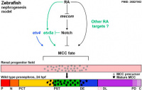
Zebrafish nephrogenesis signaling
[3]
- Epithelial cell fate in the nephron tubule is mediated by the ETS transcription factors etv5a and etv4 during zebrafish kidney development[4] "Kidney development requires the differentiation and organization of discrete nephron epithelial lineages, yet the genetic and molecular pathways involved in these events remain poorly understood. The embryonic zebrafish kidney, or pronephros, provides a simple and useful model to study nephrogenesis. The pronephros is primarily comprised of two types of epithelial cells: transportive and multiciliated cells (MCCs). Transportive cells occupy distinct tubule segments and are characterized by the expression of various solute transporters, while MCCs function in fluid propulsion and are dispersed in a "salt-and-pepper" fashion within the tubule. Epithelial cell identity is reliant on interplay between the Notch signaling pathway and retinoic acid (RA) signaling, where RA promotes MCC fate by inhibiting Notch activity in renal progenitors, while Notch acts downstream to trigger transportive cell formation and block adoption of an MCC identity. ...abrogation of Notch with the small molecule inhibitor DAPT increased the renal progenitor etv5a expression domain as well as MCC density in etv5a deficient embryos, suggesting Notch acts upstream to inhibit etv5a. In contrast, etv4 levels in renal progenitors were unaffected by changes in RA or Notch signaling levels, suggesting a possible non-cell autonomous role during pronephros formation. Taken together, these findings have revealed new insights about the genetic mechanisms of epithelial cell development during nephrogenesis." Retinoic acid | Notch
- Histone deacetylase 1 and 2 regulate Wnt and p53 pathways in the ureteric bud epithelium[5] "Histone deacetylases (HDACs) regulate a broad range of biological processes through removal of acetyl groups from histones as well as non-histone proteins. Our previous studies showed that Hdac1 and Hdac2 are bound to promoters of key renal developmental regulators and that HDAC activity is required for embryonic kidney gene expression. However, the existence of many HDAC isoforms in embryonic kidneys raises questions concerning the possible specificity or redundancy of their functions. We report here that targeted deletion of both the Hdac1 and Hdac2 genes from the ureteric bud (UB) cell lineage of mice causes bilateral renal hypodysplasia. One copy of either Hdac1 or Hdac2 is sufficient to sustain normal renal development."
- Bmp7 functions via a polarity mechanism to promote cloacal septation[6] "During normal development in human and other placental mammals, the embryonic cloacal cavity separates along the axial longitudinal plane to give rise to the urethral system, ventrally, and the rectum, dorsally. Defects in cloacal development are very common and present clinically as a rectourethral fistula in about 1 in 5,000 live human births. Yet, the cellular mechanisms of cloacal septation remain poorly understood. ...Our results strongly indicate that Bmp7/JNK signaling regulates remodeling of the cloacal endoderm resulting in a topological separation of the urinary and digestive systems. Our study points to the importance of Bmp and JNK signaling in cloacal development and rectourethral malformations." BMP
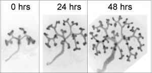
- Characterization of Mesonephric Development and Regeneration Using Transgenic Zebrafish.' [7] "The majority of previous studies have focused on the pronephros of zebrafish, which consists of only two nephrons and is structurally simpler than the mesonephros of adult fish and the metanephros of mammals. To evaluate the zebrafish system for more complex studies of kidney development and regeneration, we investigated the development and post-injury regeneration of the mesonephros in adult zebrafish." (See also Zebrafish Development)
|
| More recent papers
|
|
This table allows an automated computer search of the external PubMed database using the listed "Search term" text link.
- This search now requires a manual link as the original PubMed extension has been disabled.
- The displayed list of references do not reflect any editorial selection of material based on content or relevance.
- References also appear on this list based upon the date of the actual page viewing.
References listed on the rest of the content page and the associated discussion page (listed under the publication year sub-headings) do include some editorial selection based upon both relevance and availability.
More? References | Discussion Page | Journal Searches | 2019 References | 2020 References
Search term: Renal Embryology
<pubmed limit=5>Renal Embryology</pubmed>
|
Nephron
In humans, nephrogenesis only occurs before birth, though nephron maturation continues postnatally. Mean glomerular number shown to level at 36 weeks, increasing from about 15,000 at 15 weeks to 740,000 at 40 weeks.
Nephron development has four identifiable developmental stages:
- Vesicle (V) stage (13-19 weeks, second trimester)
- S-shaped body (S) stage ( 20-24 weeks, second trimester)
- Capillary loop (C) stage (25-29 weeks, third trimester)
- Maturation (M) stage (infants aged 1-6 months, neonatal and postnatal)
|
|
Abbreviation
(OMIM link)
|
Growth Factor
(Factor page link)
|
Renal Development
|
Expression Location
|
| BMP4
|
Bone Morphogenetic Protein 4
|
prevents ectopic ureteric bud outgrowth and extra ureteric bud divisions
|
mesenchymal cells surrounding mesonephric duct and stromal mesenchyme surrounding uteric bud stalks
|
| BMP7
|
Bone Morphogenetic Protein 7
|
survival of metanephric mesenchyme
|
metanephric mesenchyme
|
| Fgf8
|
Fibroblast Growth Factor 8
|
transition of the induced cap mesenchyme into RVs
|
cap mesenchyme
|
| GDNF
|
Glial-cell derived neurotrophic factor
|
induces uteric bud outgrowth from mesonephric duct, interacts with Ret
|
metanephric mesenchyme
|
| VEGF
|
Vascular endothelial growth factor
|
promotes endothelial cell proliferation, differentiation
|
s-shaped body
|
| Wnt4
|
Wingless-Type MMTV Integration Site Family, Member 4
|
mesenchymal-to-epithelial transition
|
cap metanephric mesenchyme, pre-tubular aggregate, nephron progenitors
|
| Wnt5a
|
Wingless-Type MMTV Integration Site Family, Member 5a
|
nephrogenesis induction, ectopic bud formation
|
uteric bud, metanephric mesenchyme
|
| Wnt9b
|
Wingless-type MMTV integration site family, Member 9B
|
renewal and differentiation of nephron progenitors and normal ureteric bud branching, mesenchymal-to-epithelial transition
|
uteric bud stalk epithelial cells
|
- Foxd1 - (Brain Factor-2) transcription factor that is a renal stroma specific gene.
- Links: OMIM Foxd1
Intermediate Mesoderm
Mesonephric Duct
Uteric Bud
Mouse E12.5 kidney in vitro
- arise near the cloacal connection of the mesonephric duct
- branch from the mesonephric duct laterally into the intermediate mesoderm
- induce the surrounding mesoderm to differentiate - metanephric blastema
- this mesoderm will in turn signal back to differentiate the uteric bud
Epithelial - mesenchymal interaction
Uteric Bud forms - ureter, pelvis, calyces, collecting ducts
Nephros Development
Three pairs appearing in sequence within intermediate mesoderm during development.
- pronephros
- mesonephros
- metanephros
Pronephros
Mesonephros
Metanephros
Renal Collecting Duct Tree
A recent in vitro experimental system and a computer model suggests that BMP7 also acts as a repelling signal to establish the branched collecting duct tree.[8]
- Links: Bone Morphogenetic Protein
References
- ↑ <pubmed>1546799</pubmed>
- ↑ <pubmed>17495859</pubmed>
- ↑ <pubmed>26827902</pubmed>
- ↑ <pubmed>26827902</pubmed>
- ↑ <pubmed>25758227</pubmed>
- ↑ <pubmed>22253716</pubmed>
- ↑ <pubmed>20810610</pubmed>
- ↑ <pubmed>25205115</pubmed>
Textbooks
- The Developing Human: Clinically Oriented Embryology (8th Edition) by Keith L. Moore and T.V.N Persaud - Moore & Persaud Chapter 13 p303-346
- Larsen’s Human Embryology by GC. Schoenwolf, SB. Bleyl, PR. Brauer and PH. Francis-West - Chapter 10 p261-306
- Before We Are Born (5th ed.) Moore and Persaud Chapter14 p289-326
- Essentials of Human Embryology, Larson Chapter 10 p173-205
- Human Embryology, Fitzgerald and Fitzgerald Chapter 21-22 p134-152
Online Textbooks
Search Bookshelf intermediate mesoderm | kidney development | renal development | ureteric bud | nephron development | bladder development
Reviews
<pubmed>25737276</pubmed>
<pubmed>25608807</pubmed>
<pubmed>20691850</pubmed>
<pubmed>19906853</pubmed>
<pubmed>19828308</pubmed>
<pubmed>19615554</pubmed>
<pubmed>18184729</pubmed>
<pubmed>17442697</pubmed>
<pubmed>9152004</pubmed>
Forefronts Symposium on Nephrogenetics: from development to physiology March 8-11, 2007 Danvers, MA A meeting to synthesize an integrated view of the normal development and function of the kidney from the genetic standpoint.
<pubmed>16916378</pubmed>
Articles
<pubmed></pubmed>
<pubmed></pubmed>
<pubmed>25205115</pubmed>
<pubmed>18846389</pubmed>
<pubmed>12783789</pubmed>
<pubmed>9690097</pubmed>
Search PubMed
Search Pubmed: Renal System Development | Renal Development | intermediate mesoderm | kidney development | renal development | ureteric bud | nephron development | bladder development
Additional Images
Terms
| Renal Terms
|
- bladder exstrophy - A congenital malformation with bladder open to ventral wall of abdomen (between umbilicus and pubic symphysis) and may have other anomolies associated with failure of closure of abdominal wall and bladder (epispadias, pubic bone anomolies).
- blastema - Term used to describe a mass of undifferentiated cells. (More? Wilm's tumour)
- Bowman's capsule - (capsula glomeruli, glomerular capsule) Surrounds the glomerulus within the nephron with a vascular and urinary pole and is the beginning of the tubular component. Named in 1842 after Sir William Bowman (1816 – 1892) an English surgeon and anatomist.
- Brenner hypothesis - a clinical hypothesis that states, individuals with a congenital reduction in nephron number have a much greater likelihood of developing adult hypertension and subsequent renal failure. Developed in the 1980's by Barry Brenner at the Brigham and Women's Hospital, this also fits with the DOHAD hypothesis. (More? PubMed 3063284 | Barry Brenner)
- capillary loop - (C stage) The third stage in nephron development between 25-29 weeks. (stage sequence: V - S - C - M)
- diabetes insipidus - The disorder is related to the hormone antidiuretic hormone (ADH, also called vasopressin) its synthesis, secretion, receptors and signaling pathway. In diabetes insipidus there is an excretion of large amounts (up to 30 litres/day) of a watery urine and an unremitting thirst.
- fenestrated capillary - Specialised capillaries containing circular pores (fenestrae) that penetrate the endothelium, may be closed by a thin diaphragm.
- glomerulus - The capillary network (tuft) within Bowman's capsule of the nephron enters at the vascular pole (afferent and efferent arteriole).
- hydronephrosis - (congenital hydronephrosis, Greek, hydro = water) A kidney abnormality due to partial or complete obstruction at the pelvi-ureteric junction. This leads to a grossly dilated renal pelvis causing extensive renal damage before birth.
- hyperplastic rests - In kidney development, embryonic blastema cells can persist and proliferate to form a pool of cells, which under either genetic or epigenetic influence can then change to become a neoplastic rest. Normally the majority of nephrogenic rests either regress or become dormant.
- juxtaglomerular cells - Cells located at the vascular pole that secrete renin and form a part of the juxtaglomerular complex.
- loop of Henle - Nephron region spanning from the proximal convoluted tubule to the distal convoluted tubule. Named after Named after Friedrich Gustav Jakob Henle (1809–1885) a German anatomist.
- macula densa - Columnar cell cluster appearing as a dense row of cell nuclei where the straight portion of the distal tubule contacts the glomerulus. Region also in close contact with the efferent and afferent arterioles of the glomerulus and involved in sodium chloride regulation. (More? image)
- maturation stage - (M stage) The forth stage in nephron development in infants aged 1-6 months. (stage sequence: V - S - C - M)
- mesangial cells - Cells in the nephron glomerulus that form the connective tissue giving structural support to podocytes and vessels.
- mesonephros - The second temporary stage of kidney development (pro-, meso-, meta-). The intermediate mesonephros develops and disappears with the exception of its duct, the mesonephric duct, which will form the male reproductive duct system. In males, the mesonephric tubules go on to form the ducts of the testis. In females, these degenerate. A few mesonephric tubules remain as efferent ductules in the male and vestigial remnants in the female.
- mesonephric duct - (= Wollfian duct) An early developing urogenital duct running the length of the embryo that will differentiate and form the male reproductive duct system. In females this duct degenerates (some remnants may remain associated in broad ligament).
- metanephros - The adult kidney, third stage of mammalian kidney (pro-, meso-, meta-) development within the intermediate mesoderm.
- metanephric cap - (metanephric blastema) The intermediate mesoderm which surrounds the ureteric bud and will contribute most of the adult nephron.
- multicystic kidney - There is no functional kidney tissue present in the kidney and it is replaced by a multilocular cyst. This is non-familial and is produced by atresia of a ureter and is always unilateral.
- neoplastic rest - In kidney development, a neoplastic rest can develop under either genetic or epigenetic influence from a hyperplastic rest, originating from an embryonic blastema cell. Normally the majority of nephrogenic rests either regress or become dormant.
- nephrin - protein of the slit diaphragm of renal filtration barrier, located at the cell surface in the area between two podocytes. NPHS1 gene location 19q13.12, mutations in this gene are associated with Congenital Nephrotic Syndrome (Nephrotic syndrome). (More? renal abnormalities)
- nephrogenic rest - Used to describe the embryonic blastema cells which persist and under either genetic or epigenetic can change to become a neoplastic rest. These neoplastic rests can develop postnatally as a benign form (adenomatous rest) or a malignant Wilm's tumour form. The rests are further characterised by the time of generation leading to different anatomical kidney locations: early intralobar nephrogenic rests (within the renal lobe) and late pelilobar nephrogenic rests (periphery of the renal lobe)
- nephron - (Greek, nephros = kidney) The functional unit of the adult kidney.
- nephros - (Greek, nephros = kidney) Term used to describe features associated with the kidney. (pronephros, mesonephros, metanephros, nephric, nephron, nephroblastoma).
- Nephrotic syndrome - (CNS, Nephrotic syndrome) rare kidney disorder characterized by heavy proteinuria, hypoproteinemia, and edema starting soon after birth. Most cases are caused by genetic abnormalities in the components of the glomerular filtration barrier, especially nephrin and podocin. (More? renal abnormalities)
- parietal layer - Cells of the outer of Bowman's capsule that form a simple squamous epithelium. The inner layer is the visceral layer.
- podocin - protein of the slit diaphragm of renal filtration barrier, located at the cell surface in the area between two podocytes. NPHS2 gene location 1q25.2, mutations in this gene are associated with Congenital Nephrotic Syndrome (Nephrotic syndrome). (More? renal abnormalities)
- podocyte - (visceral epithelial cell) kidney glomerulus cell forming the main component of the glomerular filtration barrier. (glomerular podocyte) Kidney epithelial cell type in the nephron (kidney functional unit) located in the glomerulus. Podocytes form the visceral layer of Bowman's capsule and are at the filtration barrier between capillary blood and the nephron tubular system and function to ultrafiltrate blood, and support glomerular capillary pressures. The differentiation of podocytes involves the formation of cellular foot processes and then the slit membrane. (More? image)
- podocyte specific proteins - podocalyxin, glomerular epithelial protein-1, podocin, nephrin, synaptopodin, and alpha-actinin-4), podocyte synthesized proteins (vascular endothelial growth factor and novH), transcription factors (WT1 and PAX2).
- pronephros - (Greek, pro = before) The first temporary stage of kidney development (pro-, meso-, meta-). This forms the kidney of primitive fish and lower vertebrates. Kidney development occurs within the intermediate mesoderm interacting with endoderm. In humans, this very rudimentary kidney forms very early at the level of the neck. It is rapidly replaced by the mesonephros, intermediate stage kidney, differentiating in mesoderm beneath.
- proteinuria - The abnormal presence of protein in the urine and an indicator of diesease including diabetic kidney disease (DKD, diabetic nephropathy).
- proximal tubule - Portion of the nephron duct between Bowman's capsule to the loop of Henle, divided into the proximal convoluted tubule (PCT) and the proximal straight tubule (PST).
- renal - (Latin, renes = kidney) Term used in relation to the kidney and associated structures (renal pelvis, renal artery)
- S-shaped body - (S stage) The second stage in nephron development between 20-24 weeks. (stage sequence: V - S - C - M)
- transitional epithelium - (urothelium) Histological term to describe the epithelium lining the ureters and urinary bladder. (More? image)
- trigone - refers to the urinary bladder triangular region formed by the two ureters and the urethra.
- ureter - The two ureters are hollow tubes that link the kidney and the bladder and carry urine. They develop from the ureteric bud and are lined by a transitional epithelium with an outer muscular wall.
- urethra - The single muscular tube that links and carries urine from the bladder to the exterior. In humans, the urethral length differs between the sexes (male longer, female shorter).
- vascular pole - The side of nephron Bowman's capsule where the afferent arteriole and efferent arteriole enter the glomerulus. image
- visceral layer - Cells (podocytes) of the inner of Bowman's capsule that form extremely complex shapes. Cytoplasm form a fenestrated epithelium around the fenestrated capillaries of the glomerulus. The outer layer is the parietal layer.
- vesicle stage - (V stage) The first stage in nephron development between 13-19 weeks. (stage sequence: V - S - C - M)
- urinary - Term used to describe all components of the kidney system including the bladder, ureters and urethra.
- urinary pole - The side of nephron Bowman's capsule where the proximal convoluted tubule starts. image
- urine - Term used to describe the liquid waste produced by the kidney, stored in the bladder and excreted from teh body through the urethra.
- urorectal septum - (URS) The structure which develops to separate the cloaca (common urogenital sinus) into an anterior urinary part and a posterior rectal part.
- Wilms' tumour - A form of kidney/renal cancer (nephroblastoma) named after Dr Max Wilms who first described the tumor. This childhood kidney cancer is caused by the inactivation of a tumour suppressor gene (BRCA2) or Wilms tumor-1 gene (Wt1) and is one of the most common solid tumors of childhood, occurring in 1 in 10,000 children and accounting for 8% of childhood cancers. Wt1 also required at early stages of gonadal development. (More? OMIM - Wilm's tumour | Dr Max Wilms)
- Wilms' tumor 1-associating protein - (WTAP) protein expressed in extraembryonic tissues and required for the formation of embryonic mesoderm and endoderm.
- Wolffian duct - (= mesonephric duct, preferred terminology), runs from the mesonephros to cloaca, differentiates to form the male vas deferens and in the female regresses. Named after Caspar Friedrich Wolff (1733-1794), a German scientist and early embryology researcher and is said to have established the doctrine of germ layers. (More? Caspar Friedrich Wolff)
|
|
|
External Links
External Links Notice - The dynamic nature of the internet may mean that some of these listed links may no longer function. If the link no longer works search the web with the link text or name. Links to any external commercial sites are provided for information purposes only and should never be considered an endorsement. UNSW Embryology is provided as an educational resource with no clinical information or commercial affiliation.
Glossary Links
- Glossary: A | B | C | D | E | F | G | H | I | J | K | L | M | N | O | P | Q | R | S | T | U | V | W | X | Y | Z | Numbers | Symbols | Term Link
Cite this page: Hill, M.A. (2026, April 25) Embryology Renal System - Molecular. Retrieved from https://embryology.med.unsw.edu.au/embryology/index.php/Renal_System_-_Molecular
- What Links Here?
- © Dr Mark Hill 2026, UNSW Embryology ISBN: 978 0 7334 2609 4 - UNSW CRICOS Provider Code No. 00098G


