Developmental Signals - Hippo

From Embryology
Embryology - 19 Mar 2024    Facebook link Pinterest link Twitter link  Expand to Translate  
Google Translate - select your language from the list shown below (this will open a new external page)

العربية | català | 中文 | 中國傳統的 | français | Deutsche | עִברִית | हिंदी | bahasa Indonesia | italiano | 日本語 | 한국어 | မြန်မာ | Pilipino | Polskie | português | ਪੰਜਾਬੀ ਦੇ | Română | русский | Español | Swahili | Svensk | ไทย | Türkçe | اردو | ייִדיש | Tiếng Việt    These external translations are automated and may not be accurate. (More? About Translations)

Introduction

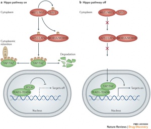
Hippo pathway on-off cartoon
Hippo pathway on-off cartoon[1]

The Hippo (Hpo) pathway, first identified in Drosophila, controls organ size by regulating cell proliferation (inhibition) and apoptosis (induction). In contrast, the TOR signalling pathway regulates organ size by stimulating cell growth, thus increasing cell size.

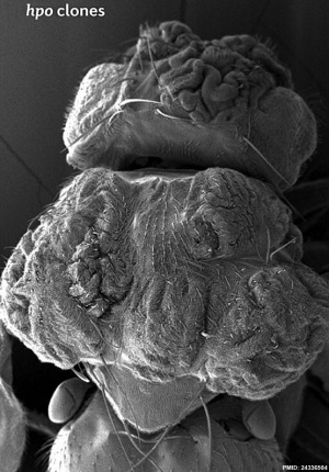
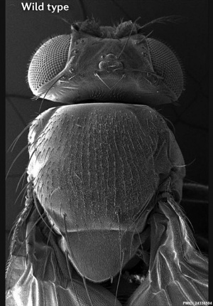
Hippo is a protein kinase cascade pathway, getting its name from the “hippopotamus”-like fly phenotype.


Current research suggests that this signaling pathways is involved with the first "patterning" decision cells make in the blastocyst, controlling trophectoderm vs inner cell mass decision making.

Fly Phenotype (dorsal view head thorax SEM)
Fly Hippo-type dorsal view head thorax SEM.jpg Fly WT dorsal view head thorax SEM.jpg
Hippo-type (hpo) Wild-type (WT)
Image source[1]
Factor Links: AMH | hCG | BMP | sonic hedgehog | bHLH | HOX | FGF | FOX | Hippo | LIM | Nanog | NGF | Nodal | Notch | PAX | retinoic acid | SIX | Slit2/Robo1 | SOX | TBX | TGF-beta | VEGF | WNT | Category:Molecular

| Category:Hippo

Some Recent Findings

  • Role of YAP in early ectodermal specification and a Huntington's Disease model of human neurulation[2] "The Hippo pathway, a highly conserved signaling cascade that functions as an integrator of molecular signals and biophysical states, ultimately impinges upon the transcription coactivator Yes-associated protein 1 (YAP). Hippo-YAP signaling has been shown to play key roles both at the early embryonic stages of implantation and gastrulation, and later during neurogenesis. To explore YAP's potential role in neurulation, we used self-organizing neuruloids grown from human embryonic stem cells on micropatterned substrates. We identified YAP activation as a key lineage determinant, first between neuronal ectoderm and nonneuronal ectoderm, and later between epidermis and neural crest, indicating that YAP activity can enhance the effect of BMP4 stimulation and therefore affect ectodermal specification at this developmental stage. Because aberrant Hippo-YAP signaling has been implicated in the pathology of Huntington's Disease (HD), we used isogenic mutant neuruloids to explore the relationship between signaling and the disease. We found that HD neuruloids demonstrate ectopic activation of gene targets of YAP and that pharmacological reduction of YAP's transcriptional activity can partially rescue the HD phenotype."
  • Deletion of Nf2 in neural crest-derived tongue mesenchyme alters tongue shape and size, Hippo signalling and cell proliferation in a region- and stage-specific manner[3] "The mammalian tongue develops from the branchial arches (1-4) and comprises highly organized tissues compartmentalized by mesenchyme/connective tissue that is largely derived from neural crest (NC). This study aimed to understand the roles of tumour suppressor Neurofibromin 2 (Nf2) in NC-derived tongue mesenchyme in regulating Hippo signalling and cell proliferation for the proper development of tongue shape and size. ...Specifically, at E12.5 Nf2cKO mutants had a decreased level of Hippo signalling transcription factor Yes-associated protein (Yap), Yap target genes and cell proliferation anteriorly, while having an increased Yap, Yap target genes and cell proliferation posteriorly, which lead to a tip-pointed and posteriorly widened tongue. At E15.5, loss of Nf2 in the NC lineage resulted in distinct changes in cell proliferation in different regions, that is, high in epithelium and mesenchyme subjacent to the epithelium, and lower in deeper layers of the mesenchyme. At E18.5, cell proliferation was reduced throughout the Nf2cKO tongue."
  • Pivotal role of the transcriptional co-activator YAP in trophoblast stemness of the developing human placenta PMAS "Defects in development and differentiation of the placenta are associated with various pregnancy disorders such as miscarriage, stillbirth, preeclampsia, and intrauterine growth restriction. However, our knowledge on critical regulators of human placentation is scarce. In the present study, we show that the Hippo signaling-dependent transcriptional coactivator YAP plays a pivotal role in the maintenance of proliferative trophoblasts, the epithelial cells of the placenta. By binding to the transcription factor TEAD4, YAP stimulates expression of genes promoting trophoblast stemness. Additionally, YAP–TEAD4 complexes actively repress genes associated with the differentiated syncytiotrophoblast, the hormone-secreting cell type of the human placenta. Hence, YAP orchestrates a complex developmental program ensuring growth and expansion of the human placenta. we herein show that YAP, the transcriptional coactivator of the Hippo signaling pathway, promotes maintenance of cytotrophoblast progenitors by different genomic mechanisms. Genetic or chemical manipulation of YAP in these cellular models revealed that it stimulates proliferation and expression of cell cycle regulators and stemness-associated genes, but inhibits cell fusion and production of syncytiotrophoblast (STB)-specific proteins, such as hCG and GDF15."
  • Cyclin-Dependent Kinase 7 CDK7 regulates organ size and tumor growth by safeguarding the Hippo pathway effector Yki/Yap/Taz in the nucleus[4] "Hippo signaling controls organ size and tumor progression through a conserved pathway leading to nuclear translocation of the transcriptional effector Yki/Yap/Taz. Most of our understanding of Hippo signaling pertains to its cytoplasmic regulation, but how the pathway is controlled in the nucleus remains poorly understood. Here we uncover an evolutionarily conserved mechanism by which CDK7 promotes Yki/Yap/Taz stabilization in the nucleus to sustain Hippo pathway outputs. We found that a modular E3 ubiquitin ligase complex CRL4DCAF12 binds and targets Yki/Yap/Taz for ubiquitination and degradation, whereas CDK7 phosphorylates Yki/Yap/Taz at S169/S128/S90 to inhibit CRL4DCAF12 recruitment, leading to Yki/Yap/Taz stabilization. As a consequence, inactivation of CDK7 reduced organ size and inhibited tumor growth, which could be reversed by restoring Yki/Yap activity. Our study identifies an unanticipated layer of Hippo pathway regulation, defines a novel mechanism by which CDK7 regulates tissue growth, and implies CDK7 as a drug target for Yap/Taz-driven cancer." OMIM - CDK7


More recent papers  
Mark Hill.jpg
PubMed logo.gif

This table allows an automated computer search of the external PubMed database using the listed "Search term" text link.

  • This search now requires a manual link as the original PubMed extension has been disabled.
  • The displayed list of references do not reflect any editorial selection of material based on content or relevance.
  • References also appear on this list based upon the date of the actual page viewing.


References listed on the rest of the content page and the associated discussion page (listed under the publication year sub-headings) do include some editorial selection based upon both relevance and availability.

More? References | Discussion Page | Journal Searches | 2019 References | 2020 References

Search term: Embryo Hippo | Images | Embryo YAP |

Older papers  
These papers originally appeared in the Some Recent Findings table, but as that list grew in length have now been shuffled down to this collapsible table.

See also the Discussion Page for other references listed by year and References on this current page.

  • Oocyte-expressed yes-associated protein is a key activator of the early zygotic genome in mouse[5] "In early mammalian embryos, the genome is transcriptionally quiescent until the zygotic genome activation (ZGA) which occurs 2-3 days after fertilization. Despite a long-standing effort, maternal transcription factors regulating this crucial developmental event remain largely elusive. Here, using maternal and paternal mouse models of Yap1 deletion, we show that maternally accumulated yes-associated protein (YAP) in oocyte is essential for ZGA. Maternal Yap1-knockout embryos exhibit a prolonged two-cell stage and develop into the four-cell stage at a much slower pace than the wild-type controls. Transcriptome analyses identify YAP target genes in early blastomeres; two of which, Rpl13 and Rrm2, are required to mediate maternal YAP's effect in conferring developmental competence on preimplantation embryos. Furthermore, the physiological YAP activator, lysophosphatidic acid, can substantially improve early development of wild-type, but not maternal Yap1-knockout embryos in both oviduct and culture."
  • Review - Position- and polarity-dependent Hippo signaling regulates cell fates in preimplantation mouse embryos[6] "During the preimplantation stage, mouse embryos establish two cell lineages by the time of early blastocyst formation: the trophectoderm (TE) and the inner cell mass (ICM). Historical models have proposed that the establishment of these two lineages depends on the cell position within the embryo (e.g., the positional model) or cell polarization along the apicobasal axis (e.g., the polarity model). Recent findings have revealed that the Hippo signaling pathway plays a central role in the cell fate-specification process: active and inactive Hippo signaling in the inner and outer cells promote ICM and TE fates, respectively. Intercellular adhesion activates, while apicobasal polarization suppresses Hippo signaling, and a combination of these processes determines the spatially regulated activation of the Hippo pathway in 32-cell-stage embryos. Therefore, there is experimental evidence in favor of both positional and polarity models. At the molecular level, phosphorylation of the Hippo-pathway component angiomotin at adherens junctions (AJs) in the inner (apolar) cells activates the Lats protein kinase and triggers Hippo signaling. In the outer cells, however, cell polarization sequesters Amot from basolateral AJs and suppresses activation of the Hippo pathway. Other mechanisms, including asymmetric cell division and Notch signaling, also play important roles in the regulation of embryonic development. In this review, I discuss how these mechanisms cooperate with the Hippo signaling pathway during cell fate-specification processes." Developmental Signals - Notch | Mouse Development
  • YAP is essential for tissue tension to ensure vertebrate 3D body shape[7] "Vertebrates have a unique 3D body shape in which correct tissue and organ shape and alignment are essential for function. For example, vision requires the lens to be centred in the eye cup which must in turn be correctly positioned in the head. Tissue morphogenesis depends on force generation, force transmission through the tissue, and response of tissues and extracellular matrix to force. Although a century ago D'Arcy Thompson postulated that terrestrial animal body shapes are conditioned by gravity, there has been no animal model directly demonstrating how the aforementioned mechano-morphogenetic processes are coordinated to generate a body shape that withstands gravity. Here we report a unique medaka fish (Oryzias latipes) mutant, hirame (hir), which is sensitive to deformation by gravity. hir embryos display a markedly flattened body caused by mutation of YAP, a nuclear executor of Hippo signalling that regulates organ size. We show that actomyosin-mediated tissue tension is reduced in hir embryos, leading to tissue flattening and tissue misalignment, both of which contribute to body flattening. By analysing YAP function in 3D spheroids of human cells, we identify the Rho GTPase activating protein ARHGAP18 as an effector of YAP in controlling tissue tension. Together, these findings reveal a previously unrecognised function of YAP in regulating tissue shape and alignment required for proper 3D body shape." Medaka Development

Early Development

Zygote

Maternally inherited Yes-associated protein (Yap), a co-activator of TEAD family transcription factors, plays a key role in activating embryonic transcription following fertilization in the mouse. Lysophosphatidic acid (LPA) in the mouse tubal fluid binds to its G-protein coupled receptor at the plasma membrane, and induces the activation of YAP by inhibiting LATS1/2. [5]

Links: Zygote | Mouse Development

Blastocyst

Current research suggests that this signaling pathways is involved with the first "patterning" decision cells make in the blastocyst, controlling trophectoderm vs inner cell mass decision making.


Mouse Blastocyst (32 cell stage) Fate
Inner cells Outer cells
Angiomotin (Amot) phosphorylation at adherens junctions Amot sequestered by cell polarization from basolateral adherens junctions
Hippo active Hippo inactive
Notch inactive Notch active
Cdx2 not expressed Cdx2 expressed
ICM - inner cell mass fate TE - trophectoderm fate

Hippo[6](TEAD4) and Notch[8](Cdx2) together appear regulate early blastocyst fate development.


Links: blastocyst

Trophoblast

Pivotal role of the transcriptional co-activator YAP in trophoblast stemness of the developing human placenta PMAS "Defects in development and differentiation of the placenta are associated with various pregnancy disorders such as miscarriage, stillbirth, preeclampsia, and intrauterine growth restriction. However, our knowledge on critical regulators of human placentation is scarce. In the present study, we show that the Hippo signaling-dependent transcriptional coactivator YAP plays a pivotal role in the maintenance of proliferative trophoblasts, the epithelial cells of the placenta. By binding to the transcription factor TEAD4, YAP stimulates expression of genes promoting trophoblast stemness. Additionally, YAP–TEAD4 complexes actively repress genes associated with the differentiated syncytiotrophoblast, the hormone-secreting cell type of the human placenta. Hence, YAP orchestrates a complex developmental program ensuring growth and expansion of the human placenta. we herein show that YAP, the transcriptional coactivator of the Hippo signaling pathway, promotes maintenance of cytotrophoblast progenitors by different genomic mechanisms. Genetic or chemical manipulation of YAP in these cellular models revealed that it stimulates proliferation and expression of cell cycle regulators and stemness-associated genes, but inhibits cell fusion and production of syncytiotrophoblast (STB)-specific proteins, such as hCG and GDF15."

Links: trophoblast

Bone

Hippo signaling pathway has recently been identified as a key regulator of osteoclast formation, see review.[9]

  • Hippo signaling pathway regulatory molecules - RASSF2, NF2, MST1/2, SAV1, LATS1/2, MOB1, YAP, and TAZ.
  • osteoclast differentiation - upon activation, MST and LAST, transcriptional co-activators YAP and TAZ bind to the members of the TEA domain (TEAD) family transcription factors
    • regulate expression of downstream target genes connective tissue growth factor (CTGF/CCN2) and cysteine-rich protein 61 (CYR61/CCN1).
  • RANKL-mediated signaling cascades including NF-κB, MAPKs, AP1, and NFATc1, Hippo-signaling molecules such as YAP/TAZ/TEAD complex, RASSF2, MST2, and Ajuba could also potentially modulate osteoclast differentiation and function.


Links: bone

Neural

During early neural development radial glia guide the migration of differentiating neural cells to the developing cortical plate (cortex). A study has shown that elimination of CEP83, in radial glial progenitor cells, activates the mechanically sensitive yes-associated protein (YAP) and promotes the excessive proliferation of these progenitor cells.

Links: radial glia | cortex

References

  1. 1.0 1.1 Johnson R & Halder G. (2014). The two faces of Hippo: targeting the Hippo pathway for regenerative medicine and cancer treatment. Nat Rev Drug Discov , 13, 63-79. PMID: 24336504 DOI.
  2. Piccolo FM, Kastan NR, Haremaki T, Tian Q, Laundos TL, De Santis R, Beaudoin AJ, Carroll TS, Luo JD, Gnedeva K, Etoc F, Hudspeth AJ & Brivanlou AH. (2022). Role of YAP in early ectodermal specification and a Huntington's Disease model of human neurulation. Elife , 11, . PMID: 35451959 DOI.
  3. Ishan M, Chen G, Yu W, Wang Z, Giovannini M, Cao X & Liu HX. (2021). Deletion of Nf2 in neural crest-derived tongue mesenchyme alters tongue shape and size, Hippo signalling and cell proliferation in a region- and stage-specific manner. Cell Prolif , 54, e13144. PMID: 34697858 DOI.
  4. Cho YS, Li S, Wang X, Zhu J, Zhuo S, Han Y, Yue T, Yang Y & Jiang J. (2020). CDK7 regulates organ size and tumor growth by safeguarding the Hippo pathway effector Yki/Yap/Taz in the nucleus. Genes Dev. , 34, 53-71. PMID: 31857346 DOI.
  5. 5.0 5.1 Yu C, Ji SY, Dang YJ, Sha QQ, Yuan YF, Zhou JJ, Yan LY, Qiao J, Tang F & Fan HY. (2016). Oocyte-expressed yes-associated protein is a key activator of the early zygotic genome in mouse. Cell Res. , 26, 275-87. PMID: 26902285 DOI.
  6. 6.0 6.1 Sasaki H. (2015). Position- and polarity-dependent Hippo signaling regulates cell fates in preimplantation mouse embryos. Semin. Cell Dev. Biol. , 47-48, 80-7. PMID: 25986053 DOI.
  7. Porazinski S, Wang H, Asaoka Y, Behrndt M, Miyamoto T, Morita H, Hata S, Sasaki T, Krens SFG, Osada Y, Asaka S, Momoi A, Linton S, Miesfeld JB, Link BA, Senga T, Shimizu N, Nagase H, Matsuura S, Bagby S, Kondoh H, Nishina H, Heisenberg CP & Furutani-Seiki M. (2015). YAP is essential for tissue tension to ensure vertebrate 3D body shape. Nature , 521, 217-221. PMID: 25778702 DOI.
  8. Rayon T, Menchero S, Nieto A, Xenopoulos P, Crespo M, Cockburn K, Cañon S, Sasaki H, Hadjantonakis AK, de la Pompa JL, Rossant J & Manzanares M. (2014). Notch and hippo converge on Cdx2 to specify the trophectoderm lineage in the mouse blastocyst. Dev. Cell , 30, 410-22. PMID: 25127056 DOI.
  9. Yang W, Han W, Qin A, Wang Z, Xu J & Qian Y. (2018). The emerging role of Hippo signaling pathway in regulating osteoclast formation. J. Cell. Physiol. , 233, 4606-4617. PMID: 29219182 DOI.

Reviews

Irvine KD & Harvey KF. (2015). Control of organ growth by patterning and hippo signaling in Drosophila. Cold Spring Harb Perspect Biol , 7, . PMID: 26032720 DOI.

Tumaneng K, Russell RC & Guan KL. (2012). Organ size control by Hippo and TOR pathways. Curr. Biol. , 22, R368-79. PMID: 22575479 DOI.

Zhao B, Tumaneng K & Guan KL. (2011). The Hippo pathway in organ size control, tissue regeneration and stem cell self-renewal. Nat. Cell Biol. , 13, 877-83. PMID: 21808241 DOI.

Kango-Singh M & Singh A. (2009). Regulation of organ size: insights from the Drosophila Hippo signaling pathway. Dev. Dyn. , 238, 1627-37. PMID: 19517570 DOI.

Articles

Bessonnard S, Mesnard D & Constam DB. (2015). PC7 and the related proteases Furin and Pace4 regulate E-cadherin function during blastocyst formation. J. Cell Biol. , 210, 1185-97. PMID: 26416966 DOI.

Yuan H, Liu H, Liu Z, Zhu D, Amos CI, Fang S, Lee JE & Wei Q. (2015). Genetic variants in Hippo pathway genes YAP1, TEAD1 and TEAD4 are associated with melanoma-specific survival. Int. J. Cancer , 137, 638-45. PMID: 25628125 DOI.

Clattenburg L, Wigerius M, Qi J, Rainey JK, Rourke JL, Muruganandan S, Sinal CJ & Fawcett JP. (2015). NOS1AP Functionally Associates with YAP To Regulate Hippo Signaling. Mol. Cell. Biol. , 35, 2265-77. PMID: 25918243 DOI.


Search Pubmed

Search Bookshelf Hippo


Search Pubmed Now: Hippo


Glossary Links

Glossary: A | B | C | D | E | F | G | H | I | J | K | L | M | N | O | P | Q | R | S | T | U | V | W | X | Y | Z | Numbers | Symbols | Term Link


Cite this page: Hill, M.A. (2024, March 19) Embryology Developmental Signals - Hippo. Retrieved from https://embryology.med.unsw.edu.au/embryology/index.php/Developmental_Signals_-_Hippo

What Links Here?
© Dr Mark Hill 2024, UNSW Embryology ISBN: 978 0 7334 2609 4 - UNSW CRICOS Provider Code No. 00098G